

The virtual cone: A novel technique to generate spherical dose distributions using a multileaf collimator and standardized control-point sequence for small target radiation surgery
- PMID: 30197943
- PMCID: PMC6127970
- DOI: 10.1016/j.adro.2018.02.011
The virtual cone: A novel technique to generate spherical dose distributions using a multileaf collimator and standardized control-point sequence for small target radiation surgery
Abstract
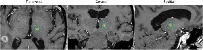
Purpose: The study aimed to develop and demonstrate a standardized linear accelerator multileaf collimator-based method of delivering small, spherical dose distributions suitable for radiosurgical treatment of small targets such as the trigeminal nerve.
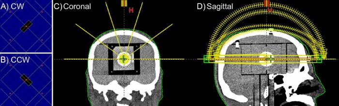
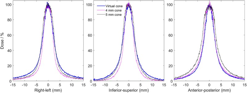
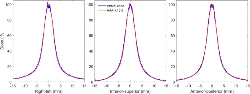
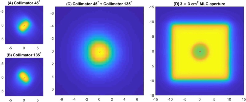
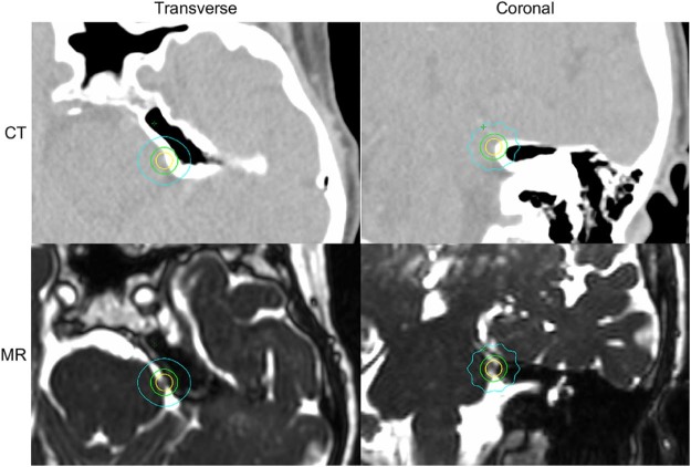
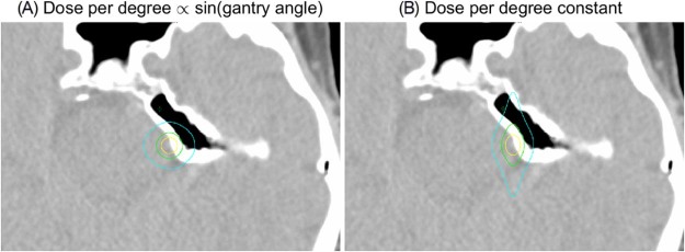
Methods and materials: The virtual cone is composed of a multileaf collimator-defined field with the central 2 leaves set to a small gap. For 5 table positions, clockwise and counter-clockwise arcs were used with collimator angles of 45 and 135 degrees, respectively. The dose per degree was proportional to the sine of the gantry angle. The dose distribution was calculated by the treatment planning system and measured using radiochromic film in a skull phantom for leaf gaps of 1.6, 2.1, and 2.6 mm. Cones with a diameter of 4 mm and 5 mm were measured for comparison. Output factor constancy was investigated using a parallel-plate chamber.
Results: The mean ratio of the measured-to-calculated dose was 0.99, 1.03, and 1.05 for 1.6, 2.1, and 2.6 mm leaf gaps, respectively. The diameter of the measured (calculated) 50% isodose line was 4.9 (4.6) mm, 5.2 (5.1) mm, and 5.5 (5.5) mm for the 1.6, 2.1, and 2.6 mm leaf gap, respectively. The measured diameter of the 50% isodose line was 4.5 and 5.7 mm for the 4 mm and 5 mm cones, respectively. The standard deviation of the parallel-plate chamber signal relative to a 10 cm × 10 cm field was less than 0.4%. The relative signal changed 32% per millimeter change in leaf gap, indicating that the parallel-plate chamber is sensitive to changes in gap width.
Conclusions: The virtual cone is an efficient technique for treatment of small spherical targets. Patient-specific quality assurance measurements will not be necessary in routine clinical use. Integration directly into the treatment planning system will make planning using this technique extremely efficient.
Figures
References
-
- Lutz W., Winston K.R., Maleki N. A system for stereotactic radiosurgery with a linear accelerator. Int J Radiat Oncol Biol Phys. 1988;14:373–381. - PubMed
-
- Pike G.B., Podgorsak E.B., Peters T.M., Pla C., Olivier A., Souhami L. Dose distributions in radiosurgery. Med Phys. 1990;17:296–304. - PubMed
-
- Schlegel W., Pastyr O., Bortfeld T. Computer systems and mechanical tools for stereotactically guided conformation therapy with linear accelerators. Int J Radiat Oncol Biol Phys. 1992;24:781–787. - PubMed
-
- Bourland J.D., McCollough K.P. Static field conformal stereotactic radiosurgery: Physical techniques. Int J Radiat Oncol Biol Phys. 1994;28:471–479. - PubMed
-
- Shiu A.S., Kooy H.M., Ewton J.R. Comparison of miniature multileaf collimation (MMLC) with circular collimation for stereotactic treatment. Int J Radiat Oncol Biol Phys. 1997;37:679–688. - PubMed
LinkOut - more resources
Full Text Sources
Other Literature Sources
Miscellaneous
