. 2018 Dec 1;526(17):2845-2855.
doi: 10.1002/cne.24530. Epub 2018 Nov 8.

Alterations in synaptic density and myelination in response to exposure to high-energy charged particles

Affiliations

Alterations in synaptic density and myelination in response to exposure to high-energy charged particles

Dara L Dickstein et al. J Comp Neurol. .

Abstract

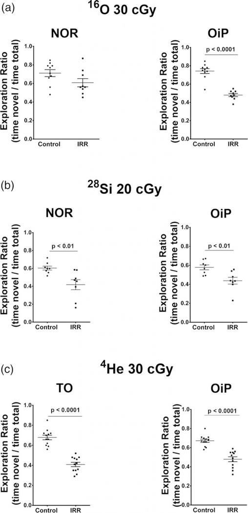
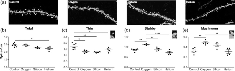
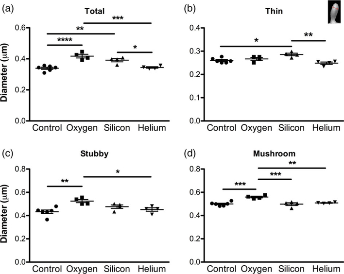
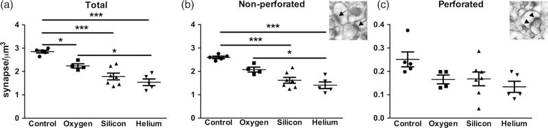
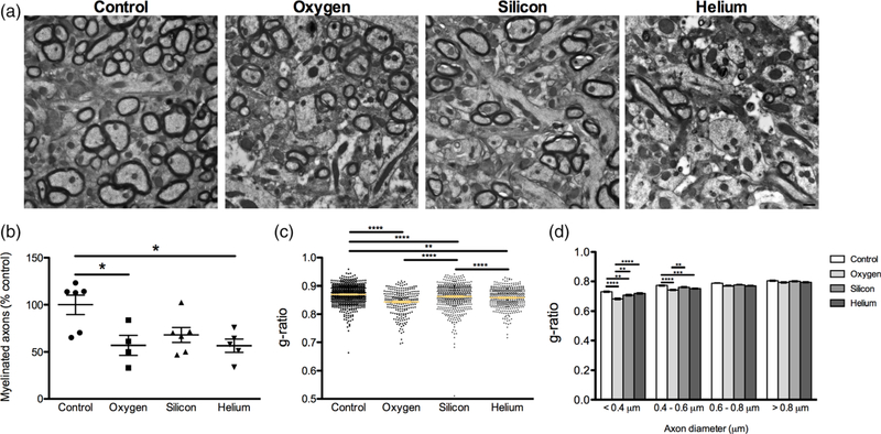
High-energy charged particles are considered particularly hazardous components of the space radiation environment. Such particles include fully ionized energetic nuclei of helium, silicon, and oxygen, among others. Exposure to charged particles causes reactive oxygen species production, which has been shown to result in neuronal dysfunction and myelin degeneration. Here we demonstrate that mice exposed to high-energy charged particles exhibited alterations in dendritic spine density in the hippocampus, with a significant decrease of thin spines in mice exposed to helium, oxygen, and silicon, compared to sham-irradiated controls. Electron microscopy confirmed these findings and revealed a significant decrease in overall synapse density and in nonperforated synapse density, with helium and silicon exhibiting more detrimental effects than oxygen. Degeneration of myelin was also evident in exposed mice with significant changes in the percentage of myelinated axons and g-ratios. Our data demonstrate that exposure to all types of high-energy charged particles have a detrimental effect, with helium and silicon having more synaptotoxic effects than oxygen. These results have important implications for the integrity of the central nervous system and the cognitive health of astronauts after prolonged periods of space exploration.

Keywords: charged particles; dendritic spines; myelin; synapses.


Conflict of interest statement

CONFLICT OF INTEREST

The authors declare they have no conflict of interest.

Figures

FIGURE 1
FIGURE 1
Schematic depicting the unbiased stereological approach taken to quantify synapses. (a) EM blocks were cut encompassing mouse CA1 SR region (black box), (b) serial sections were collected and imaged between 150 and 250 μm from the cell bodies in the pyramidal layer of CA1 (white dashed lines). (c) Example of randomized imaging protocol using physical disector approach. (d) Serial electron micrographs depicting axospinous synapses counted using the disector method. Only synapses that are present in the reference panel (green and red), but not in the look-up panel was counted. Synapses present in both panels (blue) were not included in the analysis. Scale bars: a 500 μm; b 10 μm; c 10 μm; d 500 nm
FIGURE 2
FIGURE 2
Cognitive data adapted from previously published work reveals that similar doses of the ions used in the present study elicit significant neurobehavioral deficits 6 weeks following exposure. (a) Novel object recognition (NOR) and object in place (OiP) tasks reveal significant deficits on the OiP task after exposure to energetic 16O ions (Adapted from Parihar et al., 2015a). (b) Animals subjected to similar tasks following exposure to a slightly lower dose of 28Si ions exhibit significant deficits in NOR and OIP memory (Adapted from Acharya et al., 2017). (c) Animals exposed to the 4He ions and subjected to the temporal order (TO) and OiP tasks show significant impairments in each of these tasks (Adapted from Parihar et al., 2018)
FIGURE 3
FIGURE 3
Charged particle irradiation results in altered spine density. Analysis of spine density in CA1 apical dendritic spines revealed significant changes in spine densities on apical dendrites from CA1 pyramidal neurons. (a) Representative dendritic images from all treatment groups. Scale bar = 2 mm. (b) Average total spine density, (c) thin spine density, (d) stubby spine density and (e) mushroom spine density. Inset images in c, d and e depict each spine type. Data represent means ± SEM. One-way ANOVA *p < 0.01, **p < 0.001, ****p < 0.0001
FIGURE 4
FIGURE 4
Charged particle irradiation results in changes in spine morphology. Analysis of spine head diameter in CA1 apical dendritic spines. (a) Average total spine head diameter, (b) thin spine head diameter, (c) stubby spine head diameter and (d) mushroom spine head diameter. Inset image in b shows how spine head diameter was assessed. Data represent means ± SEM. One-way ANOVA *p < 0.05, **p < 0.01, ***p < 0.001, ****p < 0.0001 [Color figure can be viewed at wileyonlinelibrary.com]
FIGURE 5
FIGURE 5
Charged particle irradiation results in a decrease in synapse density. There is a significant decrease in total (a) and nonperforated (b) synapse density in response to irradiation with 16O, 28Si, and 4He. No significant differences were observed in the density of perforated (c) synapses. Inset images depict nonperforated synapses (arrows), and perforated synapses (arrowheads). Data represent group means ± SEM, with each data point representing the average measurements from at least nine series images from a mouse (n = 4–7 mice/group). One-way ANOVA *p < 0.05, ***p < 0.0001
FIGURE 6
FIGURE 6
Alterations in myelination in response to charged particle irradiation. (a) Representative myelin images from all treatment groups. Scale bar = 500 nm. (b) There was a significant decrease in the percentage of myelinated axons in 16O, 28Si, and 4He irradiated animals compared to controls. Data represents group means ± SEM, with each data point representing the average measurements from 12 images/mouse (n = 4–7 mice/group). (c) g-ratios are lower in irradiated mice compared to controls (d) g-ratio (axon diameter/fiber diameter) of fibers grouped by axon diameter. Data represents individual measurements ± SEM, from at least six images/mouse (n = 4–7 mice/group). One-way ANOVA *p < 0.01, **p < 0.001, ***p < 0.0001 [Color figure can be viewed at wileyonlinelibrary.com]

Similar articles

Cited by

References

    1. Acharya MM, Baddour AA, Kawashita T, Allen BD, Syage AR, Nguyen TH, … Baulch JE (2017). Epigenetic determinants of space radiation-induced cognitive dysfunction. Scientific Reports, 7, 42885 10.1038/srep42885 - DOI - PMC - PubMed
    1. Allen AR, Raber J, Chakraborti A, Sharma S, & Fike JR (2015). (56) Fe irradiation alters spine density and dendritic complexity in the mouse hippocampus. Radiation Research, 184(6), 586–594. 10.1667/RR14103.1 - DOI - PubMed
    1. Attardo A, Fitzgerald JE, & Schnitzer MJ (2015). Impermanence of dendritic spines in live adult CA1 hippocampus. Nature, 523(7562), 592–596. 10.1038/nature14467 - DOI - PMC - PubMed
    1. Barker GR, Bird F, Alexander V, & Warburton EC (2007). Recognition memory for objects, place, and temporal order: A disconnection analysis of the role of the medial prefrontal cortex and perirhinal cortex. The Journal of Neuroscience, 27(11), 2948–2957. 10.1523/JNEUROSCI.5289-06.2007 - DOI - PMC - PubMed
    1. Barker GR, & Warburton EC (2011). When is the hippocampus involved in recognition memory? The Journal of Neuroscience, 31(29), 10721–10731. 10.1523/JNEUROSCI.6413-10.2011 - DOI - PMC - PubMed

Publication types