. 2019 Feb;67(2):85-97.
doi: 10.1369/0022155418798807. Epub 2018 Sep 10.

Refined Immunochemical Characterization in Healthy Dog Skin of the Epidermal Cornification Proteins, Filaggrin, and Corneodesmosin

Affiliations

Refined Immunochemical Characterization in Healthy Dog Skin of the Epidermal Cornification Proteins, Filaggrin, and Corneodesmosin

Didier Pin et al. J Histochem Cytochem. 2019 Feb.

Abstract

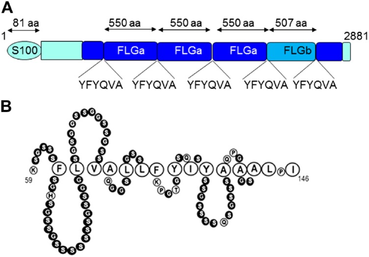
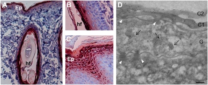
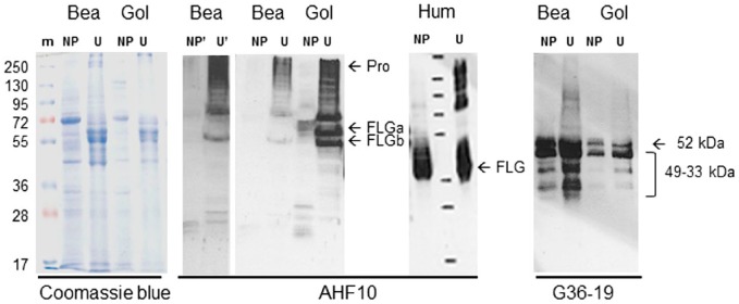
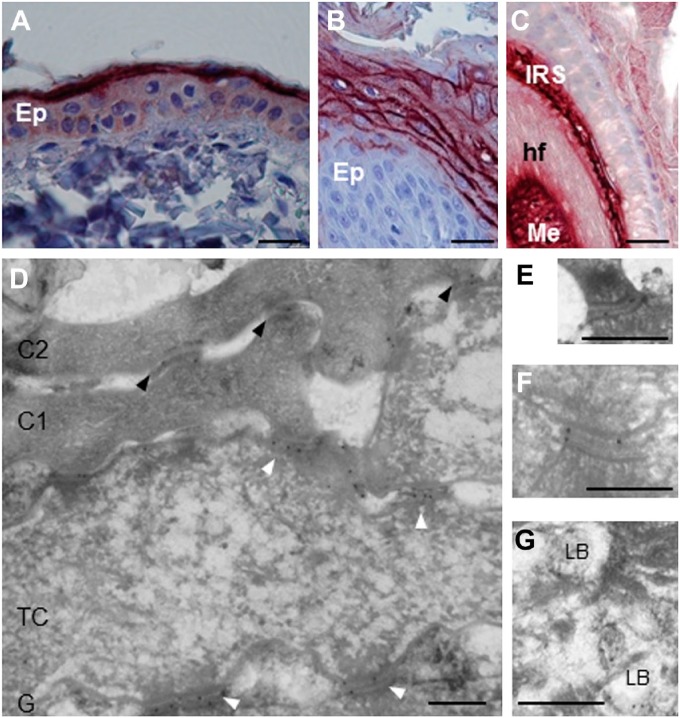
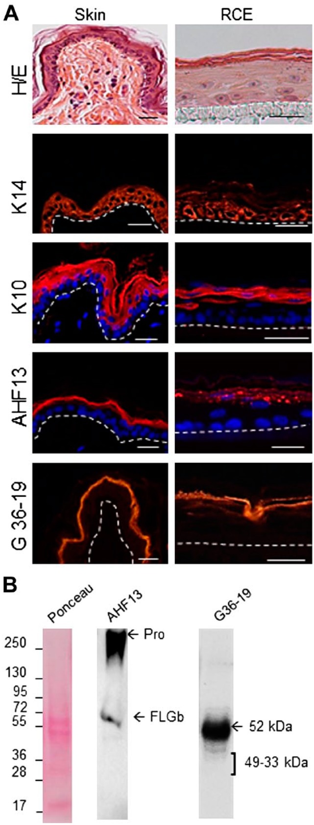
Filaggrin (FLG) and corneodesmosin (CDSN) are two key proteins of the human epidermis. FLG loss-of-function mutations are the strongest genetic risk factors for human atopic dermatitis. Studies of the epidermal distribution of canine FLG and CDSN are limited. Our aim was to better characterize the distribution of FLG and CDSN in canine skin. Using immunohistochemistry on beagle skin, we screened a series of monoclonal antibodies (mAbs) specific for human FLG and CDSN. The cross-reactive mAbs were further used using immunoelectron microscopy and Western blotting. The structure of canine CDSN and FLG was determined using publicly available databases. In the epidermis, four anti-FLG mAbs stained keratohyalin granules in the granular keratinocytes and corneocyte matrix of the lower cornified layer. In urea-extracts of dog epidermis, several bands corresponding to proFLG and FLG monomers were detected. One anti-CDSN mAb stained the cytoplasm of granular keratinocytes and cells of both the inner root sheath and medulla of hair follicles. Dog CDSN was located in lamellar bodies, in the extracellular parts of desmosomes and in corneodesmosomes. A protein of 52 kDa was immunodetected. Genomic DNA analysis revealed that the amino acid sequence and structure of canine and human CDSN were highly similar.

Keywords: atopic dermatitis; differentiation; keratinocyte; skin; veterinary medicine.


Conflict of interest statement

Competing Interests: The author(s) declared the following potential conflicts of interest with respect to the research, authorship, and/or publication of this article: V.P. and N.A. are employed by Synelvia, a private company commercially using reconstructed canine epidermis. M.S. academic research group collaborated with and received grants from Synelvia. The other authors declare that they have no conflict of interest.

Figures

Figure 1.
Figure 1.
Schematic representation of dog profilaggrin (A) and of a tentative structure of dog corneodesmosin region 59–143 (B). Serine and glycine amino acids (in black) are predicted to form the so-called glycine-loops, due to the interactions of the interspersed aromatic and aliphatic residues (enlarged). Abbreviation: FLG, Filaggrin.
Figure 2.
Figure 2.
Localization of filaggrin in the skin of healthy dogs. (A) The mAbs of the AHF family were analyzed by immunohistochemistry on sections of dog skin, from back and paw pad. AHF10, 13, 23, and 27 displayed the same pattern of reactivity, but only the data for AHF10 are shown. (A–C) AHF10 stains the upper interfollicular epidermis (A) and the intrafollicular epidermis (B) of the back skin, and the upper epidermis of paw pad (C). (D) The reactivity of AHF10 was further analyzed using immunoelectron microscopy. The mAb labels the first two layers of corneocytes (C1 and C2) and the keratohyalin granules (arrows) in a granular (G) keratinocyte. The spinous (S) keratinocytes as well as desmosomes (white arrowheads) are not labeled. Scale bar = 30 µm (A and C), 25 µm (B) and 0.25 µm (D). Abbreviations: AHF, anti-human filaggrin; Ep, epidermis; hf, hair follicle.
Figure 3.
Figure 3.
Western blotting of dog epidermis extracts. Beagle (Bea) and golden (Gol) retriever dog epidermis was sequentially extracted in Tris-EDTA buffer containing either Nonidet-P40 (TE-NP40 extracts, NP) or 8 M urea (TE-U extracts, U). The extracted proteins were separated by SDS-PAGE, Coomassie blue stained, or immunodetected with AHF10 and G36-19 mAbs, as indicated. The immunoblot corresponding to AHF10 reactivity on the extracts of the beagle dog epidermis was exposed for a longer time (NP′ and U′) to highlight the absence of FLGa-corresponding band. The migration of molecular mass markers (m) is indicated on the left in kDa. ProFLG (Pro) and FLG monomers (FLGa and FLGb) are indicated by arrows, as well as the entire CDSN and its proteolytically derived fragments. For comparison, the Western blotting with AHF10 of TE-NP40 and TE-U extracts of human epidermis (Hum) is shown. Abbreviations: AHF, anti-human filaggrin; PAGE, polyacrylamide gel electrophoresis; FLG, Filaggrin; CDSN, corneodesmosin.
Figure 4.
Figure 4.
Localization of corneodesmosin in the skin of healthy dogs. (A–C) Using immunohistochemistry, G36-19 mAb stains the upper interfollicular epidermis (Ep) of back skin (A), the upper epidermis of paw pad (B) and both the inner root sheath (IRS) and medulla (Me) of hair follicles (hf, C). (D–G) G36-19 was further used in immunoelectron microscopy. (D) The reactivity is localized in the extracellular part of corneodesmosomes (black arrowheads) in the lower cornified layer, and of desmosomes (white arrowheads) in transitional cells (TC) and granular keratinocytes (G). The first two layers of corneocytes (C1 and C2) are shown. (E, F) Enlargements of desmosomes in transitional and granular cells. (G) lamellar bodies (LB) of granular cells are also labeled. Scale bar = 30 µm (A–C), 0.3 µm (D–G).
Figure 5.
Figure 5.
Characterization of reconstructed canine epidermis. (A) Sections of fixed samples of normal dog skin and reconstructed canine epidermis were stained with Hematoxylin and eosin (H/E) and antibodies directed to keratin K14, K10, FLG, and CDSN, as indicated. The dermo-epidermal junction and the polycarbonate membrane/epidermal junction are indicated by a dotted line. Scale bars = 25 µm. In some cases nuclei are labeled with 4′,6′-diamidino-2-phénylindole (Sigma-Aldrich) (blue). (B) Total proteins were extracted from reconstructed canine epidermis, separated by SDS-PAGE, transferred to membranes and Ponceau Red-stained or immunodetected with AHF13 and G36-19 mAbs. The migration of molecular mass markers is indicated on the left in kDa. ProFLG (Pro) and FLGb monomers are indicated by arrows, as well as the entire CDSN and CDSN proteolytically derived fragments. Abbreviation: AHF, anti-human filaggrin; FLG, Filaggrin; CDSN, corneodesmosin; PAGE, polyacrylamide gel electrophoresis.

References

    1. Kezic S, Novak N, Jakasa I, Jungersted JM, Simon M, Brandner JM, Middelkamp-Hup MA, Weidinger S. Skin barrier in atopic dermatitis. Front Biosci. 2014;19:542–56. - PubMed
    1. Palmer CN, Irvine AD, Terron-Kwiatkowski A, Zhao Y, Liao H, Lee SP, Goudie DR, Sandilands A, Campbell LE, Smith FJ, O’Regan GM, Watson RM, Cecil JE, Bale SJ, Compton JG, DiGiovanna JJ, Fleckman P, Lewis-Jones S, Arseculeratne G, Sergeant A, Munro CS, El Houate B, McElreavey K, Halkjaer LB, Bisgaard H, Mukhopadhyay S, McLean WH. Common loss-of-function variants of the epidermal barrier protein filaggrin are a major predisposing factor for atopic dermatitis. Nat Genet. 2006;38:441–6. - PubMed
    1. Le Lamer M, Pellerin L, Reynier M, Cau L, Pendaries V, Leprince C, Méchin MC, Serre G, Paul C, Simon M. Defects of corneocyte structural proteins and epidermal barrier in atopic dermatitis. Biol Chem. 2015;96:1163–79. - PubMed
    1. Oyoshi MK, He R, Kumar L, Yoon J, Geha RS. Cellular and molecular mechanisms in atopic dermatitis. Adv Immunol. 2009;102:135–226. - PubMed
    1. Marsella R, De Benedetto A. Atopic dermatitis in animals and people: an update and comparative review. Vet Sci. 2017;4:37. - PMC - PubMed

Publication types

LinkOut - more resources