. 2018 Nov:524:160-171.
doi: 10.1016/j.virol.2018.08.010. Epub 2018 Sep 7.

Description and initial characterization of metatranscriptomic nidovirus-like genomes from the proposed new family Abyssoviridae, and from a sister group to the Coronavirinae, the proposed genus Alphaletovirus

Affiliations

Description and initial characterization of metatranscriptomic nidovirus-like genomes from the proposed new family Abyssoviridae, and from a sister group to the Coronavirinae, the proposed genus Alphaletovirus

Khulud Bukhari et al. Virology. 2018 Nov.

Abstract

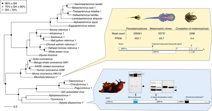
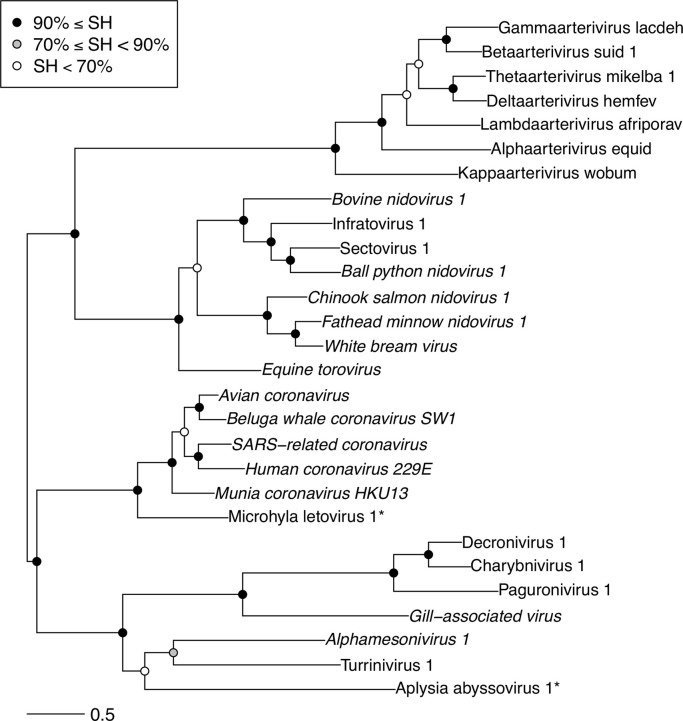
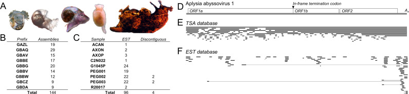
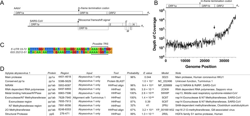
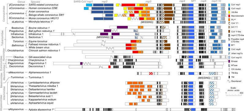
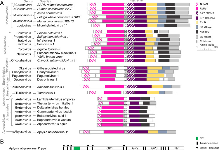
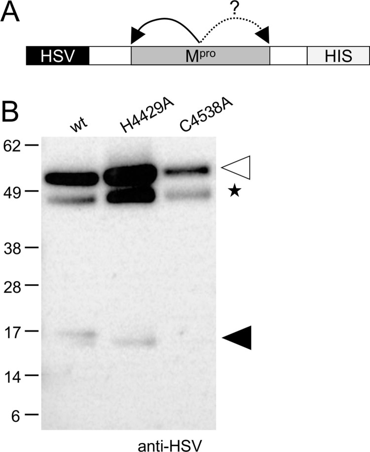
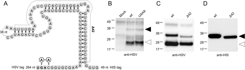
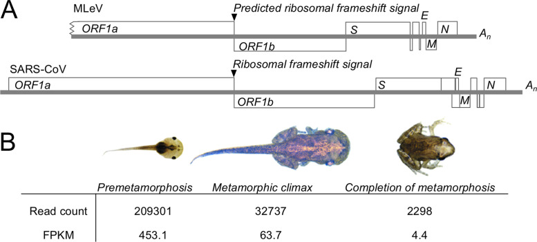
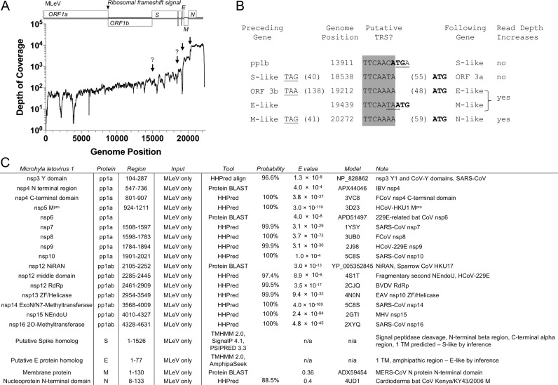
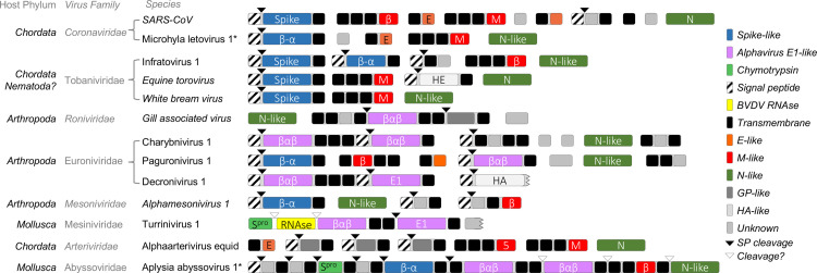
Transcriptomics has the potential to discover new RNA virus genomes by sequencing total intracellular RNA pools. In this study, we have searched publicly available transcriptomes for sequences similar to viruses of the Nidovirales order. We report two potential nidovirus genomes, a highly divergent 35.9 kb likely complete genome from the California sea hare Aplysia californica, which we assign to a nidovirus named Aplysia abyssovirus 1 (AAbV), and a coronavirus-like 22.3 kb partial genome from the ornamented pygmy frog Microhyla fissipes, which we assign to a nidovirus named Microhyla alphaletovirus 1 (MLeV). AAbV was shown to encode a functional main proteinase, and a translational readthrough signal. Phylogenetic analysis suggested that AAbV represents a new family, proposed here as Abyssoviridae. MLeV represents a sister group to the other known coronaviruses. The importance of MLeV and AAbV for understanding nidovirus evolution, and the origin of terrestrial nidoviruses are discussed.

Keywords: Nidovirales; Protease; Protein expression; Proteinase; Readthrough; Transcriptome; Translation; Virus discovery.


Figures

fx1
Graphical abstract
Fig. 1
Fig. 1
Nidovirus phylogeny reconstructed based on concatenated MSA of five replicative domains universally conserved in nidoviruses. SH-aLRT branch support values are depicted by shaded circles. Species names that are not currently recognized by ICTV are written in plain font. Asterisks designate viruses described in this study.
Fig. 2
Fig. 2
Sequence coverage of AAbV in public NCBI libraries. (A) Examples of the host organism Aplysia californica at swimming veliger, settled, metamorphic, juvenile and adult developmental stages (images not to scale, adapted from Heyland et al. (2011) and Moroz et al. (2006)). Summary of distinct sequence assemblies and reads in the TSA (B) and EST (C) matching AAbV for which the nucleotide BLAST E value was 2 × 10–70 or smaller. (D) Map of AAbV, showing the location of the replicase polyprotein genes (ORF1a, ORF1b), structural polyprotein gene (ORF2) and poly-adenosine tail (An). The position of sequences from the TSA (E) and EST (F) databases matching AAbV is shown.
Fig. 3
Fig. 3
Coding capacity, depth of coverage and bioinformatics of AAbV. (A) Genome and coding capacity of AAbV and SARS-CoV are shown to scale. (B) Total depth of coverage based on a sample of 672017 aligned spots matching AAbV from Aplysia californica RNA sequence read archives including SRR385787, SRR385788, SRR385792, SRR385793, SRR385795, SRR385800, SRR385802 and SRR385815. The putative start site of a viral subgenomic RNA species is marked with an arrow. (C) Alignment of the 5′-untranslated region and the intergenic sequence between the pp1b and pp2 genes showing a potential transcription-regulatory sequence (boxed). (D) Bioinformatic assignment of domains in AAbV. Sequence(s) used for prediction (Input) were either AAbV alone or a multiple sequence alignment containing AAbV and TurrNV. Probability score from HHPred and E value from HHPred or BLAST are shown. Accession numbers are given for sequences or protein structures identified as a match for an AAbV domain (Model).
Fig. 4
Fig. 4
Comparison of predicted domain-level organization in polyprotein 1a of new viruses to previously described nidoviruses. Gaps have been introduced so to align predicted homologous domains. Virus naming and taxonomy conventions follow the ICTV proposals in which MLeV and AAbV were first described (Gorbalenya et al., 2017b, Gorbalenya et al., 2017a, Ziebuhr et al., 2017). New viruses are marked with stars, accepted taxonomic ranks are italicized and proposed taxonomic ranks are not italicized. Polyprotein processing products from SARS-CoV are shown at top. Domains are colored to indicate predicted similarity to SARS-CoV nsp1 (CoV nsp1), SARS-CoV nsp2 (nsp2-like), ubiquitin (Ub-like), macrodomains, papain-like proteinase (PLpro), first section of the coronavirus Y domain (CoV Y1), first section of the arterivirus Y domain (ArV Y1) coronavirus-specific Y domain-like (CoV Y-like), carboxyl-terminal domain of coronavirus nsp4 (nsp4 CTD-like), region with PSIPRED predicted structural similarity to nsp4 CTD, main proteinase (Mpro), SARS-CoV nsp8-like (CoV nsp8), Equine arteritis virus nsp7α (ArV nsp7α), SARS-CoV nsp10 (CoV nsp10), protein kinase-like (Kinase), RNA methyltransferase (Mtase), potential metal ion-binding clusters with 4 cysteine or histidine residues in a 20 amino acid window (CH-cluster), transmembrane helices, hydrophobic transmembrane-like regions that may not span the membrane by analogy to coronavirus nsp4 and nsp6 (TM-like) and disordered regions (Unstructured).
Fig. 5
Fig. 5
Comparison of predicted domain-level organization in polyprotein 1b of new viruses to previously described nidoviruses. (A) Domains include the nidovirus RdRp-associated nucleotidyl transferase (NiRAN), RdRp, potential metal ion binding clusters with four cysteine or histidine residues in a window of 20 amino acids (CH cluster), homologs of the domain of unknown function in the middle of coronavirus nsp13 (CoV nsp13b), superfamily 1 helicase (SF1 Helicase), nidovirus-specific exonuclease (ExoN) and uridylate-specific endonuclease (NEndoU), RNA cap N7 methyltransferase (N7 MTase) and RNA cap 2′-O-methyltransferase (2O MTase). (B) Domains of pp2 include the structural protease (Spro), putative glycoproteins GP1, GP2 and GP3, and a nucleoprotein-like domain (N?), TMHMM-predicted transmembrane domains and SignalP-predicted signal peptidase cleavage sites.
Fig. 6
Fig. 6
Investigation of proteinase activity of AAbV Mpro. The AAbV main proteinase (Mpro; A-B) and surrounding regions were expressed as HSV and HIS-tagged constructs as shown in panel A. A white triangle marks the expected size of the 52.5 kDa uncleaved Mpro constructs. Black triangles mark the size of approximately 16 kDa amino-terminal cleavage products. Non-specific bands that were also present in control lanes are indicated with a star.
Fig. 7
Fig. 7
Mutational analysis of the termination-suppression signal (TSS) at the ORF1a/b junction. (A) Schematic view of the TSS expression construct and introduced HSV and HIS tags, showing only predicted RNA secondary structures that were consistent in the best six models generated by Mfold. Mutations around the stop codon (bold, producing the UAAA construct) or removing one side of the predicted stem-loops (Δ42) are shown. (B-D) Western blots showing translation of mutant TSS expression constructs in a coupled T7 polymerase rabbit reticulocyte lysate expression system. Blots were probed with anti-HSV (B, D) to detect both 25 kDa terminated and 32–35 kDa readthrough products, or with anti-HIS (C) to detect only readthrough products.
Fig. 8
Fig. 8
Coding capacity and prevalence of MLeV (A) Schematic representation of the coding capacity of MLeV compared to SARS-CoV, showing the similarities in genome organization. (B) Prevalence of MLeV transcripts in Microhyla fissipes by age, by total number of reads and fragments per kilobase of transcript per million mapped reads (FPKM).
Fig. 9
Fig. 9
Depth of coverage and bioinformatics of MLeV. (A) Total depth of coverage is based on 275503 aligned spots matching MLeV from Microhyla fissipes RNA sequence read archives SRR2418812, SRR2418623 and SRR2418554. The putative start sites of a viral subgenomic RNA species are marked with an arrow. Potential subgenomic RNA start sites not marked by a sharp rise in read depth are indicated with question marks. (B) Positions and usage of putative transcription-regulatory sequences. Termination codons from the preceding gene are underlined, initiation codons of the following gene are in bold. (C) Bioinformatic assignment of domains in MLeV.
Fig. 10
Fig. 10
Speculative annotation of nidovirus structural proteins. Where structures or functions were not known, proteins were categorized according to general PSIPRED secondary structure profile. Marked domains include coronavirus spike protein homologs (Spike) and structurally similar regions (β-α), alphavirus E1 homologs (E1) and structurally similar regions (βαβ), coronavirus envelope-like proteins (E-like), coronavirus membrane proteins (M-like) and structurally similar proteins (β), potential nucleoprotein (N-like), chymotrypsin-like structural proteinase (Spro), similar to the bovine viral diarrhea virus structural RNAse (BVDV RNAse), proteins related to influenza A virus hemagglutinin (HA) or torovirus hemagglutinin-esterase (HE), other viral surface glycoproteins (GP-like), domains of no known function (Unknown), SignalP-predicted signal peptidase cleavage sites (SP cleavage), and potential sites cleaved by unknown proteinases by analogy to other nidovirus structural proteins.

References

    1. Altschul S.F., Gish W., Miller W., Myers E.W., Lipman D.J. Basic local alignment search tool. J. Mol. Biol. 1990;215:403–410. doi: 10.1016/S0022-2836(05)80360-2. - DOI - PubMed
    1. Anand K., Palm G.J., Mesters J.R., Siddell S.G., Ziebuhr J., Hilgenfeld R. Structure of coronavirus main proteinase reveals combination of a chymotrypsin fold with an extra α-helical domain. EMBO J. 2002;21:3213–3224. doi: 10.1093/emboj/cdf327. - DOI - PMC - PubMed
    1. Bailey-Elkin B.A., Knaap R.C.M., Johnson G.G., Dalebout T.J., Ninaber D.K., Van Kasteren P.B., Bredenbeek P.J., Snijder E.J., Kikkert M., Mark B.L. Crystal structure of the middle east respiratory syndrome coronavirus (MERS-CoV) papain-like protease bound to ubiquitin facilitates targeted disruption of deubiquitinating activity to demonstrate its role in innate immune suppression. J. Biol. Chem. 2014;289:34667–34682. doi: 10.1074/jbc.M114.609644. - DOI - PMC - PubMed
    1. Birktoft J.J., Blow D.M. Structure of crystalline α-chymotrypsin. V. The atomic structure of tosyl-α-chymotrypsin at 2 Å resolution. J. Mol. Biol. 1972;68:187–240. doi: 10.1016/0022-2836(72)90210-0. - DOI - PubMed
    1. Brinton, M.A., Gulyaeva, A., Balasuriya, U.B.R., Dunowska, M., Faaberg, K.S., Goldberg, T., Leung, F.-C., Nauwynck, H.J., Snijder, E.J., Stadejek, T., Gorbalenya, A.E., 2017. ICTV Pending proposal 2017.012S. Expansion of the rank structure of the family Arteriviridae and renaming its taxa.

Publication types