Review
. 2018 Sep 1;10(9):297.
doi: 10.3390/cancers10090297.

The p53 Pathway in Glioblastoma

Affiliations
Review

The p53 Pathway in Glioblastoma

Ying Zhang et al. Cancers (Basel). .

Abstract

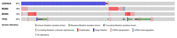
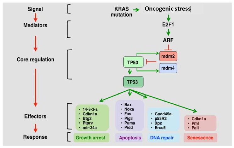
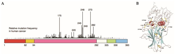
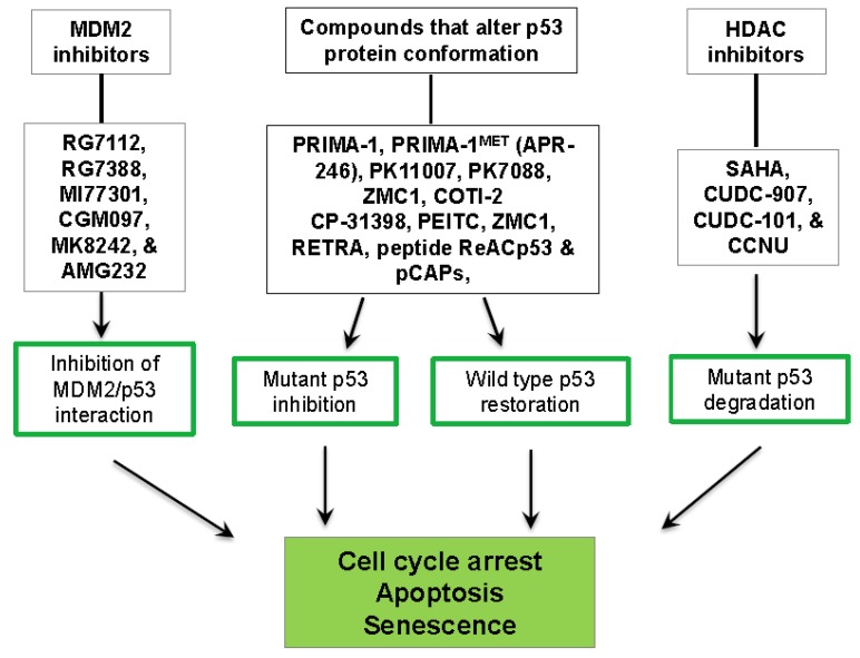
The tumor suppressor and transcription factor p53 plays critical roles in tumor prevention by orchestrating a wide variety of cellular responses, including damaged cell apoptosis, maintenance of genomic stability, inhibition of angiogenesis, and regulation of cell metabolism and tumor microenvironment. TP53 is one of the most commonly deregulated genes in cancer. The p53-ARF-MDM2 pathway is deregulated in 84% of glioblastoma (GBM) patients and 94% of GBM cell lines. Deregulated p53 pathway components have been implicated in GBM cell invasion, migration, proliferation, evasion of apoptosis, and cancer cell stemness. These pathway components are also regulated by various microRNAs and long non-coding RNAs. TP53 mutations in GBM are mostly point mutations that lead to a high expression of a gain of function (GOF) oncogenic variants of the p53 protein. These relatively understudied GOF p53 mutants promote GBM malignancy, possibly by acting as transcription factors on a set of genes other than those regulated by wild type p53. Their expression correlates with worse prognosis, highlighting their potential importance as markers and targets for GBM therapy. Understanding mutant p53 functions led to the development of novel approaches to restore p53 activity or promote mutant p53 degradation for future GBM therapies.

Keywords: gain-of-function; glioblastoma; mutant p53; wild type p53.


Conflict of interest statement

The authors declare no conflict of interest.

Figures

Figure 1
Figure 1
The p53 pathway is highly deregulated in GBM (adapted from cBioportal) [22,23]. The most common mutations of the p53 pathway are missense mutations in TP53, deletions of CDKN2A/ARF, and/or amplifications of MDM2 and MDM4. These mutations often lead to diminished tumor suppressor activity.
Figure 2
Figure 2
The p53-ARF-MDM2/4 pathway (adapted from the p53 website) [24]. In response to hyperproliferative stress and/or DNA damage, pathway mediators, such as ARF, are activated. MDM2 and MDM4 mark p53 for degradation, and are subsequently degraded when upstream mediators are activated. This releases p53 from degradation, and leads to increased cell cycle arrest, apoptosis, DNA Repair, and cellular senescence.
Figure 3
Figure 3
Most TP53 mutations are in the DNA binding domain. (A) The schematic view of the domains of p53 protein. The protein has 393 residues with numerous different domains including the N-terminus (red), proline rich domain (orange), DNA binding domain (purple), tetramerization domain (green) and negative regulatory domain/C-terminus (blue). The peaks represent the frequency of mutations in cancer with the DNA binding domain containing the six hot spot mutations. (B) Ribbon diagram of DNA-bound p53 (PBD ID 2AHI). The residues highlighted in yellow represent residues that are hot spot mutations as well as other residues of interest. Modified and reprinted with permission from Springer Nature: [Springer Nature] [Oncogene] [112].
Figure 4
Figure 4
Summary of select strategies for the therapeutic targeting of mutant p53 in GBM. Approaches include restoration of wild type p53 activity or degradation of mutant p53.

References

    1. Jeffrey P.D., Gorina S., Pavletich N.P. Crystal structure of the tetramerization domain of the p53 tumor suppressor at 1.7 angstroms. Science. 1995;267:1498–1502. doi: 10.1126/science.7878469. - DOI - PubMed
    1. Rajagopalan S., Huang F., Fersht A.R. Single-Molecule characterization of oligomerization kinetics and equilibria of the tumor suppressor p53. Nucleic Acids Res. 2011;39:2294–2303. doi: 10.1093/nar/gkq800. - DOI - PMC - PubMed
    1. El-Deiry W.S., Kern S.E., Pietenpol J.A., Kinzler K.W., Vogelstein B. Definition of a consensus binding site for p53. Nat. Genet. 1992;1:45–49. doi: 10.1038/ng0492-45. - DOI - PubMed
    1. Wei C.L., Wu Q., Vega V.B., Chiu K.P., Ng P., Zhang T., Shahab A., Yong H.C., Fu Y., Weng Z., et al. A global map of p53 transcription-factor binding sites in the human genome. Cell. 2006;124:207–219. doi: 10.1016/j.cell.2005.10.043. - DOI - PubMed
    1. Wade M., Li Y.C., Wahl G.M. MDM2, MDMX and p53 in oncogenesis and cancer therapy. Nat. Rev. Cancer. 2013;13:83–96. doi: 10.1038/nrc3430. - DOI - PMC - PubMed