. 2018 Sep 1;11(9):1580.
doi: 10.3390/ma11091580.

Demineralized Bone Matrix Coating Si-Ca-P Ceramic Does Not Improve the Osseointegration of the Scaffold

Affiliations

Demineralized Bone Matrix Coating Si-Ca-P Ceramic Does Not Improve the Osseointegration of the Scaffold

Andrés Parrilla-Almansa et al. Materials (Basel). .

Abstract

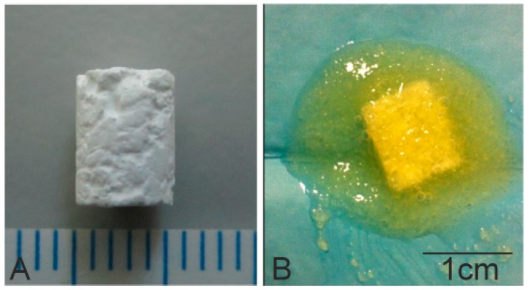
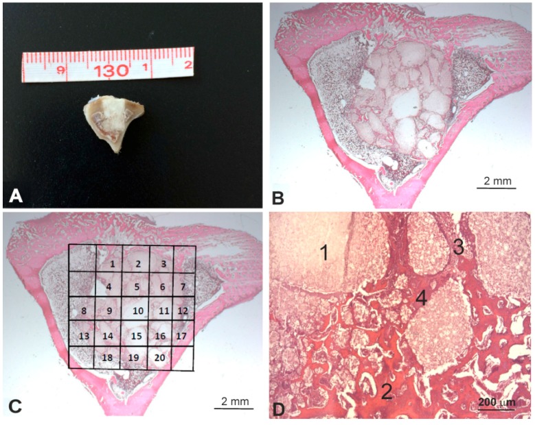
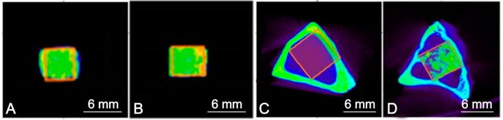
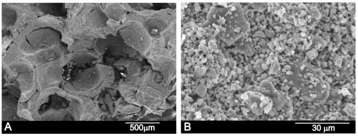
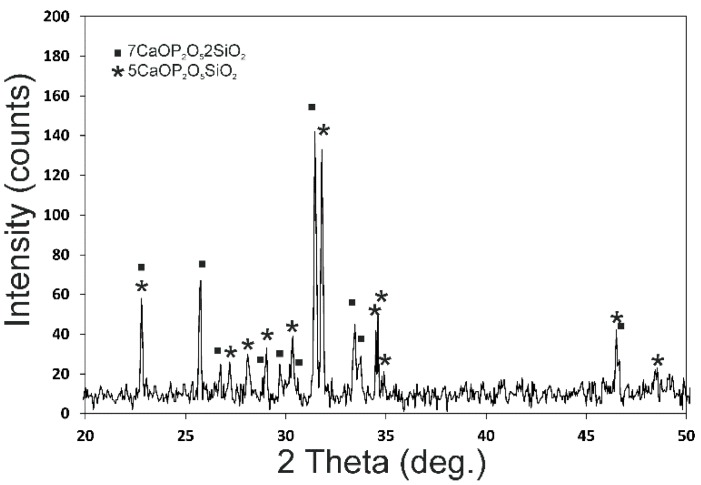
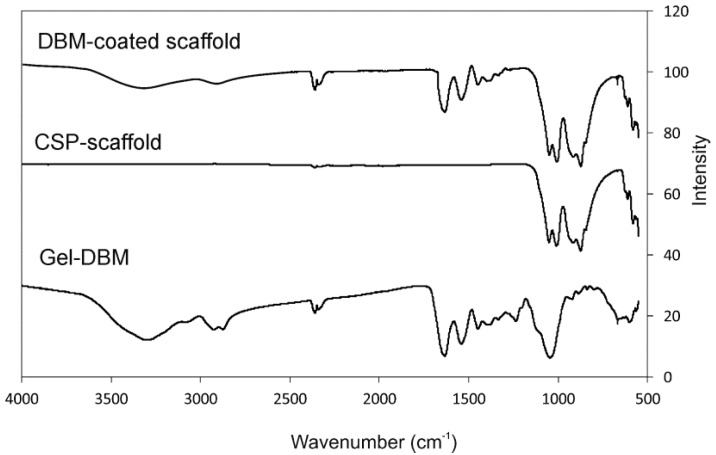
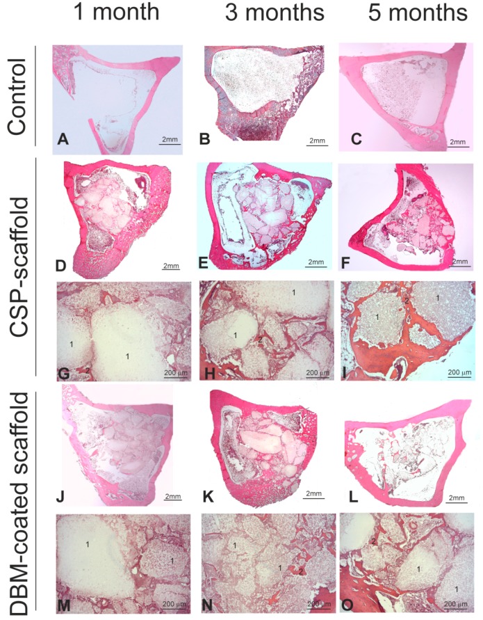
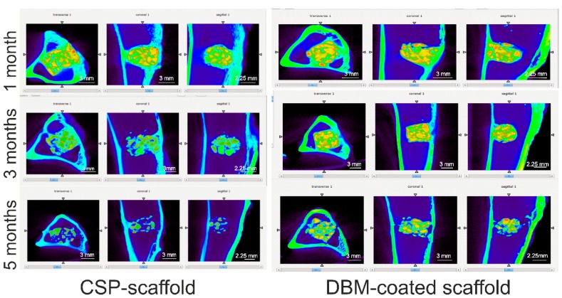
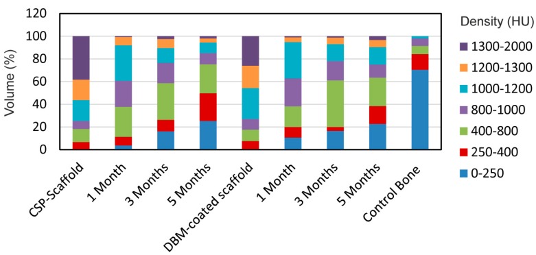
The aim of this study was to manufacture and evaluate the effect of a biphasic calcium silicophosphate (CSP) scaffold ceramic, coated with a natural demineralized bone matrix (DBM), to evaluate the efficiency of this novel ceramic material in bone regeneration. The DBM-coated CSP ceramic was made by coating a CSP scaffold with gel DBM, produced by the partial sintering of different-sized porous granules. These scaffolds were used to reconstruct defects in rabbit tibiae, where CSP scaffolds acted as the control material. Micro-CT and histological analyses were performed to evaluate new bone formation at 1, 3, and 5 months post-surgery. The present research results showed a correlation among the data obtained by micro-CT and the histomorphological results, the gradual disintegration of the biomaterial, and the presence of free scaffold fragments dispersed inside the medullary cavity occupied by hematopoietic bone marrow over the 5-month study period. No difference was found between the DBM-coated and uncoated implants. The new bone tissue inside the implants increased with implantation time. Slightly less new bone formation was observed in the DBM-coated samples, but it was not statistically significant. Both the DBM-coated and the CSP scaffolds gave excellent bone tissue responses and good osteoconductivity.

Keywords: bone regeneration; demineralized bone matrix; dicalcium silicate; histomorphometry; micro-CT; tricalcium phosphate.


Conflict of interest statement

The authors declare no conflict of interest.

Figures

Figure 1
Figure 1
Photographs of the (A) calcium silicophosphate (CSP) scaffold and (B) the demineralized bone matrix (DBM)-coated scaffold.
Figure 2
Figure 2
(A) Representative image of the macroscopic appearance of the implanted scaffold, (B) microscopic appearance of the implant stained with hematoxylin and eosin, (C) manually traced individual regions of interest (ROIs) by including only the scaffold, and (D) the four different components analyzed in the histomorphometric analysis (1, residual scaffold; 2, neoformed bone; 3, connective tissue; and 4, fibroblastic tissue). Hematoxylin and eosin stain (BD).
Figure 3
Figure 3
ROIs of (A) the CSP scaffold, (B) the DBM-coated CSP, (C) the rabbit tibiae before implantation, and (D) the scaffold after implantation as being representative of the two implanted materials.
Figure 4
Figure 4
(A) SEM and (B) high-resolution SEM images of the manufactured CSP scaffold.
Figure 5
Figure 5
X-ray diffraction pattern of the synthesized CSP scaffold.
Figure 6
Figure 6
The FTIR of the CSP scaffold, the DBM-coated scaffold, and gel DBM for comparison purposes.
Figure 7
Figure 7
(AC) Representative images of the tibia samples from the control, (DI) implanted CSP and (JO) DBM-coated scaffolds. In the controls, the bone defect (A) was repaired by physiological bone repair at months 3 and 5 (B,C, respectively). In the CSP group, the scaffold surface progressively decreased (DI, number 1) with increasing neoformed bone organization (GI, number 2). Similarly, the DBM-coated scaffold surface progressively decreased from month 1 to month 5 (J,K,MO, number 1) but, in this case, the neoformed bone organization slightly diminished (M,N, number 2) compared with the CSP scaffold. Hematoxylin and eosin stain. (1, residual scaffold; 2, neoformed bone).
Figure 8
Figure 8
The micro-CT images of both materials implanted in tibiae at 1, 3, and 5 months post-surgery. Orange color corresponds to residual biomaterial. Yellow color corresponds to biomaterial partially reabsorbed. Green color corresponds to the cortical bone and represents the normal cortex of the host bone. Green color located within the biomaterial represents the reabsorbed biomaterial and new bone tissue. Blue color corresponds normal medullary tissue of the host bone. Blue color located within the biomaterial represents neoformed fibrous connective tissue.
Figure 9
Figure 9
The density regions displayed as a percentage of the total volume of the ROIs after implantation months 1, 3, and 5, and in control, CSP scaffold, and DBM-coated scaffold bone specimens with densities given in HU.

Similar articles

Cited by

References

    1. Johansson P., Jimbo R., Kozai Y., Sakurai T., Kjellin P., Currie F., Wennerberg A. Nanosized Hydroxyapatite Coating on PEEK Implants Enhances Early Bone Formation: A Histological and Three-Dimensional Investigation in Rabbit Bone. Materials. 2015;8:3815–3830. doi: 10.3390/ma8073815. - DOI - PMC - PubMed
    1. Mate-Sanchez de Val J.E., Mazon P., Calvo-Guirado J.L., Delgado-Ruiz R.A., Ramirez-Fernandez M.P., Negri B., Abboud M., De Aza P.N. Comparison of three hydroxyapatite/β-tricalcium phosphate/collagen ceramic scaffolds: An in vivo study. J. Biomed. Mater. Res. A. 2014;102:1037–1046. doi: 10.1002/jbm.a.34785. - DOI - PubMed
    1. Neacsu P., Staras A.I., Voicu S.I., Ionascu I., Soare T., Uzun S., Cojocaru V.D., Andreea Pandele M., Croitoru S.M., Miculescu F., et al. Characterization and In Vitro and In Vivo Assessment of a Novel Cellulose Acetate-Coated Mg-Based Alloy for Orthopedic Applications. Materials. 2017;10:686. doi: 10.3390/ma10070686. - DOI - PMC - PubMed
    1. Rabadan-Ros R., Velásquez P., Meseguer-Olmo L., De Aza P.N. Morphological and structural study of a novel porous Nurse´s A ceramic with osteoconductive properties for tissue engineering. Materials. 2016;9:474. doi: 10.3390/ma9060474. - DOI - PMC - PubMed
    1. Fernandez-Pradas J.M., Serra P., Morenza J.M., De Aza P.N. Pulser laser deposition of pseudowollastonite coatings. Biomaterials. 2002;23:2057–2061. doi: 10.1016/S0142-9612(01)00335-0. - DOI - PubMed

LinkOut - more resources