

Regulation of Ovarian Cancer Prognosis by Immune Cells in the Tumor Microenvironment
- PMID: 30200478
- PMCID: PMC6162424
- DOI: 10.3390/cancers10090302
Regulation of Ovarian Cancer Prognosis by Immune Cells in the Tumor Microenvironment
Abstract
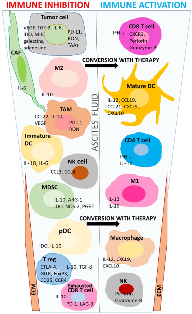
It is estimated that in the United States in 2018 there will be 22,240 new cases of ovarian cancer and 14,070 deaths due to this malignancy. The most common subgroup of this disease is high-grade serous ovarian cancer (HGSOC), which is known for its aggressiveness, high recurrence rate, metastasis to other sites, and the development of resistance to conventional therapy. It is important to understand the ovarian cancer tumor microenvironment (TME) from the viewpoint of the function of pre-existing immune cells, as immunocompetent cells are crucial to mounting robust antitumor responses to prevent visible tumor lesions, disease progression, or recurrence. Networks consisting of innate and adaptive immune cells, metabolic pathways, intracellular signaling molecules, and a vast array of soluble factors, shape the pathogenic nature of the TME and are useful prognostic indicators of responses to conventional therapy and immunotherapy, and subsequent survival rates. This review highlights key immune cells and soluble molecules in the TME of ovarian cancer, which are important in the development of effective antitumor immunity, as well as those that impair effector T cell activity. A more insightful knowledge of the HGSOC TME will reveal potential immune biomarkers to aid in the early detection of this disease, as well as biomarkers that may be targeted to advance the design of novel therapies that induce potent antitumor immunity and survival benefit.
Keywords: antitumor immunity; dendritic cells; immune inhibition; immunotherapy; tumor microenvironment; tumor-associated macrophages; tumor-infiltrating lymphocytes.
Conflict of interest statement
The authors declare no conflict of interest.
Figures
References
-
- Colombo N., Peiretti M., Parma G., Lapresa M., Mancari R., Carinelli S., Sessa C., Castiglione M. Newly Diagnosed and Relapsed Epithelial Ovarian Carcinoma: ESMO Clinical Practice Guidelines for Diagnosis, Treatment and Follow-Up. Ann. Oncol. 2010;21:v23–v30. doi: 10.1093/annonc/mdq244. - DOI - PubMed
-
- Perets R., Wyant G., Muto K., Bijron J., Poole B., Chin K., Chen J., Ohman A., Stepule C., Kwak S., et al. Transformation of the Fallopian Tube Secretory Epithelium Leads to High-Grade Serous Ovarian Cancer in Brca;Tp53;Pten Models. Cancer Cell. 2013;24:751–765. doi: 10.1016/j.ccr.2013.10.013. - DOI - PMC - PubMed
-
- Cole A.J., Dwight T., Gill A.J., Dickson K.A., Zhu Y., Clarkson A., Gard G.B., Maidens J., Valmadre S., Clifton-Bligh R., et al. Assessing Mutant p53 in Primary High-Grade Serous Ovarian Cancer using Immunohistochemistry and Massively Parallel Sequencing. Sci. Rep. 2016;6:26191. doi: 10.1038/srep26191. - DOI - PMC - PubMed
Publication types
LinkOut - more resources
Full Text Sources
Other Literature Sources
