. 2018 Sep 1;10(9):1208.
doi: 10.3390/nu10091208.

Ginsenoside Rh2 Ameliorates Lipopolysaccharide-Induced Acute Lung Injury by Regulating the TLR4/PI3K/Akt/mTOR, Raf-1/MEK/ERK, and Keap1/Nrf2/HO-1 Signaling Pathways in Mice

Affiliations

Ginsenoside Rh2 Ameliorates Lipopolysaccharide-Induced Acute Lung Injury by Regulating the TLR4/PI3K/Akt/mTOR, Raf-1/MEK/ERK, and Keap1/Nrf2/HO-1 Signaling Pathways in Mice

Yung-Hung Hsieh et al. Nutrients. .

Abstract

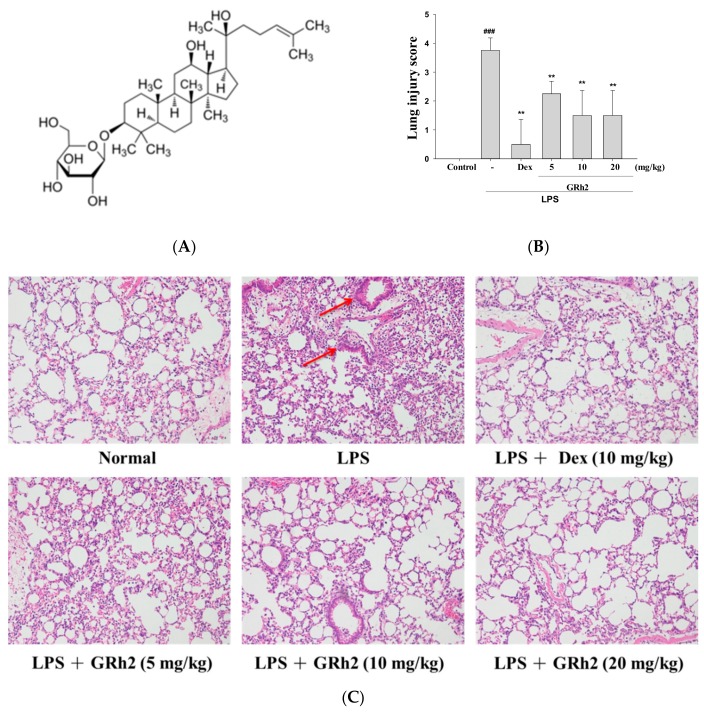
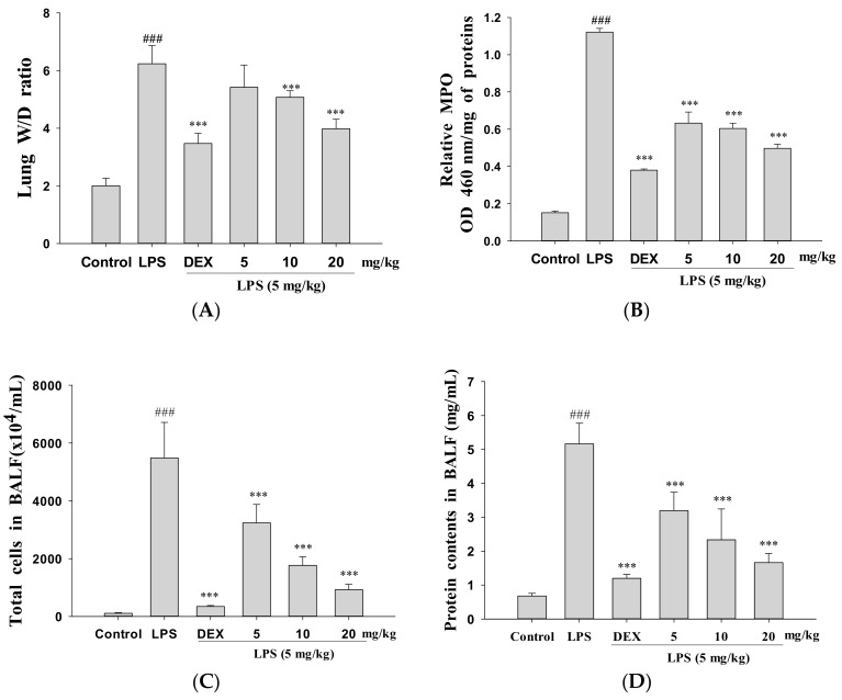
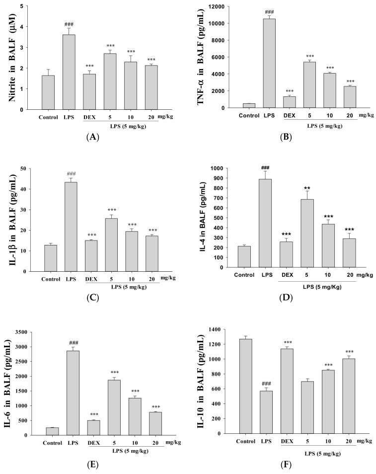
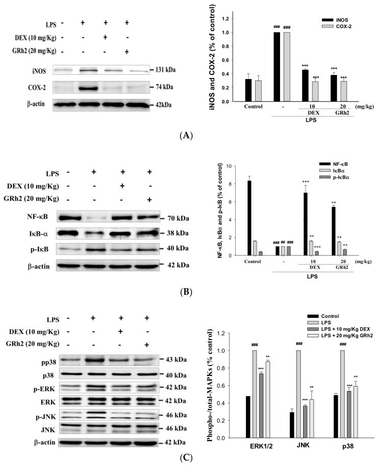
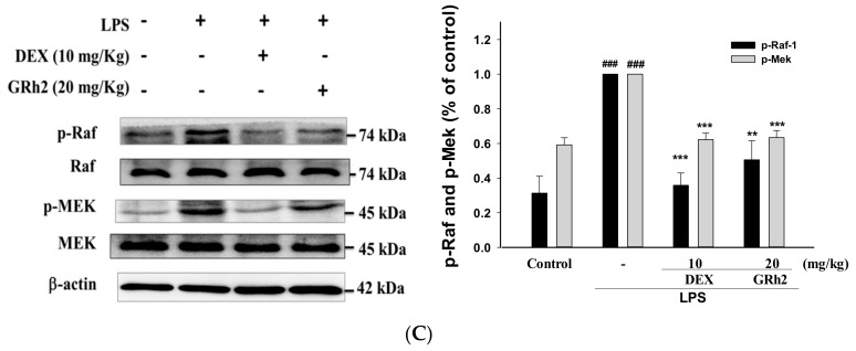
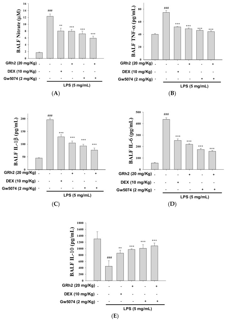
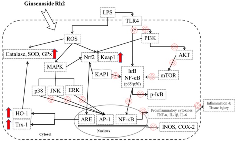
The anti-inflammatory effect of ginsenoside Rh2 (GRh2) has labeled it as one of the most important ginsenosides. The purpose of this study was to identify the anti-inflammatory and antioxidant effects of GRh2 using a lipopolysaccharide (LPS) challenge lung-injury animal model. GRh2 reduced LPS-induced proinflammatory mediator nitric oxide (NO), tumor necrosis factor-alpha, interleukin (IL)-1β, and anti-inflammatory cytokines (IL-4, IL-6, and IL-10) production in lung tissues. GRh2 treatment decreased the histological alterations in the lung tissues and bronchoalveolar lavage fluid (BALF) protein content; total cell number also reduced in LPS-induced lung injury in mice. Moreover, GRh2 blocked iNOS, COX-2, the phosphorylation of IκB-α, ERK, JNK, p38, Raf-1, and MEK protein expression, which corresponds with the growth of HO-1, Nrf-2, catalase, SOD, and GPx expression in LPS-induced lung injury. An in vivo experimental study suggested that GRh2 has anti-inflammatory effects, and has potential therapeutic efficacy in major anterior segment lung diseases.

Keywords: MEK; Nrf-2; acute lung injury; ginsenoside Rh2; lipopolysaccharide.


Conflict of interest statement

All authors have no conflicts of interest with respect to the data collected and procedures used within this study.

Figures

Figure 1
Figure 1
(A) The chemical structure of ginsenoside Rh2 (GRh2); (B) the lung-injury scores were determined; and (C) the effects of GRh2 (5, 10, and 20 mg/mL) on lipopolysaccharide (LPS)-induced lung histopathologic changes in mice. At 6 h after LPS challenge, lungs in each group were prepared for histological evaluation. Representative histological section of the lungs was stained by hematoxylin and eosin (H and E) staining, magnification (400×). The data are presented as the means ± S.E.M (n = 5). ### denotes p < 0.001 compared with sample of control group. ** p < 0.01 compared with the LPS-alone group. The red arrow indicates the symptoms of bleeding and inflammatory cell infiltration. Dexamethasone (Dex).
Figure 2
Figure 2
GRh2 improved (A) pulmonary edema (wet-dry (W/D) ratio); (B) myeloperoxidase (MPO) activity in vivo; and reduced (C) cellular counts and (D) total protein in bronchoalveolar lavage fluid (BALF). Lung tissues were weighed and calculated the W/D ratio. BALF was harvested to investigate at 6 h after LPS treatment. Total cells and total proteins in BALF were analyzed. Data are presented as mean ± S.E.M. (n = 5). ### denotes p < 0.001 compared with sample of control group. *** denotes p < 0.001 compared with the LPS-only group.
Figure 3
Figure 3
GRh2 downregulated (A) nitric oxide (NO); (B) tumor necrosis factor-alpha (TNF-α); (C) IL-1β; (D) IL-4; and (E) IL-6; and (F) increased IL-10 in BALF. BALF was collected. NO, TNF-α, IL-1β, IL-4, IL-6, and IL-10 were detected at 6 h after LPS challenge by enzyme-linked immunosorbent assay (ELISA). Data are represented as mean ± S.E.M. (n = 5). ### denotes p < 0.001 compared with sample of control group. ** p < 0.01 and *** p < 0.001 compared with LPS-only group.
Figure 4
Figure 4
Effects of GRh2 on LPS-induced (A) iNOs, COX-2; (B) IκB-α, NF-κB; and (C) mitogen-activated protein kinase (MAPK) phosphorylation signaling expression in lungs. Protein levels of iNOs, COX-2, IκB-α, NF-κB, and MAPK phosphorylation protein expression in lung homogenates were evaluated by western blot analysis after LPS challenge 6 h later. Densitometric analysis of the relevant bands was performed. Data are represented as mean ± S.E.M. (n = 2). ## p < 0.01 and ### p < 0.001 compared with the control group. ** p < 0.01 and *** p < 0.001 compared with LPS-only group.
Figure 5
Figure 5
Effects of GRh2 on (A) LPS-induced antioxidative enzymes (catalase, superoxide dismutase (SOD), and glutathione peroxidase (GPx)); heme oxygenase-1 (HO-1), Trx-1, NF-E2-related factor 2 (Nrf2)/Keap1 and KAP1; (B) TLR4, PI3K, Akt, and mTOR; and (C) Raf-1, p-Raf-1, Mek and p-Mek protein expression in the lungs. Protein levels of catalase, SOD, GPx, HO-1, Trx-1, Nrf2/Keap1, KAP1, TLR4, PI3K, Akt, mTOR, Raf-1, p-Raf-1, Mek and p-Mek protein expression in lung homogenates were evaluated by western blot analysis after LPS challenge 6 h hours later. Densitometric analysis of the relevant bands was performed. Data are represented as mean ± S.E.M. (n = 2). ## p < 0.05 and ### p < 0.001 compared with sample of control group. ** p < 0.01 and *** p < 0.001 compared with LPS-only group.
Figure 5
Figure 5
Effects of GRh2 on (A) LPS-induced antioxidative enzymes (catalase, superoxide dismutase (SOD), and glutathione peroxidase (GPx)); heme oxygenase-1 (HO-1), Trx-1, NF-E2-related factor 2 (Nrf2)/Keap1 and KAP1; (B) TLR4, PI3K, Akt, and mTOR; and (C) Raf-1, p-Raf-1, Mek and p-Mek protein expression in the lungs. Protein levels of catalase, SOD, GPx, HO-1, Trx-1, Nrf2/Keap1, KAP1, TLR4, PI3K, Akt, mTOR, Raf-1, p-Raf-1, Mek and p-Mek protein expression in lung homogenates were evaluated by western blot analysis after LPS challenge 6 h hours later. Densitometric analysis of the relevant bands was performed. Data are represented as mean ± S.E.M. (n = 2). ## p < 0.05 and ### p < 0.001 compared with sample of control group. ** p < 0.01 and *** p < 0.001 compared with LPS-only group.
Figure 6
Figure 6
(A) GRh2 and Raf-1 inhibitor (GW-5074) reduced NO; (B) TNF-α; (C) IL-1β; (D) IL-6; and (E) IL-10 in BALF. BALF was collected. NO, TNF-α, IL-1β, IL-6, and IL-10 were detected at 6 h after LPS challenge by ELISA. Data are represented as mean ± S.E.M. (n = 5). ### p < 0.001 compared with sample of control group. ** p < 0.01 and *** p < 0.001 compared with LPS-only group.
Figure 7
Figure 7
The schemes of the mechanism for the protective effect of GRh2 on LPS-induced inflammation.

Similar articles

Cited by

References

    1. Li K.C., Ho Y.L., Hsieh W.T., Huang S.S., Chang Y.S., Huang G.J. Apigenin-7-glycoside prevents LPS-induced acute lung injury via downregulation of oxidative enzyme expression and protein activation through inhibition of MAPK phosphorylation. Int. J. Mol. Sci. 2015;16:1736–1754. doi: 10.3390/ijms16011736. - DOI - PMC - PubMed
    1. Li K.C., Ho Y.L., Chen C.Y., Hsieh W.T., Chang Y.S., Huang G.J. Lobeline improve acute lung injury via nuclear factor-kB-signaling pathway and oxidative stress. Respirator. Physiol. Neurobiol. 2016;225:19–30. doi: 10.1016/j.resp.2015.12.003. - DOI - PubMed
    1. Huang G.J., Deng J.S., Chen C.C., Huang C.J., Sung P.J., Huang S.S., Kuo Y.H. Methanol extract of Antrodia camphorata protects against lipopolysaccharide-induced acute lung injury by suppressing NF-κB and MAPK pathways in mice. J. Agric. Food Chem. 2014;62:5321–5329. doi: 10.1021/jf405113g. - DOI - PubMed
    1. Chang J.S., Lin H.J., Deng J.S., Wu W.T., Huang S.S., Huang G.J. Preventive effects of Velvet Antler (Cervus elaphus) against lipopolysaccharide induced acute lung injury in mice by inhibiting MAPK/NF-κB activation and inducing AMPK/Nrf2 pathways. Evid. Based Complement. Alternat. Med. 2018;2018:2870503. doi: 10.1155/2018/2870503. - DOI - PMC - PubMed
    1. Hsieh Y.H., Deng J.S., Pan H.P., Liao J.C., Huang S.S., Huang G.J. Sclareol ameliorate lipopolysaccharide-induced acute lung injury through inhibition of MAPK and induction of HO-1 signaling. Int. Immunopharmacol. 2017;44:16–25. doi: 10.1016/j.intimp.2016.12.026. - DOI - PubMed

MeSH terms

Substances