. 2018 Sep 6;19(9):2637.
doi: 10.3390/ijms19092637.

Peiminine Protects against Lipopolysaccharide-Induced Mastitis by Inhibiting the AKT/NF-κB, ERK1/2 and p38 Signaling Pathways

Affiliations

Peiminine Protects against Lipopolysaccharide-Induced Mastitis by Inhibiting the AKT/NF-κB, ERK1/2 and p38 Signaling Pathways

Qian Gong et al. Int J Mol Sci. .

Abstract

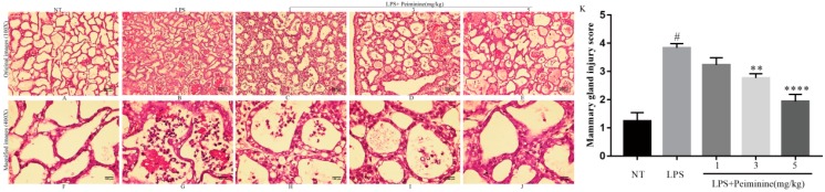
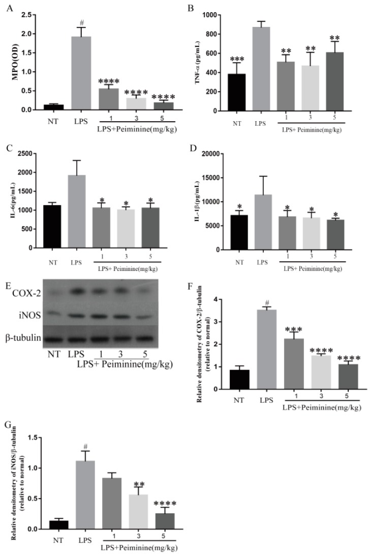
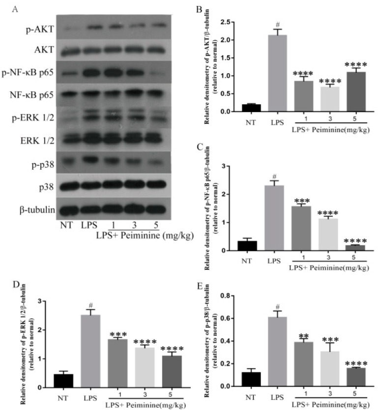
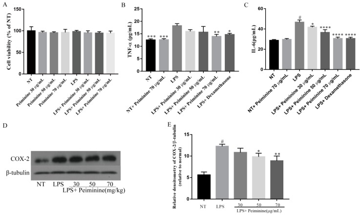
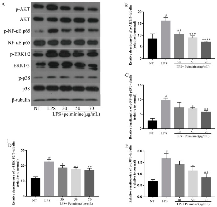
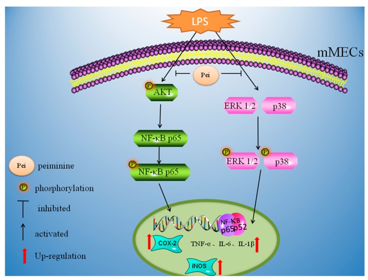
Peiminine, an alkaloid extracted from Fritillaria plants, has been reported to have potent anti-inflammatory properties. However, the anti-inflammatory effect of peiminine on a mouse lipopolysaccharide (LPS)-induced mastitis model remains to be elucidated. The purpose of this experiment was to investigate the effect of peiminine on LPS-induced mastitis in mice. LPS was injected through the canals of the mammary gland to generate the mouse LPS-induced mastitis model. Peiminine was administered intraperitoneally 1 h before and 12 h after the LPS injection. In vitro, mouse mammary epithelial cells (mMECs) were pretreated with different concentrations of peiminine for 1 h and were then stimulated with LPS. The mechanism of peiminine on mastitis was studied by hematoxylin-eosin staining (H&E) staining, western blotting, and enzyme-linked immunosorbent assay (ELISA). The results showed that peiminine significantly decreased the histopathological impairment of the mammary gland in vivo and reduced the production of pro-inflammatory mediators in vivo and in vitro. Furthermore, peiminine inhibited the phosphorylation of the protein kinase B (AKT)/ nuclear factor-κB (NF-κB), extracellular regulated protein kinase (ERK1/2), and p38 signaling pathways both in vivo and in vitro. All the results suggested that peiminine exerted potent anti-inflammatory effects on LPS-induced mastitis in mice. Therefore, peiminine might be a potential therapeutic agent for mastitis.

Keywords: AKT/NF-κB; ERK1/2; LPS; mastitis; p38; peiminine.


Conflict of interest statement

The authors declare no conflict of interest.

Figures

Figure 1
Figure 1
Chemical structure of peiminine.
Figure 2
Figure 2
Histopathological sections of the mammary tissues (H&E, 100× and 400×). The mammary tissues (n = 6 in each group) from each group were processed for histological evaluation. Representative histological changes in the mammary tissues from each group were as follows: (A,F) no-treatment (NT) group; (B,G) lipopolysaccharide (LPS) group; (C,H) LPS + 1 mg/kg peiminine group; (D,I) LPS + 3 mg/kg peiminine group; and (E,J) LPS + 5 mg/kg peiminine group. Histologic grade of the mammary gland (K). The histological morphology and pathology results showed that peiminine treatment alleviated the LPS-induced pathological changes. The number sign (#) indicates a significant difference from the NT group at p < 0.0001. The tetrad asterisks (****) indicate p < 0.0001 and the double asterisks (**) indicate p < 0.01 vs. the LPS group.
Figure 3
Figure 3
Effect of peiminine on (A) the myeloperoxidase (MPO) activity in the mammary gland of LPS-induced mastitis, the levels of (B) tumor necrosis factor alpha (TNF-α), (C) interleukin-6 (IL-6), and (D) interleukin-1β (IL-1β) in the homogenate of the mouse mammary tissues, including the NT group, the LPS group, and the groups treated with peiminine (1, 3, and 5 mg/kg), and the protein levels of (E,F) COX-2 and (E,G) iNOS were measured by western blotting. The data are presented as the mean ± SD (n = 6). The number sign (#) indicates p < 0.0001, which was significantly different from the NT group. The tetrad asterisks (****) indicate p < 0.0001, the triple asterisks (***) indicate p < 0.0005, the double asterisks (**) indicate p < 0.01, and the single asterisk (*) indicates p < 0.05 vs. the LPS group.
Figure 4
Figure 4
Peiminine inhibits the phosphorylation of the AKT, nuclear factor-κB (NF-κB) p65, ERK1/2 and p38 signaling pathways in the mammary tissues. The results for the protein levels of (A,B) p-AKT, (A,C) p-NF-κB p65, (A,D) p-ERK1/2, and (A,E) p-p38 were measured by western blotting. The data are presented as the mean ± SD (n = 6). The number sign (#) indicates a significant difference from the NT group at p < 0.0001. The tetrad asterisks (****) indicate p < 0.0001, the triple asterisks (***) indicate p < 0.0005, and the double asterisks (**) indicate p < 0.01 vs. the LPS group.
Figure 5
Figure 5
Effect of peiminine on (A) cell viability. The cells were cultured with different concentrations of peiminine (30, 50, or 70 μg/mL) for 4 h. Cell viability was determined by the CCK8 assay. The levels of (B) TNF-α and (C) IL-6 in the mouse mammary epithelial cells (mMECs), including the NT group, the NT + peiminine 70 μg/mL group, the LPS group, and the groups treated with peiminine (30, 50, or 70 μg/mL), and the protein level of (D,E) COX-2 were measured by western blotting. The data are presented as the mean ± SD (n = 6). The number sign (#) indicates a significant difference from the NT group at p < 0.0001. The tetrad asterisks (****) indicate p < 0.0001, the triple asterisks (***) indicate p < 0.0005, the double asterisks (**) indicate p < 0.01, and the single asterisk (*) indicates p < 0.05 vs. the LPS group.
Figure 6
Figure 6
Peiminine inhibits the phosphorylation of the AKT, NF-κB p65, ERK1/2, and p38 signaling pathways in mMECs. The results for the protein levels of (A,B) p-AKT, (A,C) p-NF-κB p65, (A,D) p-ERK1/2, and (A,E) p-p38 were measured by western blotting. The data are presented as the mean ± SD (n = 6). The number sign (#) indicates a significant difference from the NT group at p < 0.0001. The tetrad asterisks (****) indicate p < 0.0001, the triple asterisks (***) indicate p < 0.0005, the double asterisks (**) indicate p < 0.01, and the single asterisk (*) indicates p < 0.05 vs. the LPS group.
Figure 7
Figure 7
Peiminine inhibits LPS-induced inflammation by suppressing the phosphorylation of the AKT, NF-κB p65, ERK1/2, and p38 signaling pathways.

Similar articles

Cited by

References

    1. Zadoks R.N., Middleton J.R., McDougall S., Katholm J., Schukken Y.H. Molecular epidemiology of mastitis pathogens of dairy cattle and comparative relevance to humans. J. Mammary Gland Biol. Neoplasia. 2011;16:357–372. doi: 10.1007/s10911-011-9236-y. - DOI - PMC - PubMed
    1. Kauf A.C., Vinyard B.T., Bannerman D.D. Effect of intramammary infusion of bacterial lipopolysaccharide on experimentally induced Staphylococcus aureus intramammary infection. Res. Vet. Sci. 2007;82:39–46. doi: 10.1016/j.rvsc.2006.05.006. - DOI - PubMed
    1. Hartlage-Rubsamen M., Lemke R., Schliebs R. Interleukin-1β, inducible nitric oxide synthase, and nuclear factor-κB are induced in morphologically distinct microglia after rat hippocampal lipopolysaccharide/interferon-γ injection. J. Neurosci. Res. 1999;57:388–398. doi: 10.1002/(SICI)1097-4547(19990801)57:3<388::AID-JNR11>3.0.CO;2-2. - DOI - PubMed
    1. Takeuchi H., Jin S.J., Wang J.Y., Zhang G.Q., Kawanokuchi J., Kuno R., Sonobe Y., Mizuno T., Suzumura A. Tumor necrosis factor-α induces neurotoxicity via glutamate release from hemichannels of activated microglia in an autocrine manner. J. Biol. Chem. 2006;281:21362–21368. doi: 10.1074/jbc.M600504200. - DOI - PubMed
    1. Toubiana M., Gerdol M., Rosani U., Pallavicini A., Venier P., Roch P. Toll-like receptors and MyD88 adaptors in Mytilus: Complete CDS and gene expression levels. Dev. Comp. Immunol. 2013;40:158–166. doi: 10.1016/j.dci.2013.02.006. - DOI - PubMed

MeSH terms

LinkOut - more resources