

Evolution of Human Respiratory Syncytial Virus (RSV) over Multiple Seasons in New South Wales, Australia
- PMID: 30200580
- PMCID: PMC6164696
- DOI: 10.3390/v10090476
Evolution of Human Respiratory Syncytial Virus (RSV) over Multiple Seasons in New South Wales, Australia
Abstract
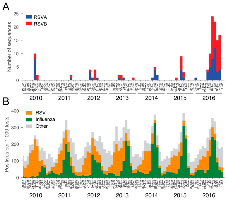
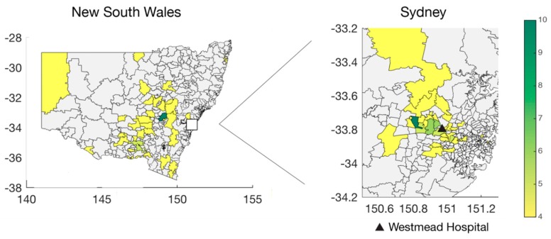
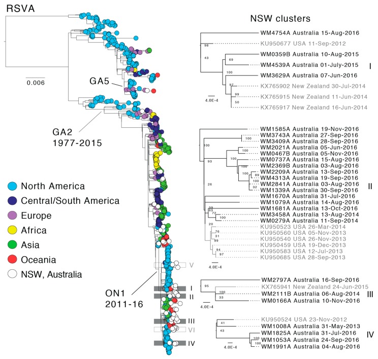
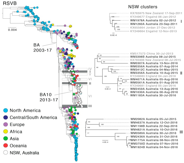
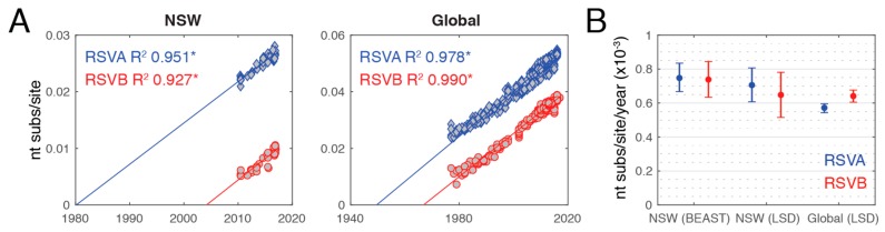
There is an ongoing global pandemic of human respiratory syncytial virus (RSV) infection that results in substantial annual morbidity and mortality. In Australia, RSV is a major cause of acute lower respiratory tract infections (ALRI). Nevertheless, little is known about the extent and origins of the genetic diversity of RSV in Australia, nor the factors that shape this diversity. We have conducted a genome-scale analysis of RSV infections in New South Wales (NSW). RSV genomes were successfully sequenced for 144 specimens collected between 2010⁻2016. Of these, 64 belonged to the RSVA and 80 to the RSVB subtype. Phylogenetic analysis revealed a wide diversity of RSV lineages within NSW and that both subtypes evolved rapidly in a strongly clock-like manner, with mean rates of approximately 6⁻8 × 10⁻⁴ nucleotide substitutions per site per year. There was only weak evidence for geographic clustering of sequences, indicative of fluid patterns of transmission within the infected population and no evidence of any clustering by patient age such that viruses in the same lineages circulate through the entire host population. Importantly, we show that both subtypes circulated concurrently in NSW with multiple introductions into the Australian population in each year and only limited evidence for multi-year persistence.
Keywords: evolution; multi-year persistence; phylogenetics; respiratory syncytial virus.
Conflict of interest statement
The authors declare no conflicts of interest.
Figures
References
-
- Nair H., Nokes D.J., Gessner B.D., Dherani M., Madhi S.A., Singleton R.J., O’Brien K.L., Roca A., Wright P.F., Bruce N., et al. Global burden of acute lower respiratory infections due to respiratory syncytial virus in young children: A systematic review and meta-analysis. Lancet. 2010;375:1545–1555. doi: 10.1016/S0140-6736(10)60206-1. - DOI - PMC - PubMed
-
- Caram L.B., Chen J., Taggart E.W., Hillyard D.R., She R., Polage C.R., Twersky J., Schmader K., Petti C.A., Woods C.W. Respiratory syncytial virus outbreak in a long-term care facility detected using reverse transcriptase polymerase chain reaction: An argument for real-time detection methods. J. Am. Geriatr. Soc. 2009;57:482–485. doi: 10.1111/j.1532-5415.2008.02153.x. - DOI - PMC - PubMed
-
- Shi T., McAllister D.A., O’Brien K.L., Simoes E.A.F., Madhi S.A., Gessner B.D., Polack F.P., Balsells E., Acacio S., Aguayo C., et al. Global, regional, and national disease burden estimates of acute lower respiratory infections due to respiratory syncytial virus in young children in 2015: A systematic review and modelling study. Lancet. 2017;390:946–958. doi: 10.1016/S0140-6736(17)30938-8. - DOI - PMC - PubMed
-
- Nolan T., Borja-Tabora C., Lopez P., Weckx L., Ulloa-Gutierrez R., Lazcano-Ponce E., Kerdpanich A., Weber M.A.R., de Los Santos A.M., Tinoco J.C., et al. Prevalence and incidence of respiratory syncytial virus and other respiratory viral infections in children aged 6 months to 10 years with influenza-like illness enrolled in a randomized trial. Clin. Infect. Dis. 2015;60:E80–E89. doi: 10.1093/cid/civ065. - DOI - PMC - PubMed
-
- Ranmuthugala G., Brown L., Lidbury B.A. Respiratory syncytial virus—The unrecognised cause of health and economic burden among young children in Australia. Commun. Dis. Intell. 2011;35:177–184. - PubMed
Publication types
MeSH terms
Grants and funding
LinkOut - more resources
Full Text Sources
Other Literature Sources
Medical
