Mechanism of Lakoochin A Inducing Apoptosis of A375.S2 Melanoma Cells through Mitochondrial ROS and MAPKs Pathway
- PMID: 30200660
- PMCID: PMC6164788
- DOI: 10.3390/ijms19092649
Mechanism of Lakoochin A Inducing Apoptosis of A375.S2 Melanoma Cells through Mitochondrial ROS and MAPKs Pathway
Abstract
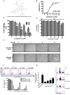
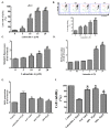
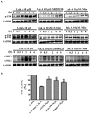
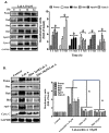
Malignant melanoma is developed from pigment-containing cells, melanocytes, and primarily found on the skin. Malignant melanoma still has a high mortality rate, which may imply a lack of therapeutic agents. Lakoochin A, a compound isolated from Artocarpus lakoocha and Artocarpus xanthocarpus, has an inhibitory function of tyrosinase activity and melanin production, but the anti-cancer effects are still unclear. In the current study, the therapeutic effects of lakoochin A with their apoptosis functions and possible mechanisms were investigated on A375.S2 melanoma cells. Several methods were applied, including 3-(4,5-Dimethylthiazol-2-yl)-2,5- diphenyltetrazolium bromide (MTT), flow cytometry, and immunoblotting. Results suggest that lakoochin A attenuated the growth of A375.S2 melanoma cells through an apoptosis mechanism. Lakoochin A first increase the production of cellular and mitochondrial reactive oxygen species (ROSs); mitochondrial ROSs then promote mitogen-activated protein kinases (MAPKs) pathway activation and raise downstream apoptosis-related protein and caspase expression. This is the first study to demonstrate that lakoochin A, through ROS-MAPK, apoptosis-related proteins, caspases cascades, can induce melanoma cell apoptosis and may be a potential candidate compound for treating malignant melanoma.
Keywords: MAPKs; apoptosis; lakoochin A; melanoma cells; mitochondria; pro-oxidation.
Conflict of interest statement
The authors declare no conflict of interest.
Figures


Similar articles
-
Cudraflavone C Induces Apoptosis of A375.S2 Melanoma Cells through Mitochondrial ROS Production and MAPK Activation.Int J Mol Sci. 2017 Jul 13;18(7):1508. doi: 10.3390/ijms18071508. Int J Mol Sci. 2017. PMID: 28703746 Free PMC article.
-
Plumbagin induces cell cycle arrest and apoptosis through reactive oxygen species/c-Jun N-terminal kinase pathways in human melanoma A375.S2 cells.Cancer Lett. 2008 Jan 18;259(1):82-98. doi: 10.1016/j.canlet.2007.10.005. Epub 2007 Nov 19. Cancer Lett. 2008. PMID: 18023967
-
Quinalizarin induces cycle arrest and apoptosis via reactive oxygen species-mediated signaling pathways in human melanoma A375 cells.Drug Dev Res. 2019 Dec;80(8):1040-1050. doi: 10.1002/ddr.21582. Epub 2019 Aug 20. Drug Dev Res. 2019. PMID: 31432559
-
Casticin Induced Apoptosis in A375.S2 Human Melanoma Cells through the Inhibition of NF-[Formula: see text]B and Mitochondria-Dependent Pathways In Vitro and Inhibited Human Melanoma Xenografts in a Mouse Model In Vivo.Am J Chin Med. 2016;44(3):637-61. doi: 10.1142/S0192415X1650035X. Epub 2016 Apr 24. Am J Chin Med. 2016. PMID: 27109154
-
Benzyl isothiocyanate (BITC) induces G2/M phase arrest and apoptosis in human melanoma A375.S2 cells through reactive oxygen species (ROS) and both mitochondria-dependent and death receptor-mediated multiple signaling pathways.J Agric Food Chem. 2012 Jan 18;60(2):665-75. doi: 10.1021/jf204193v. Epub 2012 Jan 6. J Agric Food Chem. 2012. PMID: 22148415
Cited by
-
Pharmacological effects of Artocarpus lakoocha methanol extract on inhibition of squalene synthase and other downstream enzymes of the cholesterol synthesis pathway.Pharm Biol. 2022 Dec;60(1):840-845. doi: 10.1080/13880209.2022.2063346. Pharm Biol. 2022. PMID: 35588395 Free PMC article.
-
The protective effect and mechanism of catalpol on high glucose-induced podocyte injury.BMC Complement Altern Med. 2019 Sep 5;19(1):244. doi: 10.1186/s12906-019-2656-8. BMC Complement Altern Med. 2019. PMID: 31488111 Free PMC article.
-
Resveratrol reduces RVLM neuron activity via activating the AMPK/Sirt3 pathway in stress-induced hypertension.J Biol Chem. 2025 Apr;301(4):108394. doi: 10.1016/j.jbc.2025.108394. Epub 2025 Mar 10. J Biol Chem. 2025. PMID: 40074082 Free PMC article.
References
MeSH terms
Substances
Grants and funding
LinkOut - more resources
Full Text Sources
Other Literature Sources
Medical

