. 2018 Oct 10;140(40):12808-12818.
doi: 10.1021/jacs.8b05777. Epub 2018 Oct 2.

Implications of Pyran Cyclization and Pterin Conformation on Oxidized Forms of the Molybdenum Cofactor

Affiliations

Implications of Pyran Cyclization and Pterin Conformation on Oxidized Forms of the Molybdenum Cofactor

Douglas R Gisewhite et al. J Am Chem Soc. .

Abstract

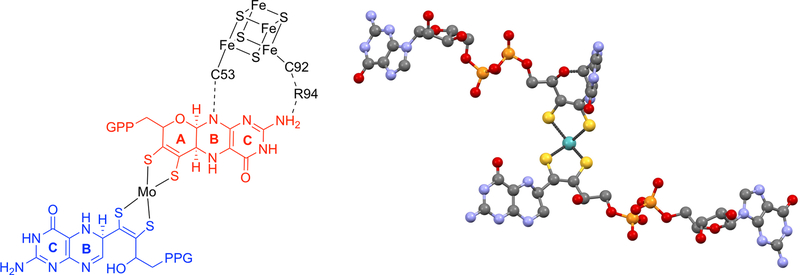
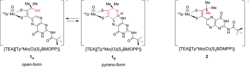
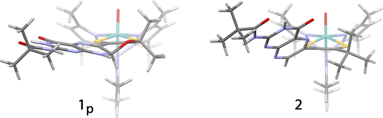
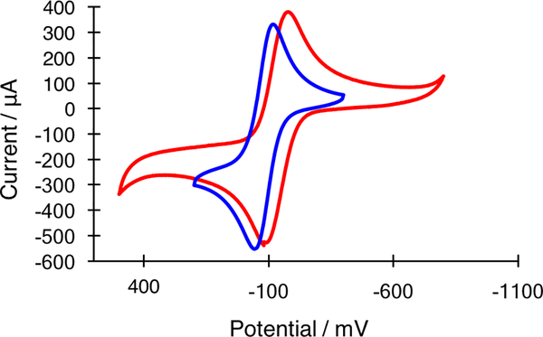
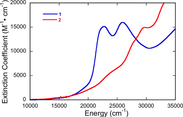
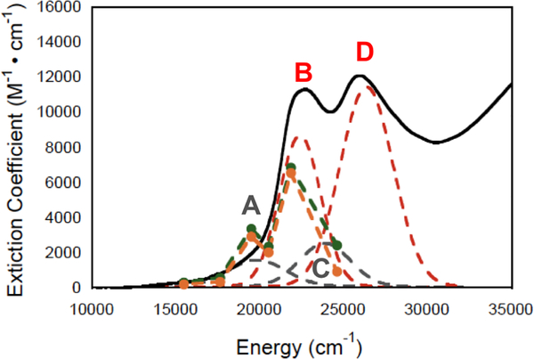
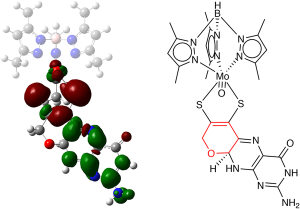
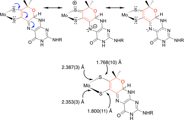
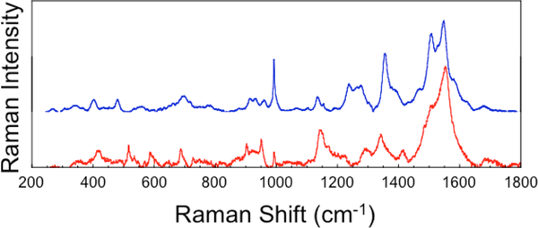
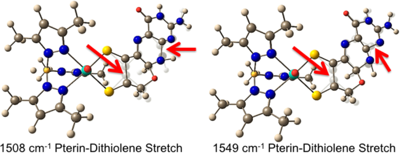
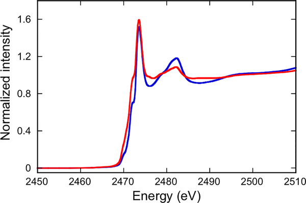
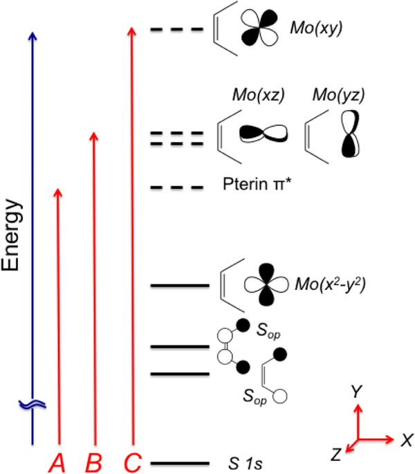
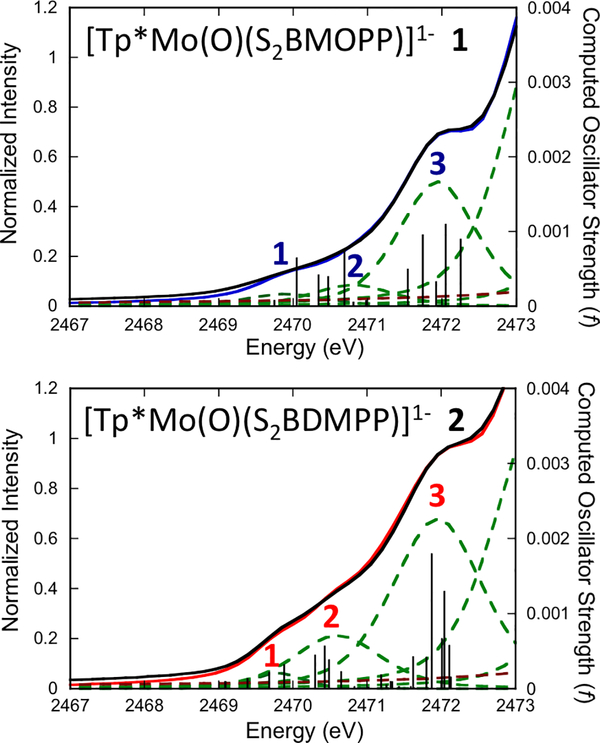
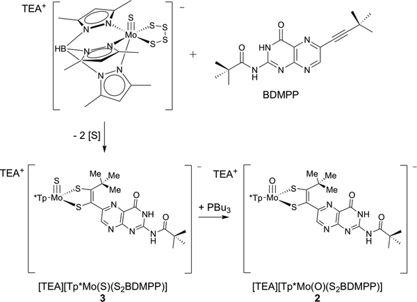
The large family of mononuclear molybdenum and tungsten enzymes all possess the special ligand molybdopterin (MPT), which consists of a metal-binding dithiolene chelate covalently bound to a pyranopterin group. MPT pyran cyclization/scission processes have been proposed to modulate the reactivity of the metal center during catalysis. We have designed several small-molecule models for the Mo-MPT cofactor that allow detailed investigation into how pyran cyclization modulates electronic communication between the dithiolene and pterin moieties and how this cyclization alters the electronic environment of the molybdenum catalytic site. Using a combination of cyclic voltammetry, vibrational spectroscopy (FT-IR and rR), electronic absorption spectroscopy, and X-ray absorption spectroscopy, distinct changes in the Mo≡O stretching frequency, Mo(V/IV) reduction potential, and electronic structure across the pterin-dithiolene ligand are observed as a function of pyran ring closure. The results are significant, for they reveal that a dihydropyranopterin is electronically coupled into the Mo-dithiolene group due to a coplanar conformation of the pterin and dithiolene units, providing a mechanism for the electron-deficient pterin to modulate the Mo environment. A spectroscopic signature identified for the dihydropyranopterin-dithiolene ligand on Mo is a strong dithiolene → pterin charge transfer transition. In the absence of a pyran group bridge between pterin and dithiolene, the pterin rotates out of plane, largely decoupling the system. The results support a hypothesis that pyran cyclization/scission processes in MPT may function as a molecular switch to electronically couple and decouple the pterin and dithiolene to adjust the redox properties in certain pyranopterin molybdenum enzymes.


Figures

Figure 1.
Figure 1.
(left) The two pterin dithiolene ligands of the periplasmic nitrate reductase NarGHI from E. coli. The pyranopterin dithiolene is comprised of pyrimidine (C), pyrazine (B), and pyran (A) rings of the proximal MPT (red) adjacent to the [4Fe-4S] cluster FS0, and the distal MPT (blue) is bicyclic. Dashed lines represent hydrogen bonding interactions between Moco and FS0. A bidentate carboxylate interaction from Asp222 completes the Mo coordination sphere. PPG denotes a guanosine dinucleotide group. (right) Detailed view of Moco from NarGHI illustrating the markedly different conformations of the proximal vs distal pterin regions.
Figure 2.
Figure 2.
(Left) Equilibrium of [TEA][Tp*Mo(O)(S2BMOPP)] (1) between the open and pyran forms 1o and 1p. (Right) [TEA][Tp*Mo(O)(S2BDMPP)] (2) synthesized by isosteric replacement of a hydroxyl by a methyl group to preclude pyran ring formation.
Figure 3.
Figure 3.
(Left) X-ray structure of 1p shows that pyran formation enforces a nearly coplanar arrangement of the dithiolene and pterin systems, with the angle between the dithiolene chelate and the pterin rings 40° out of planarity being τ = 9°. (Right) The DFT optimized structure of 2 shows the pterin rotated τ ≈ with the dithiolene due to a steric repulsion between the t-butyl group and the pterin.
Figure 4.
Figure 4.
Cyclic voltammograms of the Mo (V/IV) couple of 1 (blue) and 2 (red). The voltammograms are plotted versus the potential of reference electrode Ag/AgCl in (n-Bu4N)(ClO4)/CH3CN at a scan rate of 100 mV/sec using a Pt working electrode. Mo(V/IV) potentials: 1 −520 mV, 2 −574 mV, vs Fc+/Fc.
Figure 5.
Figure 5.
Room temperature electronic absorption spectra of 1 (blue) and 2 (red), 3.00 × 10⁻⁵ M, in DMSO.
Figure 6.
Figure 6.
Gaussian resolved electronic absorption spectrum of 1 in DMSO. Resonance Raman profiles for two high frequency S2BMOPP ligand C=C stretches (green and orange dots) are included with Gaussian-resolved peaks corresponding to transitions A (grey), B (red), C (grey), and D (red). Resonance enhancement of these modes is fully consistent with our assignents of Bands A and B, which possess pterin LUMO acceptor character.
Figure 7.
Figure 7.
Electron density difference map (EDDM; isovalue = 0.001) for Band B in 1 derived from TD-DFT pterin calculations showing dominant (74%) Sdithiolene → pterin character. Red regions represent a loss in electron density for the transition and green regions represent a gain in electron density for the transition. The molecule is oriented with the Mo≡O bond out of the plane and toward the reader. The bond line drawing illustrates the computational model, where the two methyl groups on pyran ring are replaced by protons and the pivaloyl group was removed from the amino group.
Figure 8.
Figure 8.
(Top) Contributing resonance structures for 1, with bond line drawing depicting the resultant extended π conjugation. (Bottom) Contributing resonance structures result from an admixture of ILCT excited states into the electronic ground state, and create bond asymmetry in the dithiolene chelate, which is observed in the bond metrics of the X-ray structure of 1.
Figure 9.
Figure 9.
Solid-state rR spectra (488 nm/20,492 cm−1 excitation) of 1 (blue) (50 mW) and 2 (red) (40 mW).
Figure 10.
Figure 10.
DFT computed normal mode descriptions for the most resonantly enhanced pterin-dithiolene stretching vibrations in 1.
Figure 11.
Figure 11.
S K-edge XAS spectra for 1 (blue) and 2 (red). The spectral range is limited to 2510 eV due to the presence of the Mo L3 absorption at 2520 eV. Note the greater pre-edge intensity of 2 relative to 1, indicating a greater degree of S orbital character in the valence molecular orbitals of 2.
Figure 12.
Figure 12.
Energy level diagram for S K-edge XAS analysis that is consistent with bonding calculations for 1 and 2. Solid horizontal lines represent doubly occupied orbitals and dashed horizontal lines represent empty (virtual) orbitals. For 1, the HOMO is the Mo(x2-y2) orbital and the LUMO is a pterin π* orbital. For 2, there are two pterin π* orbitals. Higher energy acceptor orbitals for the S K-edge transitions are to the Mo(xz,yz) orbitals that are Mo≡O π* in nature, and the Mo(x2-y2) orbital which is σ* with the dithiolene S donors. Transitions A-C describe the nature of the S K-edge peaks observed in Figure 12. Note that the z-axis is orthogonal to the plane of the paper.
Figure 13.
Figure 13.
Pseudo-Viogt fits (black) and individual fitted peaks (green) to the S K-edge pre-edge region for 1 (blue) and 2 (red). TDDFT computed transition energies and oscillator strengths for the individual S(1s) to valence orbital transitions are depicted as stick spectra for comparison. The TDDFT computed energies are shifted by +39.4 eV.
Scheme 1.
Scheme 1.
Synthetic route to [TEA][Tp*Mo(O)(S2BDMPP)] 2.

Similar articles

Cited by

References

    1. Kirk ML, Spectroscopic and Electronic Structure Studies of Mo Model Compounds and Enzymes In Molybdenum and Tungsten Enzymes: Spectroscopic and Theoretical Investigations, The Royal Society of Chemistry: Cambridge, UK, 2016.
    1. Stein BW; Kirk ML, Electronic structure contributions to reactivity in xanthine oxidase family enzymes. Journal of Biological Inorganic Chemistry 2015, 20 (2), 183–194. - PMC - PubMed
    1. Kirk ML; Stein B, The Molybdenum Enzymes In Comprehensive Inorganic Chemistry II (Second Edition), Editors-in-Chief: Jan R; Kenneth P, Eds. Elsevier: Amsterdam, 2013; pp 263–293.
    1. Maia LB; Moura I; Moura JJG, Molybdenum and Tungsten-Containing Enzymes: An Overview. 2017; Vol. 5, p 1–80.
    1. Hille R; Hall J; Basu P, The Mononuclear Molybdenum Enzymes. Chemical Reviews 2014, 114 (7), 3963–4038. - PMC - PubMed

Publication types

LinkOut - more resources