

Phase contrast mapping MRI measurements of global cerebral blood flow across different perfusion states - A direct comparison with 15O-H2O positron emission tomography using a hybrid PET/MR system
- PMID: 30200799
- PMCID: PMC6890999
- DOI: 10.1177/0271678X18798762
Phase contrast mapping MRI measurements of global cerebral blood flow across different perfusion states - A direct comparison with 15O-H2O positron emission tomography using a hybrid PET/MR system
Abstract
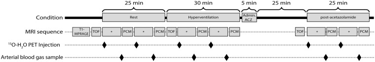
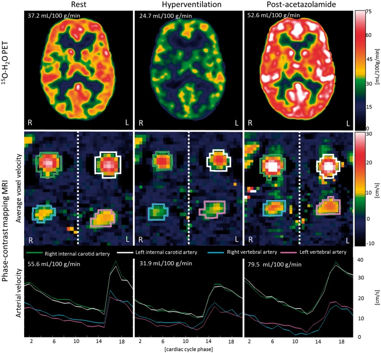
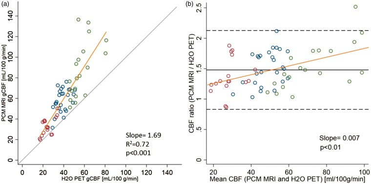
Phase-contrast mapping (PCM) magnetic resonance imaging (MRI) provides easy-access non-invasive quantification of global cerebral blood flow (gCBF) but its accuracy in altered perfusion states is not established. We aimed to compare paired PCM MRI and 15O-H2O positron emission tomography (PET) measurements of gCBF in different perfusion states in a single scanning session. Duplicate combined gCBF PCM-MRI and 15O-H2O PET measurements were performed in the resting condition, during hyperventilation and after acetazolamide administration (post-ACZ) using a 3T hybrid PET/MR system. A total of 62 paired gCBF measurements were acquired in 14 healthy young male volunteers. Average gCBF in resting state measured by PCM-MRI and 15O-H2O PET were 58.5 ± 10.7 and 38.6 ± 5.7 mL/100 g/min, respectively, during hyperventilation 33 ± 8.6 and 24.7 ± 5.8 mL/100 g/min, respectively, and post-ACZ 89.6 ± 27.1 and 57.3 ± 9.6 mL/100 g/min, respectively. On average, gCBF measured by PCM-MRI was 49% higher compared to 15O-H2O PET. A strong correlation between the two methods across all states was observed (R2 = 0.72, p < 0.001). Bland-Altman analysis suggested a perfusion dependent relative bias resulting in higher relative difference at higher CBF values. In conclusion, measurements of gCBF by PCM-MRI in healthy volunteers show a strong correlation with 15O-H2O PET, but are associated with a large and non-linear perfusion-dependent difference.
Keywords: 15O-H2O; cerebral blood flow; magnetic resonance imaging; phase contrast mapping; positron emission tomography.
Figures
References
-
- Wintermark M, Sesay M, Barbier E, et al. Comparative overview of brain perfusion imaging techniques. Stroke 2005; 36: e86–99. - PubMed
-
- Spilt A, Box FM, Van Der Geest RJ, et al. Reproducibility of total cerebral blood flow measurements using phase contrast magnetic resonance imaging. J Magn Reson Imaging 2002; 16: 1–5. - PubMed
-
- Vernooij MW, Van Der Lugt A, Ikram MA, et al. Total cerebral blood flow and total brain perfusion in the general population: the Rotterdam Scan Study. J Cereb Blood Flow Metab 2008; 28: 412–419. - PubMed
-
- Appelman AP, Van Der Graaf Y, Vincken KL, et al. Total cerebral blood flow, white matter lesions and brain atrophy: the SMART-MR study. J Cereb Blood Flow Metab 2008; 28: 633–639. - PubMed
-
- Coverdale NS, Gati JS, Opalevych O, et al. Cerebral blood flow velocity underestimates cerebral blood flow during modest hypercapnia and hypocapnia. J Appl Physiol 2014; 117: 1090–1096. - PubMed
Publication types
MeSH terms
Substances
LinkOut - more resources
Full Text Sources
Other Literature Sources
