. 2018 Sep 10;9(1):40.
doi: 10.1186/s13293-018-0202-x.

Sex-specific differences in hepatic steatosis in obese spontaneously hypertensive (SHROB) rats

Affiliations

Sex-specific differences in hepatic steatosis in obese spontaneously hypertensive (SHROB) rats

Qingming Dong et al. Biol Sex Differ. .

Abstract

Background: Patients with metabolic syndrome, who are characterized by co-existence of insulin resistance, hypertension, hyperlipidemia, and obesity, are also prone to develop non-alcoholic fatty liver disease (NAFLD). Although the prevalence and severity of NAFLD is significantly greater in men than women, the mechanisms by which gender modulates the pathogenesis of hepatic steatosis are poorly defined. The obese spontaneously hypertensive (SHROB) rats represent an attractive model of metabolic syndrome without overt type 2 diabetes. Although pathological manifestation caused by the absence of a functional leptin receptor has been extensively studied in SHROB rats, it is unknown whether these animals elicited sex-specific differences in the development of hepatic steatosis.

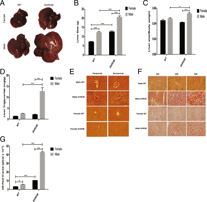
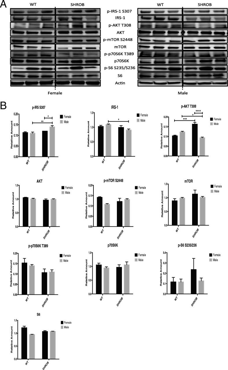
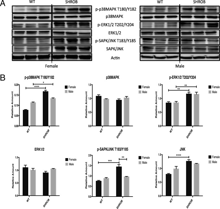
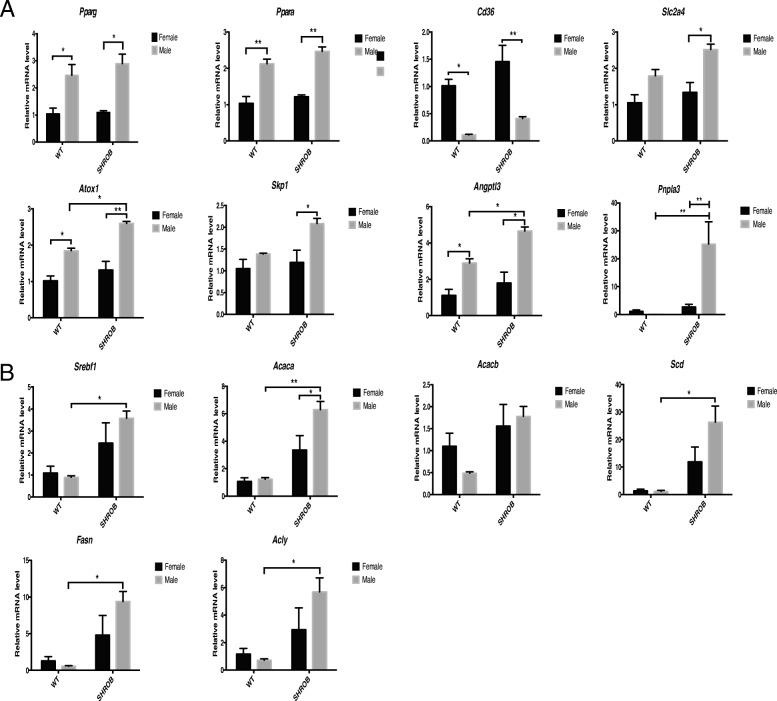
Methods: We compared hepatic pathology in male and female SHROB rats. Additionally, we examined key biochemical and molecular parameters of signaling pathways linked with hyperinsulinemia and hyperlipidemia. Finally, using methods of quantitative polymerase chain reaction (qPCR) and western blot analysis, we quantified expression of 45 genes related to lipid biosynthesis and metabolism in the livers of male and female SHROB rats.

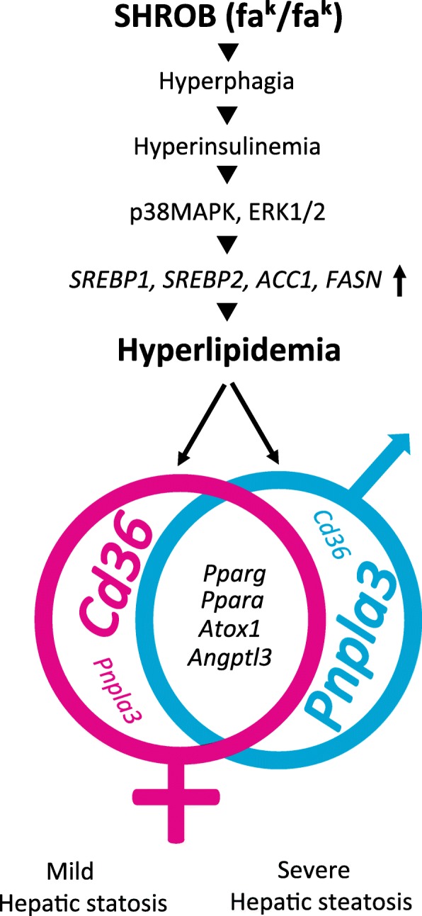
Results: We show that all SHROB rats developed hepatic steatosis that was accompanied by enhanced expression of SREBP1, SREBP2, ACC1, and FASN proteins. The livers of male rats also elicited higher induction of Pparg, Ppara, Slc2a4, Atox1, Skp1, Angptl3, and Pnpla3 mRNAs. In contrast, the livers of female SHROB rats elicited constitutively higher levels of phosphorylated JNK and AMPK and enhanced expression of Cd36.

Conclusion: Based on these data, we conclude that the severity of hepatic steatosis in male and female SHROB rats was mainly driven by increased de novo lipogenesis. Moreover, male and female SHROB rats also elicited differential severity of hepatic steatosis that was coupled with sex-specific differences in fatty acid transport and esterification.


Conflict of interest statement

Ethics approval and consent to participate

All animal procedures were approved by the Institutional Animal Care and Use Committee (IACUC) of University of Tennessee Health Science Center (UTHSC).

Competing interests

The authors declare that they have no competing interests.

Publisher’s Note

Springer Nature remains neutral with regard to jurisdictional claims in published maps and institutional affiliations.

Figures

Fig. 1
Fig. 1
Gender-specific comparison of body composition in WT and SHROB rats. a Representative gross morphology of 10-week-old female and male WT and SHROB rats. b Body weight curves of various groups between week 3 and week 10. c Body weight at week 10. d Fat mass at week 10. e Percent body fat at week 10. f Lean mass at week 10. g Percent lean mass at week 10. Data are presented as mean ± SEM. n = 5 to 12 for each group. Statistical significance: *p < 0.05, **p < 0.01, ***p < 0.001, and ****p < 0.0001
Fig. 2
Fig. 2
Morphological and histological comparisons of the livers from WT and SHROB rats. a Representative gross morphology of the livers from male and female WT and SHROB rats at week 10 of age. Liver weight (b) and liver weight normalized against total body weight (c) in WT and SHROB rats at week 10 is shown. Each group had between 5 and 12 rats. d, e Representative microphotographs of female and male WT and SHROB rat livers stained with H&E (d) and Oil Red O (e). f Quantification Oil Red O staining. g Results are presented as the means ± SEM of three separate experiments; × 20 magnification is used for quantification. n = 3 for each group. Statistical significance: **p < 0.01, ****p < 0.0001
Fig. 3
Fig. 3
Serum chemistries for 10-week-old female and male WT and SHROB rats. Data are mean ± SEM. n = 5 to 12 for each group. a AST, aspartate aminotransferase. b ALT, alanine aminotransferase. c Amylase. d BUN, blood urea nitrogen. e Creatinine. f Glucose. g Cholesterol. h Triglyceride. i Insulin. Statistical significance: *p < 0.05, **p < 0.01, ***p < 0.001, and ****p < 0.0001
Fig. 4
Fig. 4
Analysis of canonical insulin and mTOR signaling pathways in the livers of female and male WT and SHROB rats. a Western blots of total and phosphorylated proteins from three rat livers for each group are shown. b Densitometry of polypeptide band (panel a) was used to calculate the amount of target proteins and was normalized against actin. Statistical significance: *p < 0.05, **p < 0.01, ***p < 0.001, and ****p < 0.0001
Fig. 5
Fig. 5
Western blots and densitometry for MAPK pathway in female and male WT and SHROB rat livers. a Western blots. b Densitometry data from panel a showing relative amount of total proteins, p-ERK1/2T202/Y204 and p-SAPK/JNK T183/Y185 after their normalization against actin. The relative amount of p-p38MAPK T180/Y182 was normalized against p38MAPK level and actin. Statistical significance: *p < 0.05, **p < 0.01, ***p < 0.001, and ****p < 0.0001
Fig. 6
Fig. 6
Western blot analysis and quantification of lipogenesis proteins in the livers of WT and SHROB rats. a Western blots for female/male WT and SHROB rat livers sacrificed at week 10, n = 3 for each group. b Densitometry data from (panel a). The relative amount of total proteins and p-AMPK T172 is based on target gene expression level normalized by actin. Statistical significance: *p < 0.05, **p < 0.01, ***p < 0.001, and ****p < 0.0001
Fig. 7
Fig. 7
Quantification of gender-specific differences in hepatic gene expression by qPCR in SHROB rats. The relative mRNA levels were normalized against Rn18s RNA and compared with WT female set as 1. N = 3 for each group. Statistical significance: *p < 0.05, **p < 0.01. a Genes related to gender differences including peroxisome proliferator-activated receptor gamma (Pparg), peroxisome proliferator-activated receptor alpha (Ppara), cluster of differentiation 36 (Cd36), solute carrier family 2 member 4 (Slc2a4), antioxidant protein 1 (Atox1), S-phase kinase-associated protein 1 (Skp1), angiopoietin-like 3 (Angptl3), and papatin-like phospholipase domain-containing protein 3 (Pnpla3). b Genes related to lipogenesis including sterol regulatory element-binding transcription factor 1 (Srebf1), acetyl-CoA carboxylase alpha (Acaca), acetyl-CoA carboxylase beta (Acacb), stearoyl-CoA desaturase (Scd), fatty acid synthesis (Fasn), and ATP citrate lyase (Acly)
Fig. 8
Fig. 8
Hypothetical scheme showing major steps involved in sex-specific differences in the pathogenesis of hepatic steatosis in SHROB rats. Both female and male SHROB rats elicit hyperphagia, hyperinsulinemia, and hyperlipidemia. Activation of hyperinsulinemia-induced signaling pathways leads to induction of genes that regulate de novo lipogenesis. Several genes that control lipid metabolism are highly expressed in male and female SHROB rat livers. Expression of Cd36 and Pnpla3 genes was differentially regulated in female and male SHROB rats, leading to milder and severe hepatic steatosis, respectively

Similar articles

Cited by

References

    1. Reaven GM. Banting lecture 1988. Role of insulin resistance in human disease. Diabetes. 1988;37:1595–1607. doi: 10.2337/diab.37.12.1595. - DOI - PubMed
    1. Aguilar M, Bhuket T, Torres S, Liu B, Wong RJ. Prevalence of the metabolic syndrome in the United States, 2003–2012. JAMA. 2015;313:1973–1974. doi: 10.1001/jama.2015.4260. - DOI - PubMed
    1. Zhang Y, Proenca R, Maffei M, Barone M, Leopold L, Friedman JM. Positional cloning of the mouse obese gene and its human homologue. Nature. 1994;372:425–432. doi: 10.1038/372425a0. - DOI - PubMed
    1. Chen H, Charlat O, Tartaglia LA, Woolf EA, Weng X, Ellis SJ, Lakey ND, Culpepper J, Moore KJ, Breitbart RE, et al. Evidence that the diabetes gene encodes the leptin receptor: identification of a mutation in the leptin receptor gene in db/db mice. Cell. 1996;84:491–495. doi: 10.1016/S0092-8674(00)81294-5. - DOI - PubMed
    1. Chua SC, Jr, Chung WK, Wu-Peng XS, Zhang Y, Liu SM, Tartaglia L, Leibel RL. Phenotypes of mouse diabetes and rat fatty due to mutations in the OB (leptin) receptor. Science. 1996;271:994–996. doi: 10.1126/science.271.5251.994. - DOI - PubMed

Publication types