. 2018 Sep 10;20(1):207.
doi: 10.1186/s13075-018-1701-1.

Osteoporosis of the vertebra and osteochondral remodeling of the endplate causes intervertebral disc degeneration in ovariectomized mice

Affiliations

Osteoporosis of the vertebra and osteochondral remodeling of the endplate causes intervertebral disc degeneration in ovariectomized mice

Zhi-Feng Xiao et al. Arthritis Res Ther. .

Abstract

Background: Studies on the relationship between osteoporosis and intervertebral disc degeneration (IVDD) are inconsistent. Therefore, we assessed whether IVDD is affected by vertebral osteoporosis in ovariectomized mice and investigated the underlying pathogenesis of IVDD related to osteoporosis.

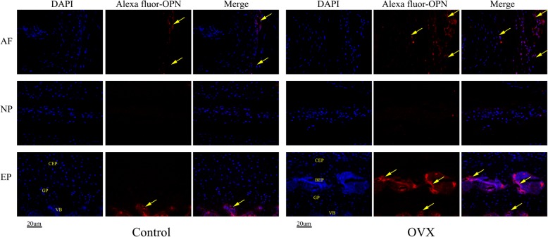
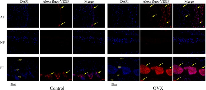
Methods: Thirty healthy female C57BL/6 J mice aged 8 weeks were randomly divided into two groups: a control group (sham operation, n = 15) and an ovariectomy group (OVX; bilateral ovariectomy, n = 15). At 12 weeks after surgery, the bone quantity and microstructure in the lumbar vertebra and endplate as well as the volume of the L4/5 disc space were evaluated by microcomputed tomography (micro-CT). The occurrence and characteristic alterations of IVDD were identified via histopathological staining. The osteoclasts were detected using tartrate-resistant acid phosphatase (TRAP) staining. Type II collagen (Col II), osterix (OSX), osteopontin (OPN), and vascular endothelial growth factor (VEGF) expression in the intervertebral disc were detected by immunohistochemical analysis.

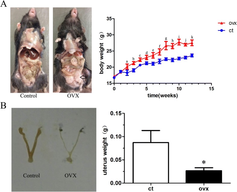
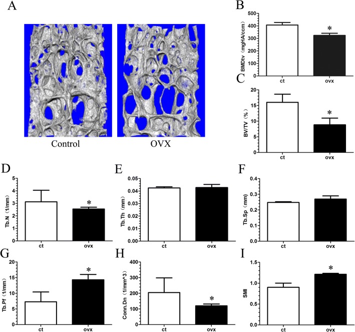
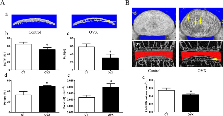
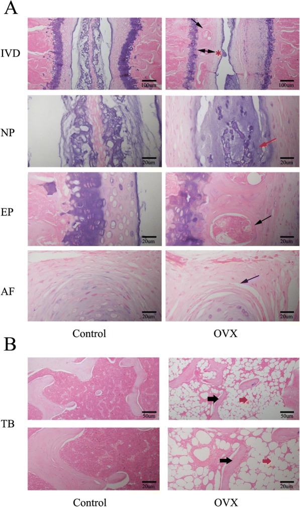
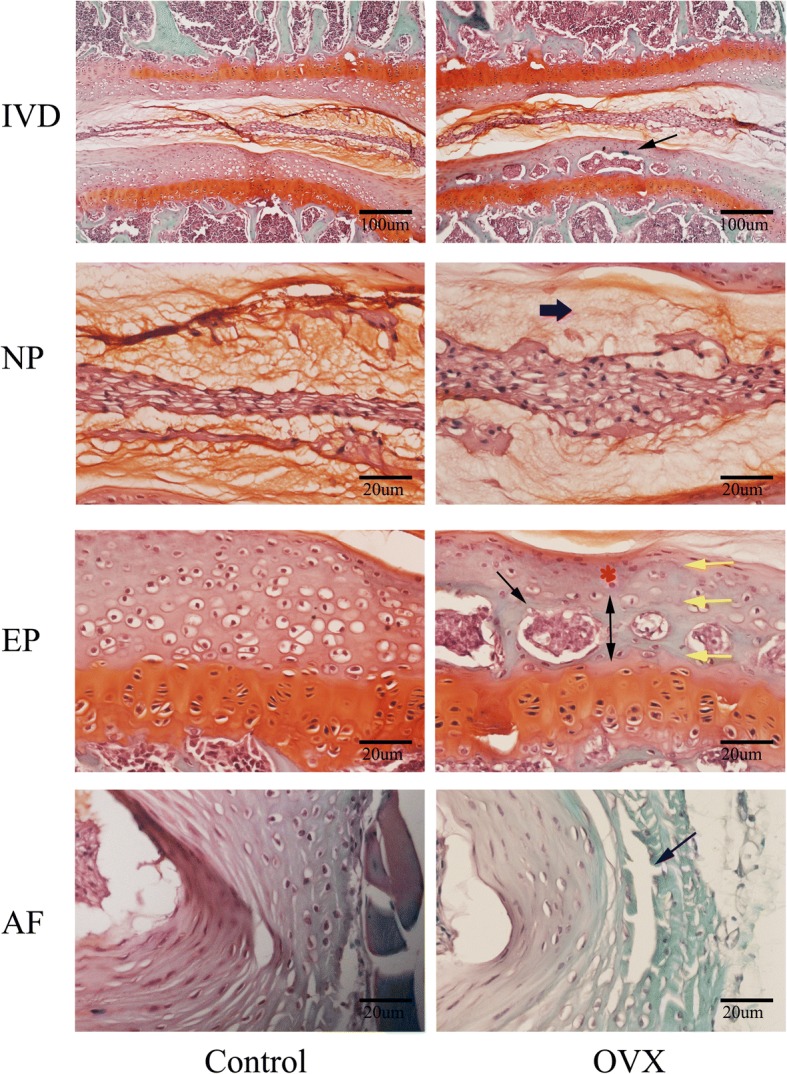
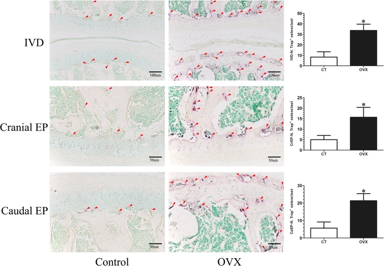
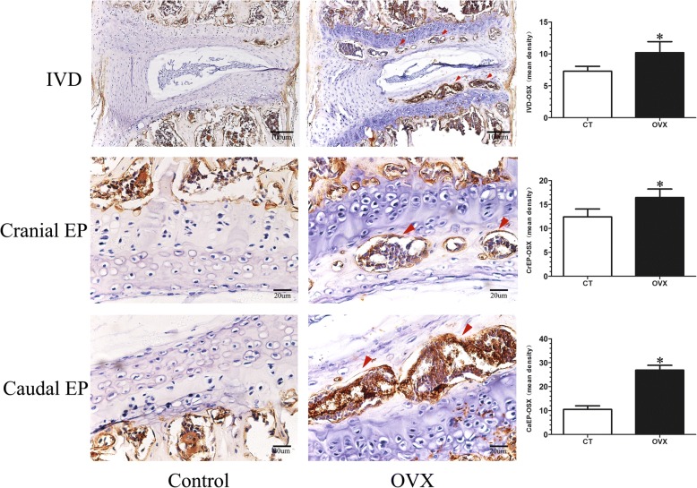
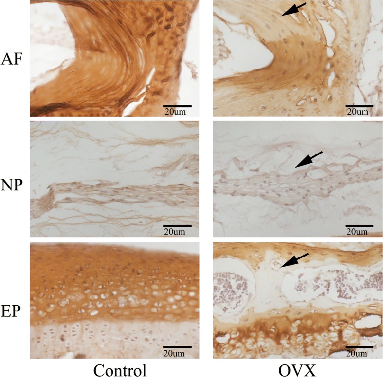
Results: OVX significantly increased the body weight and decreased the uterus weight. Micro-CT analysis showed that osteoporosis of the vertebra and osteochondral remodeling of the endplate were accompanied by an increase in the endplate porosity and a decrease in the disc volume in the OVX group. Likewise, histological evaluation revealed that IVDD occurred at 12 weeks after ovariectomy, with features of endochondral ossification of the endplate, loose and broken annulus fibrosus, and degeneration of nucleus pulposus. TRAP staining showed that numerous active osteoclasts appeared in the subchondral bone and cartilaginous endplate of OVX mice, whereas osteoclasts were rarely detected in control mice. Immunohistochemical analysis demonstrated that the expression of osterix was significantly increased, notably in the endplate of OVX mice. In addition, Col II was decreased in the ossification endplate and the degenerative annulus fibrosus, where OPN and VEGF expressions were elevated in OVX mice.

Conclusions: OVX induced vertebral osteoporosis and osteochondral remodeling of the cartilaginous endplate contributing to the angiogenesis and an increase in porosity of the bone-cartilage surface, and also affected the matrix metabolism which consequently had detrimental effects on the intervertebral disc. Our study suggests that preserving the structural integrity and the function of the adjacent structures, including the vertebrae and endplates, may protect the disc against degeneration.

Keywords: Endplate; Intervertebral disc degeneration; Microcomputed tomography; Osteochondral remodeling; Osteoporosis; Ovariectomy.


Conflict of interest statement

Ethics approval

Animal studies were performed under institutional guidelines and in accordance with protocols approved by the Ethics Committee, Guangzhou University of Chinese Medicine.

Consent for publication

Not applicable.

Competing interests

The authors declare that they have no competing interests.

Publisher’s Note

Springer Nature remains neutral with regard to jurisdictional claims in published maps and institutional affiliations.

Figures

Fig. 1
Fig. 1
Gross macrographs and body and uterus weight changes of mice after ovariectomy. a There is a remarkable accumulation of fat in the abdominal cavity (black open arrow) and the body weight increase significantly over time in ovariectomized (OVX) mice. b The uterus is visibly atrophied in the OVX group and is significant lighter in weight compared with the control (ct) group. Data are shown as mean ± SD; n = 15 per group. a–kp < 0.05 vs control group at 2–12 weeks for body weight; *p < 0.05 vs control group for uterus weight
Fig. 2
Fig. 2
Changes in L5 total volume of bone mineral density (BMDtv) and microarchitecture parameters of the trabecular bone determined by micro-CT. a Representative three-dimensional images of trabecular bone. bi The parameters of L5 trabecular bone. There is markedly decreased BMDtv, bone volume (BV)/total volume (TV), trabecular number (Tb.N; 1/mm), and connectivity density (CONN.D, 1/mm3), and increased trabecular thickness (Tb.Th; mm), and structural model index (SMI) in the ovariectomized (OVX) group when compared with the control (ct) group. Mice were analyzed at 12 weeks post-sham or OVX surgery, n = 6 per group. Data are shown as mean ± SD. *p < 0.05. Tb.Pf trabecular pattern factor, Tb.Sp trabecular separation
Fig. 3
Fig. 3
Changes in microarchitecture, porosity of L4/5 caudal endplate, and disc volume quantified by micro-CT analysis. A a Three-dimensional images and parameters of caudal endplate. Results showing that increased cavities in OVX mice (yellow arrow) indicate osteochondral remodeling of the endplate. b–e Markedly decreased bone volume (BV)/total volume (TV) and number of closed pores (Po.N(cl)) and increased open porosity (Po(op)) and total volume of pore space (Po.V(tot)) are shown in the ovariectomized (OVX) group. B a The top view of the caudal endplate showing a higher surface porosity in OVX mice (yellow arrows). b,c Quantification of disc volume by micro-CT showing a significant decrease in OVX mice. The ROI of the disc is indicated by the red color. Data are shown as mean ± SD, n = 6 per group. *p < 0.05. CT control, IVD intervertebral disc
Fig. 4
Fig. 4
Representative images of H&E staining of the intervertebral disc (IVD) and tibia (TB). a Panoramic images of IVD pathology and higher magnification of the endplate (EP), nucleus pulposus (NP), and annulus fibrosus (AF). Ossific nodules (black arrows) in the endplate along with thickening of bony endplate (double arrow) and thinning of the cartilage endplate (red asterisk) were indicated in ovariectomized (OVX) mice. In addition, reduction of notochord cells, degeneration of nucleus pulposus (red arrow), and cleft formation within the annulus fibrosus (blue arrow) appeared in OVX mice. b Images of tibial pathology. Black arrows demonstrate slender trabecular bone and red arrows indicate fat droplets in the bone marrow of OVX mice. n = 9 per group; scale bars = 100 μm, 50 μm, and 20 μm as indicated
Fig. 5
Fig. 5
Representative images of safranin O and fast green staining of the intervertebral disc (IVD). This showed consistent results with H&E staining. Specially, thickening of the bony endplate (EP; double arrow) accompanied by accelerated osteochondral remodeling (black arrows) and duplication of tidemarks (yellow arrow) were displayed more clearly in the EP of ovariectomized (OVX) mice by safranin O fast green staining. Moreover, reduction of aggrecan in the nucleus pulposus (NP), as indicated by a paucity of safranin O staining (blue thick arrow), cleft/crack formation in the annulus fibrosus (AF) (blue arrow), and loss of cells indicate intervertebral disc degeneration in OVX mice. n = 9 per group; scale bars = 100 μm and 20 μm as indicated
Fig. 6
Fig. 6
Tartrate acid phosphatase (TRAP) staining of L4/5 coronal sections. The osteoclasts were obtained by counting the number of TRAP-positive staining cells (N. Trap+). A few TRAP-positive cells (purple; red arrows) were distributed on the surface of the trabecular of the subchondral bone and were rarely detected in the endplate of control (CT) mice. However, the TRAP-positive cells (purple; red arrows) significantly increased in the subchondral bone and were obviously noted in the endplate of ovariectomized (OVX) mice, suggesting osteoclast activity increases after OVX. Data are shown as mean ± SD, n = 6 per group. *p < 0.05; scale bars = 100 μm and 50 μm as indicated. CaEP caudal endplate, CrEP cranial endplate, IVD intervertebral disc
Fig. 7
Fig. 7
Representative sections of immunohistochemistry of osterix in the mid-sagittal plane. The mean integrated optical density (mean density) was obtained to assess the expression of osterix (OSX). Little osterix expression could be observed in the intervertebral disc (IVD) of control (CT) mice, whereas a significant increased osterix expression (red arrows) was found in the subchondral bone and endplate of ovariectomized (OVX) mice. Data are shown as mean ± SD, n = 6 per group. *p < 0.05; scale bars = 100 μm and 20 μm as indicated. CaEP caudal endplate, CrEP cranial endplate
Fig. 8
Fig. 8
Representative sections of immunohistochemistry of Col II. Positive immunostaining was noted as brown staining. Black arrows indicate loss of Col II, especially in the ossification area of the cartilaginous endplate (EP) and the outer layer of the annulus fibrosus (AF) in ovariectomized (OVX) mice. n = 9 per group; scale bars = 20 μm as indicated. NP nucleus pulposus
Fig. 9
Fig. 9
Immunohistochemical staining of osteopontin (OPN). DAPI stains nuclei blue and OPN expression was detected as red. Both control and ovariectomized (OVX) mice have OPN expression in the vertebral body (VB) and outer annulus fibrosus (AF), but it was almost undetectable in the nucleus. However, remarkable expression of OPN was found in the bony endplate (BEP) and outer AF in OVX mice. Meanwhile, the expression of OPN was also detected in the inner AF. n = 9 per group; scale bar = 20 μm as indicated. CEP cartilaginous endplate, EP endplate, GP growth plate, NP nucleus pulposus
Fig. 10
Fig. 10
Immunohistochemical staining of vascular endothelial growth factor (VEGF). Consistent with OPN expression, a large amount of VEGF expression was observed in the outer layer of the annulus fibrosus (AF) and the bone marrow of the bony endplate (BEP) indicating that vasculogenesis and angiogenesis are associated with ossification of soft tissue and play an important role in the process of IVDD. n = 9 per group; scale bars = 20um as indicated. CEP cartilaginous endplate, EP endplate, GP growth plate, NP nucleus pulposus, VB vertebral body

References

    1. Luoma K, Vehmas T, Kerttula L, Grönblad M, Rinne E. Chronic low back pain in relation to modic changes, bony endplate lesions, and disc degeneration in a prospective MRI study. Eur Spine J. 2016;25(9):2873–2881. doi: 10.1007/s00586-016-4715-x. - DOI - PubMed
    1. Izzo R, Popolizio T, D'Aprile P, Muto M. Spinal pain. Eur J Radiol. 2015;84(5):746–756. doi: 10.1016/j.ejrad.2015.01.018. - DOI - PubMed
    1. Wenig CM, Schmidt CO, Kohlmann T, Schweikert B. Costs of back pain in Germany. Eur J Pain. 2009;13(3):280–286. doi: 10.1016/j.ejpain.2008.04.005. - DOI - PubMed
    1. Paesold G, Nerlich AG, Boos N. Biological treatment strategies for disc degeneration: potentials and shortcomings. Eur Spine J. 2007;16(4):447–468. doi: 10.1007/s00586-006-0220-y. - DOI - PMC - PubMed
    1. Humzah MD, Soames RW. Human intervertebral disc: structure and function. Anat Rec. 1988;220(4):337–356. doi: 10.1002/ar.1092200402. - DOI - PubMed

Publication types

LinkOut - more resources