. 2019 Apr:77:117-128.
doi: 10.1016/j.matbio.2018.09.004. Epub 2018 Sep 7.

Adamts10 inactivation in mice leads to persistence of ocular microfibrils subsequent to reduced fibrillin-2 cleavage

Affiliations

Adamts10 inactivation in mice leads to persistence of ocular microfibrils subsequent to reduced fibrillin-2 cleavage

Lauren W Wang et al. Matrix Biol. 2019 Apr.

Abstract

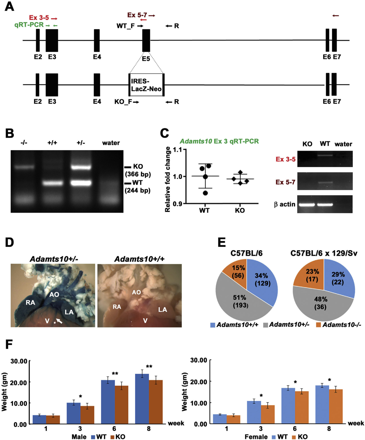
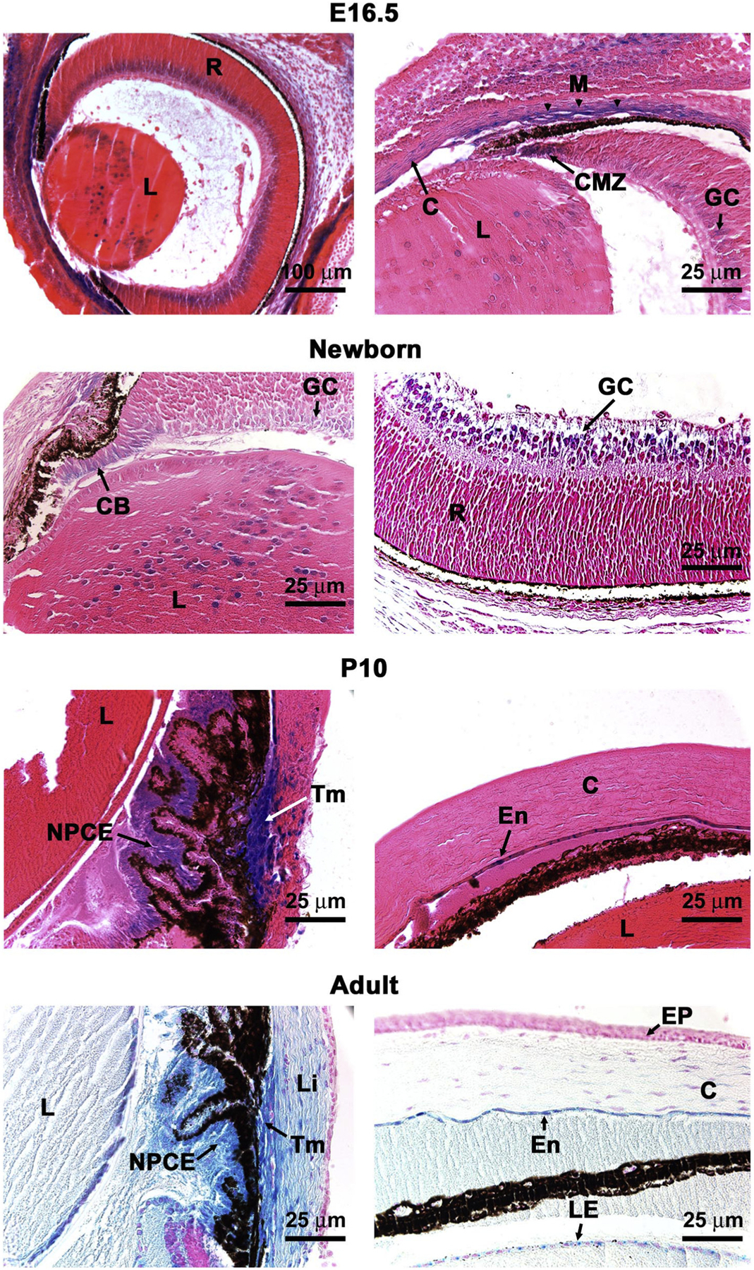
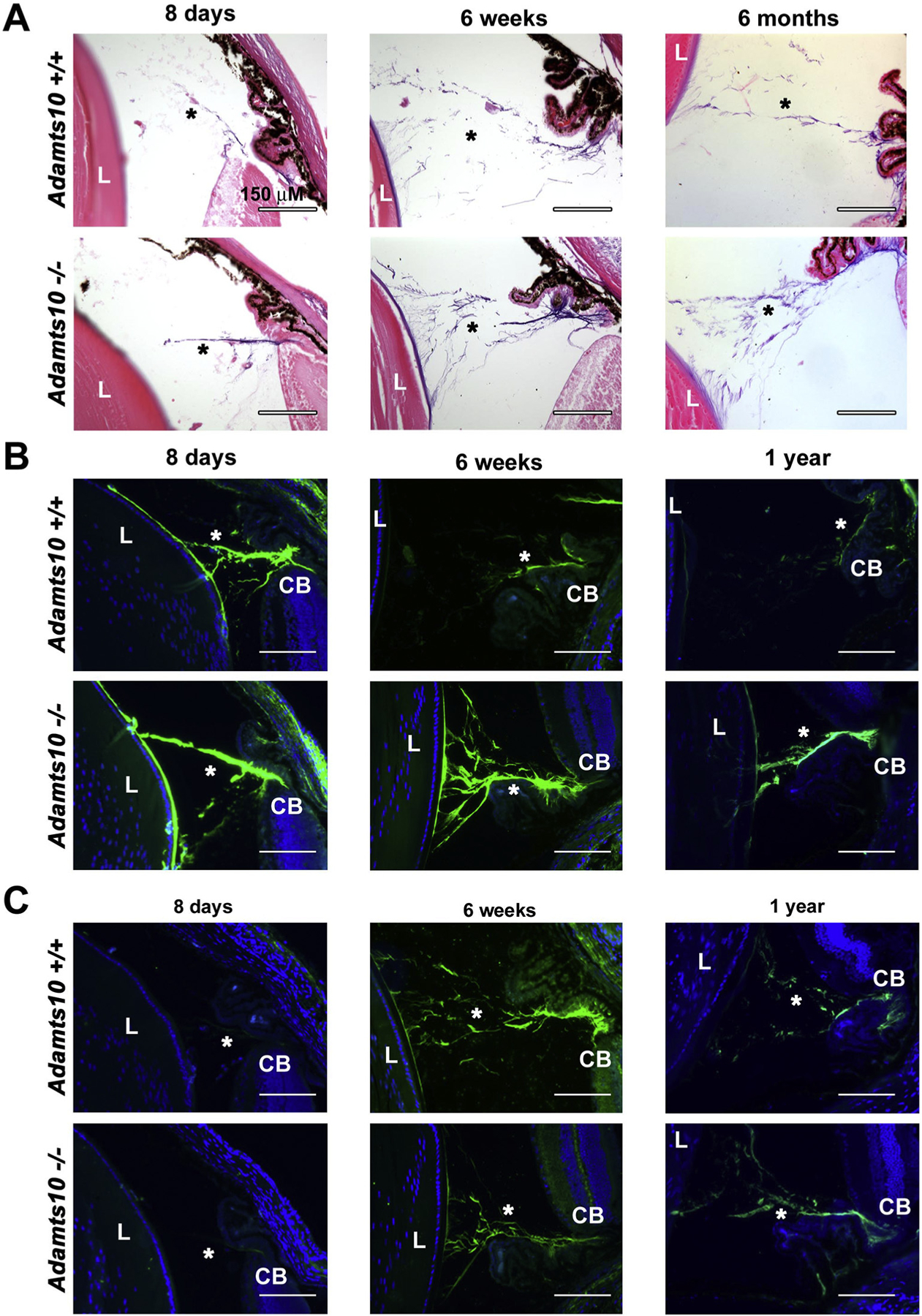
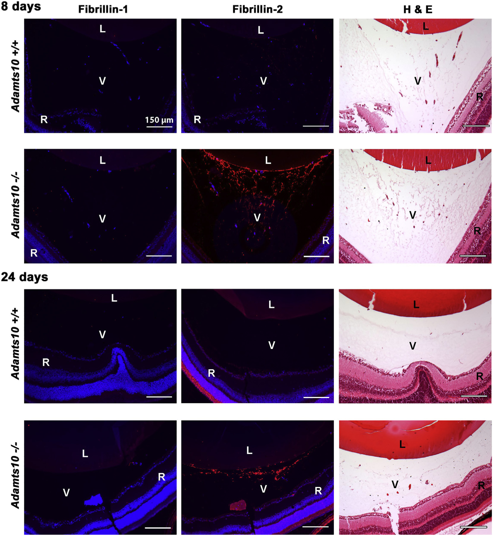
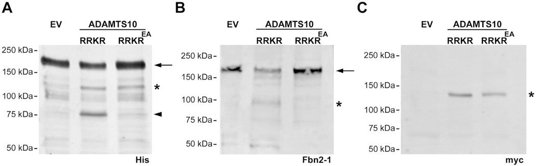
Mutations in the secreted metalloproteinase ADAMTS10 cause recessive Weill-Marchesani syndrome (WMS), comprising ectopia lentis, short stature, brachydactyly, thick skin and cardiac valve anomalies. Dominant WMS caused by FBN1 mutations is clinically similar and affects fibrillin-1 microfibrils, which are a major component of the ocular zonule. ADAMTS10 was previously shown to enhance fibrillin-1 assembly in vitro. Here, Adamts10 null mice were analyzed to determine the impact of ADAMTS10 deficiency on fibrillin microfibrils in vivo. An intragenic lacZ reporter identified widespread Adamts10 expression in the eye, musculoskeletal tissues, vasculature, skin and lung. Adamts10-/- mice had reduced viability on the C57BL/6 background, and although surviving mice were slightly smaller and had stiff skin, they lacked brachydactyly and cardiovascular defects. Ectopia lentis was not observed in Adamts10-/- mice, similar to Fbn1-/- mice, most likely because the mouse zonule contains fibrillin-2 in addition to fibrillin-1. Unexpectedly, in contrast to wild-type eyes, Adamts10-/- zonule fibers were thicker and immunostained strongly with fibrillin-2 antibodies into adulthood, whereas fibrillin-1 staining was reduced. Furthermore, fibrillin-2 staining of hyaloid vasculature remnants persisted post-natally in Adamts10-/- eyes. ADAMTS10 was found to cleave fibrillin-2, providing an explanation for persistence of fibrillin-2 at these sites. Thus, analysis of Adamts10-/- mice led to identification of fibrillin-2 as a novel ADAMTS10 substrate and defined a proteolytic mechanism for clearance of ocular fibrillin-2 at the end of the juvenile period.

Keywords: Acromelic dysplasia; Ectopia lentis; Extracellular matrix; Metalloprotease; Weill-Marchesani syndrome; Zonule.


Figures

Fig. 1.
Fig. 1.
Generation and characterization of Adamts10−/− mice. A. Gene targeting strategy: the structure of the wild-type Adamts10 allele inclusive of exon 2 through exon 7 (top) and the targeted Adamts10 allele (bottom) are illustrated. The primers used for genotyping are shown by black arrows (WT_F/R and KO_F/R). The primers used for RT-PCR are shown as pairs of red, maroon or green arrows indicating their location. B. Agarose gel electrophoresis of genomic PCR products shows the distinction between wild type (WT) and knockout (KO) alleles. Water was used as the control target. C. Left: q-RT-PCR shows comparable upstream RNA levels in wild-type and Adamts10−/− mice (KO). Right: RT-PCR (exons 3–5) shows interruption of exon 5 by insertion of the IRES-lacZ-neo cassette and loss of RNA downstream of the targeting site (exons 5–7). β-actin PCR shows comparable source RNA. D. Consistent with generation of an Adamts10-IRES-lacZ transcript, aorta (AO), right atrium (RA), left atrium (LA) and coronary vessels (arrow), but not the ventricular myocardium (V) from an Adamts10+/− mouse, but not a wild-type mouse demonstrated robust β-gal staining (blue). E. Pie chart of the genotypes detected at 10 days of age from intercrosses of Adamts10+/− mice in the indicated strains. Note reduced viability of Adamts10−/− mice in the C57BL/6 strain. F. Adamts10−/− mice segregated by sex, as shown, weighed significantly less than wild-type littermates. *p < 0.01, **p < 0.001, n = 7 male wild-type mice, 6 female wild type mice; 13 male Adamts10−/− mice and 11 female Adamts10−/− mice, error bars indicate S.D.
Fig. 2.
Fig. 2.
An Adamts10 reporter is expressed at specific sites in the mouse eye. β-gal staining (blue) of Adamts10+/− eyes indicates sites of Adamts10 mRNA expression in the 16.5-day old embryo (E16.5), newborn, and post-natal day 10 (P10) or adult (8 week) eyes as labeled. C, cornea; CB, ciliary body; CMZ, ciliary margin zone; En, corneal endothelium; EP, corneal epithelium; GC, ganglion cell layer of retina; L, lens; LE, lens epithelium; Li, limbus; M, primordial trabecular meshwork; NPCE, non-pigmented ciliary epithelium; Tm, trabecular meshwork; R, retina. Scale bars are 100 μm at top left and 25 μm in other panels.
Fig. 3.
Fig. 3.
Increased microfibrils and stronger fibrillin-2 staining in the Adamts10−/− zonule. A. Oxytalan stain at various ages indicated showing consistently stronger staining (purple) of the Adamts10−/− zonule. B. Fibrillin-2 immunostaining is stronger post-natally in the Adamts10−/− zonule. C. Fibrillin-1 staining was reduced in the 6 week Adamts10−/− zonule, but was comparable thereafter to the wild-type. Asterisks indicate the zonule. L, lens, CB, ciliary body. Scale bars = 150 μm.
Fig. 4.
Fig. 4.
Persistent postnatal fibrillin-2 stained extracellular matrix strands in the Adamts10−/− vitreous. The figure shows fibrillin-1 and fibrillin-2 immunofluorescence staining (red, left-hand and center column) and H&E staining of an adjacent section (right-hand column) from juvenile wild-type and Adamts10−/− eyes. Note the relative abundance of H&E stained strands in the Adamts10−/− vitreous (V) at 8 days and their persistence at 24 days accompanied by increased fibrillin-2 staining but no change in fibrillin-1 staining. DAPI staining (blue) showed no intra-vitreal nuclei at 24 days, indicating that Adamts10−/− eyes had persistence of microfibrils but not hyaloid vasculature. Images are representative of staining observed in 5 wild-type and 5 Adamts10−/− eyes. L, lens, R, retina Scale bar = 150 μm.
Fig. 5.
Fig. 5.
ADAMTS10 cleaves fibrillin-2. A. Representative Western blot using anti-His6 shows a major 185-kDa band in conditioned medium that corresponds to the intact fibrillin-2 C-terminal half (FBN2-1 (arrow)) and a 120-kDa band corresponding to ADAMTS10 (asterisk), which like FBN2-1, also has a C-terminal His6 tag. The arrowhead indicates a 75-kDa band corresponding to cleaved FBN2-1 in the presence of active ADAMTS10 (ADAMTS10 RRKR), but not a catalytically inactive form (ADAMTS10 RRKREA). B. Western blot using polyclonal antibody Fbn2-1 shows a major 185-kDa band in conditioned medium that corresponds to FBN2-1 (arrow) and a 100-kDa fragment (asterisk) that appears specifically in the presence of active ADAMTS10 but not ADAMTS10 RRKREA. C. Anti-myc Western blot confirming expression of active and inactive ADAMTS10 (120-kDa). EV, empty vector.

Similar articles

Cited by

References

    1. Davis EC, Roth RA, Heuser JE, Mecham RP, Ultrastructural properties of ciliary zonule microfibrils, J. Struct. Biol 139 (2) (2002) 65–75. - PubMed
    1. Reinhardt DP, Keene DR, Corson GM, Poschl E, Bachinger HP, Gambee JE, Sakai LY, Fibrillin-1: organization in microfibrils and structural properties, J. Mol. Biol 258 (1) (1996) 104–116. - PubMed
    1. Visconti RP, Barth JL, Keeley FW, Little CD, Codistribution analysis of elastin and related fibrillar proteins in early vertebrate development, Matrix Biol 22 (2) (2003) 109–121. - PubMed
    1. Shi Y, Tu Y, De Maria A, Mecham RP, Bassnett S, Development, composition, and structural arrangements of the ciliary zonule of the mouse, Invest. Ophthalmol. Vis. Sci 54 (4) (2013) 2504–2515. - PMC - PubMed
    1. Corson GM, Charbonneau NL, Keene DR, Sakai LY, Differential expression of fibrillin-3 adds to microfibril variety in human and avian, but not rodent, connective tissues, Genomics 83 (3) (2004) 461–472. - PubMed

Publication types

MeSH terms

LinkOut - more resources