. 2018 Oct 2;145(19):dev164319.
doi: 10.1242/dev.164319.

Lineage tracing of axial progenitors using Nkx1-2CreERT2 mice defines their trunk and tail contributions

Affiliations

Lineage tracing of axial progenitors using Nkx1-2CreERT2 mice defines their trunk and tail contributions

Aida Rodrigo Albors et al. Development. .

Abstract

The vertebrate body forms by continuous generation of new tissue from progenitors at the posterior end of the embryo. The study of these axial progenitors has proved to be challenging in vivo largely because of the lack of unique molecular markers to identify them. Here, we elucidate the expression pattern of the transcription factor Nkx1-2 in the mouse embryo and show that it identifies axial progenitors throughout body axis elongation, including neuromesodermal progenitors and early neural and mesodermal progenitors. We create a tamoxifen-inducible Nkx1-2CreERT2 transgenic mouse and exploit the conditional nature of this line to uncover the lineage contributions of Nkx1-2-expressing cells at specific stages. We show that early Nkx1-2-expressing epiblast cells contribute to all three germ layers, mostly neuroectoderm and mesoderm, excluding notochord. Our data are consistent with the presence of some self-renewing axial progenitors that continue to generate neural and mesoderm tissues from the tail bud. This study identifies Nkx1-2-expressing cells as the source of most trunk and tail tissues in the mouse and provides a useful tool to genetically label and manipulate axial progenitors in vivo.

Keywords: Axial progenitors; Body axis elongation; Genetic lineage tracing; Mouse embryo; Neuromesodermal progenitors; Nkx1-2.


Conflict of interest statement

Competing interestsThe authors declare no competing or financial interests.

Figures

Fig. 1.
Fig. 1.
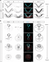
Expression of Nkx1-2 in the developing mouse embryo. (A-C) Nkx1-2 RNA in situ hybridisation of an E6.0 embryo (n=4) (A) and lateral (B) and posterior (C) views of an E7.5 embryo (early head fold, EHF) (n=4). (Ca-Cc) Transverse sections through the regions indicated in C. (D,E) Nkx1-2 expression in an E8.5 embryo (D; 8-10 somites) and higher magnification of the posterior end of the embryo (boxed area; E) (n=4). (Ea-Ec) Transverse sections through the regions indicated in E. (F) Dorsal view of the posterior end of an E9.5 embryo (n=4). (Fa-Fd) Transverse sections through the regions indicated in F. (G) Dorsal view of the tail end of an E10.5 embryo (n=9). (Ga-Gd) Transverse sections through the regions indicated in G. (H) Dorsal view of the tail end of an E11.5 embryo (n=4). (Ha-Hc) Transverse sections through the regions indicated in H. (I) Dorsal view of the tail end of an E12.5 embryo (n=4). Arrowheads in Fc, Fd, Hb and Hc indicate the mesenchymal cell group expressing Nkx1-2 in the tail bud. Asterisks in G, H and I indicate the last-formed somite. cle, caudal lateral epiblast; cnh, chordoneural hinge; hf, headfolds; hg, hindgut; n, node; not, notochord; np, neural plate; nt, neural tube; pnp, posterior neuropore; ps, primitive streak; psm, presomitic mesoderm; tbm, tail bud mesenchyme. Scale bars: 100 µm (whole-mount embryos); 50 µm (transverse sections).
Fig. 2.
Fig. 2.
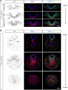
SOX2 and T co-expression within Nkx1-2 regions. (A) Transverse sections across the rostral node, NSB and CLE of an E8.5 embryo immunolabelled for SOX2 and T (n=4). (B) Transverse sections across the tail end of an E10.5 embryo immunolabelled for SOX2 and T (n=7). The cartoons in (A) and (B) depict the expression pattern of Nkx1-2 (as shown in Fig. 1). The different levels of Nkx1-2 expression (based on in situ hybridisation signal) are represented by different grey intensities (light grey, low; dark grey, high). The dashed lines delineate regions not limited by basement membrane. Abbreviations are as in Fig. 1. nml, neuromesodermal lip; not*, notochord end; som, somite. Scale bars: 50 µm.
Fig. 3.
Fig. 3.
Strategy to knock-in CreERT2 into the Nkx1-2 locus. Nkx1-2 locus and targeting vector designed to replace Nkx1-2 exon 1 and the splice donor site at the junction between exon 1 and intron 1 with a cassette containing the open reading frame of CreERT2. Recombinant clones were injected into mouse blastocysts and transferred to mice. The resulting chimeric mice were bred to Flp deleter mice, which ubiquitously express Flp recombinase, to remove the puromycin selection marker to generate the Nkx1-2CreERT2 line. The detailed map of the targeting vector and vector sequence can be found in Fig. S5 and Table S1. pA, polyadenylation site; puro, puromycin; tk, thymidine kinase.
Fig. 4.
Fig. 4.
A subset of Nkx1-2-expressing cells and/or their progeny express SOX2 and T. (A) Transverse sections through the rostral node, NSB and CLE of an E8.5 Nkx1-2CreERT2 embryo that was exposed to tamoxifen at E7.5 and immunolabelled for SOX2, T and YFP (n=7). (B) Transverse sections through the tail end of an E10.5 Nkx1-2CreERT2 embryo that was exposed to tamoxifen at E9.5 and immunolabelled for SOX2, T and YFP (n=9). Abbreviations are as in Fig. 1. nml, neuromesodermal lip; not*, notochord end. Scale bars: 100 µm.
Fig. 5.
Fig. 5.
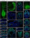
Lineage tracing of cells expressing Nkx1-2 at E7.5. Timed-pregnant Nkx1-2CreERT2 mice received tamoxifen at E7.5 and the contribution of YFP+ cells to developing embryos was assessed at E8.5, E9.5 and E10.5. (A) Maximum intensity projection (MIP) of an E8.5 embryo immunolabelled for YFP on whole-mount (n=7). The arrowhead marks the presumptive midbrain/anterior hindbrain boundary. (Aa-Ag) Transverse sections through the regions indicated in A (n=8). (B) Composite MIP of a E9.5 embryo immunolabelled for YFP on whole-mount (n=16, 14 of 16 embryos analysed had YFP+ cells in the eye). (Ba-Bf) Transverse sections through the regions indicated in B (n=8). (Ba) At the level of the otic vesicles (ov) and rhombomere 5 (rh5) scattered YFP+ cells were found in the neural tube (arrowhead). A few YFP+ cells populated the branchial arches (ba). The foregut (fg) was, however, always unlabelled. (Bb) Section comprising the anterior (left) and posterior (right) levels of the trunk. YFP+ cells were found scattered across the more anterior neural tube, including the floor plate (fp), and spanned the dorsoventral extent of the posterior neural tube. YFP+ cells generated neural crest cells (arrowheads), contributed to posterior somites, and to limb bud (lb) mesenchyme. YFP+ cells were absent from the notochord (not) and midgut (mg). (Bb′) Higher magnification of limb bud mesenchyme in Bb (dashed box). (Bc) YFP+ cells contributed to the neural tube (nt), somites (som), intermediate mesoderm (im), lateral plate mesoderm (lpm) and surface ectoderm (se). (Bd, Be) YFP+ cells were found frequently in the hindgut (hg) (arrowheads). (Bf) YFP+ cells extended to the caudal epiblast and underlying mesenchyme. (C) Wide-field fluorescence image of an E10.5 embryo that received tamoxifen at E7.5. Most of the posterior body derived from YFP+ cells at this stage (n=7). (Ca) YFP+ cells made most of the neural tube and somites (som) and also contributed to hindgut (hg) endoderm and surface ectoderm (arrowhead), but were absent from the notochord (not). (Cb) Posterior to Ca, most presomitic mesoderm (psm) is YFP+. (Cc) In addition to the neural tube and paraxial mesoderm, YFP+ cells were found in the neuromesodermal lip in the CNH (cnh) region. YFP+ cells were also present in the VER (ver, arrowhead). (Cd) The tail bud mesenchyme (tbm) except the ventral-medial cell group was YFP+. The images are representative images of each stage and anterioposterior level. al, allantois; cle, caudal lateral epiblast; np, neural plate; nsb, node-streak border; ps, primitive streak. Scale bars: 100 µm (whole-mount embryos); 50 µm (transverse sections).
Fig. 6.
Fig. 6.
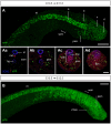
Lineage tracing of cells expressing Nkx1-2 at E10.5. (A,B) Timed-pregnant Nkx1-2CreERT2 mice received tamoxifen at E10.5 and the contribution of YFP+ cells to developing embryos was assessed at E11.5 (A) and E12.5 (B). (A) Dorsal view (composite MIP) of the tail of an E11.5 embryo immunolabelled for YFP on whole-mount (n=7). (Aa-Ad) Transverse sections representative of the levels indicated in A and immunolabelled for SOX2, T and YFP (n=9). YFP+ cells contributed to the secondary neural tube (Aa-Ad), presomitic mesoderm (Ab-Ad) and tail bud mesenchyme (Ad). (B) Side view (MIP) of the tail of an E12.5 embryo immunolabelled for YFP on whole-mount (n=9). Abbreviations are as in Fig. 1. vmes, ventral tail bud mesoderm. Scale bars: 100 µm (whole-mount embryos); 50 µm (transverse sections).

References

    1. Aires R., Jurberg A. D., Leal F., Nóvoa A., Cohn M. J. and Mallo M. (2016). Oct4 is a key regulator of vertebrate trunk length diversity. Dev. Cell 38, 262-274. 10.1016/j.devcel.2016.06.021 - DOI - PubMed
    1. Bae Y.-K., Shimizu T., Muraoka O., Yabe T., Hirata T., Nojima H., Hirano T. and Hibi M. (2004). Expression of sax1/nkx1.2 and sax2/nkx1.1 in zebrafish. Gene Expr. Patterns 4, 481-486. 10.1016/j.modgep.2003.12.001 - DOI - PubMed
    1. Beck C. W. (2015). Development of the vertebrate tailbud. WIREs Dev. Biol. 4, 33-44. 10.1002/wdev.163 - DOI - PubMed
    1. Beddington R. S. (1994). Induction of a second neural axis by the mouse node. Development 120, 613-620. - PubMed
    1. Brennan J., Norris D. P. and Robertson E. J. (2002). Nodal activity in the node governs left-right asymmetry. Genes Dev. 16, 2339-2344. 10.1101/gad.1016202 - DOI - PMC - PubMed

Publication types

MeSH terms

LinkOut - more resources