. 2018 Nov 20;86(12):e00431-18.
doi: 10.1128/IAI.00431-18. Print 2018 Dec.

Limited Pulmonary Mucosal-Associated Invariant T Cell Accumulation and Activation during Mycobacterium tuberculosis Infection in Rhesus Macaques

Affiliations

Limited Pulmonary Mucosal-Associated Invariant T Cell Accumulation and Activation during Mycobacterium tuberculosis Infection in Rhesus Macaques

Keith D Kauffman et al. Infect Immun. .

Abstract

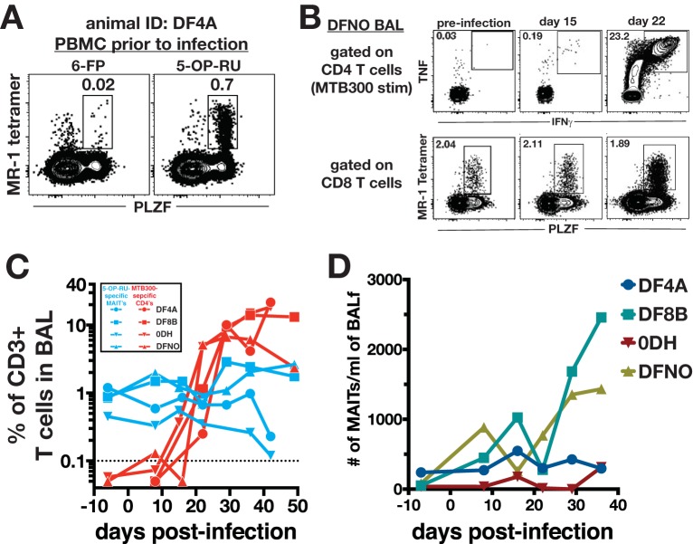
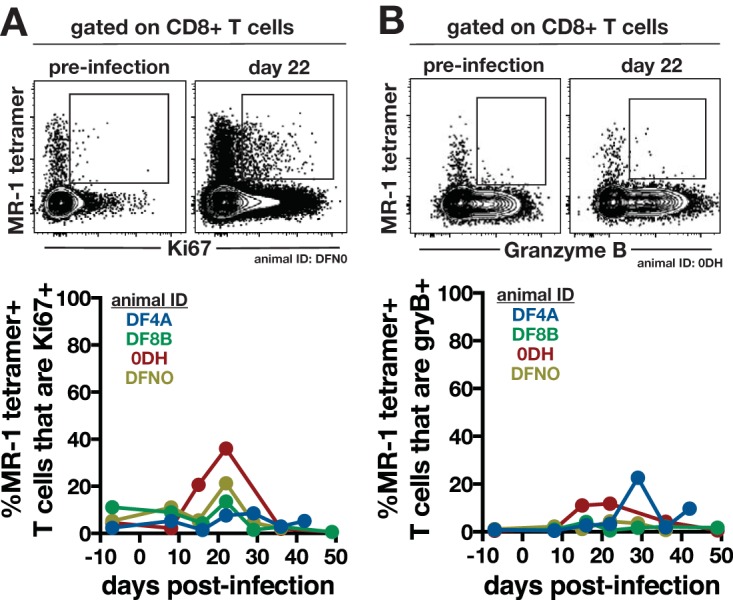
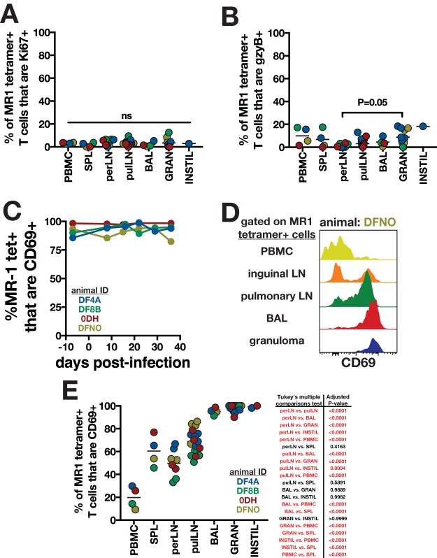
Mucosal-associated invariant T cells (MAITs) are positioned in airways and may be important in the pulmonary cellular immune response against Mycobacterium tuberculosis infection, particularly prior to priming of peptide-specific T cells. Accordingly, there is interest in the possibility that boosting MAITs through tuberculosis (TB) vaccination may enhance protection, but MAIT responses in the lungs during tuberculosis are poorly understood. In this study, we compared pulmonary MAIT and peptide-specific CD4 T cell responses in M. tuberculosis-infected rhesus macaques using 5-OP-RU-loaded MR-1 tetramers and intracellular cytokine staining of CD4 T cells following restimulation with an M. tuberculosis-derived epitope megapool (MTB300), respectively. Two of four animals showed a detectable increase in the number of MAIT cells in airways at later time points following infection, but by ∼3 weeks postexposure, MTB300-specific CD4 T cells arrived in the airways and greatly outnumbered MAITs thereafter. In granulomas, MTB300-specific CD4 T cells were ∼20-fold more abundant than MAITs. CD69 expression on MAITs correlated with tissue residency rather than bacterial loads, and the few MAITs found in granulomas poorly expressed granzyme B and Ki67. Thus, MAIT accumulation in the airways is variable and late, and MAITs display little evidence of activation in granulomas during tuberculosis in rhesus macaques.

Keywords: MAIT cells; Mycobacterium tuberculosis; T cells.


Figures

FIG 1
FIG 1
Kinetics of MAIT versus conventional M. tuberculosis-specific CD4 T cell responses in the airways following M. tuberculosis infection. (A) Sample fluorescence-activated cell sorting (FACS) plots of MR-1 tetramer staining specificity. PBMCs from an animal prior to infection were stained with 5-OP-RU-loaded rhesus macaque MR-1 tetramers. 6-FP-loaded tetramers served as a negative control. MAITs were identified as CD3+ CD8+ PLZF+ tetramer-binding T cells. (B) Sample FACS plots of MAITs and peptide-specific Th1 cells following infection with ∼40 to 80 CFU of M. tuberculosis Erdman-mCherry. M. tuberculosis-specific CD4 T cells were identified by intracellular cytokine staining for IFN-γ and TNF after a 6-h stimulation with the MTB300 peptide megapool. CD4 T cells were identified as CD3+ CD4+ CD8 cells. MAITs were identified as for panel A. (C) Summary graphs of the kinetics of MAIT and MTB300-specific CD4 T cells in the airways following M. tuberculosis infection. In order to allow for direct comparison of their relative abundances, the frequencies of both populations are expressed as a percentage of total live CD3+ cells. (D) Summary graph of the number of MAIT cells per milliliter of bronchoalveolar lavage fluid.
FIG 2
FIG 2
Expression of Ki67 and granzyme B by airway MAITs following M. tuberculosis infection. MR-1–5-OP-RU-specific MAITs in the airways were analyzed for Ki67 (A) and granzyme B (B) expression following M. tuberculosis infection. Sample FACS plots from preinfection and day 22 postinfection (top) and summary graphs of entire infection kinetics (bottom) are shown.
FIG 3
FIG 3
Abundance of MAIT versus peptide-specific Th1 cells in M. tuberculosis-infected tissues of rhesus macaques. Animals were euthanized 6 to 7 weeks postinfection for analysis of MAIT and M. tuberculosis-specific CD4 T cells in infected tissues. Animals were intravenously injected with biotinylated anti-CD45 just prior to euthanasia to allow for identification of parenchymal T cells. (A) Sample FACS plots of MAITs and MTB300-specific CD4 T cells in different tissues of a rhesus macaque 6 weeks postinfection. Both MAITs and peptide-specific CD4 T cells are gated on intravascular stain-negative cells. (B) The frequencies of MAIT and MTB300-specific CD4 T cells among total CD3+ cells were compared in various tissues. Connecting lines indicate that both MAITs and MTB300-specific CD4 T cells were measured in the same samples from spleen, peripheral lymph nodes, pulmonary lymph nodes, PBMCs, BAL, and large instillation site lesions. Paired t tests were used to calculate statistical difference for these tissues. For individual granulomas, either MAITs or MTB300-specific T cells were measured in different lesions, so an unpaired t test was used to calculate statistical difference. (C and D) Bacterial burdens in individual granulomas (C) or lymph nodes (D) from each animal in this study. (E and F) Correlation between bacterial burdens and MAITs (E) or MTB300-specific CD4 T cells (F) in individual granulomas.
FIG 4
FIG 4
MAIT activation in tissues of rhesus macaques following M. tuberculosis infection. The percentages of MR-1 tetramer+ T cells in various tissues of M. tuberculosis-infected rhesus macaques that were positive for Ki67 (A) and granzyme B (B) are shown. P values are for one-way ANOVA with Fisher's LSD test for multiple comparisons. (C) CD69 expression on tetramer+ MAITs in BAL. (D and E) Sample FACS plots (D) and summary graphs (E) of CD69 expression on tetramer+ MAITs in various tissues. MAITs in lymph nodes, BAL, granulomas, and instillation site lesions are gated on intravascular stain-negative cells. P values for one-way ANOVA with Fisher's LSD test for multiple comparisons are shown to the right of the graph, with significant differences highlighted in red.

Similar articles

Cited by

References

    1. World Health Organization. 2016. Global tuberculosis report. World Health Organization, Geneva, Switzerland.
    1. Houben RM, Dodd PJ. 2016. The global burden of latent tuberculosis infection: a re-estimation using mathematical modelling. PLoS Med 13:e1002152. doi:10.1371/journal.pmed.1002152. - DOI - PMC - PubMed
    1. Tameris MD, Hatherill M, Landry BS, Scriba TJ, Snowden MA, Lockhart S, Shea JE, McClain JB, Hussey GD, Hanekom WA, Mahomed H, McShane H, the MVA85A 020 Trial Study Team. 2013. Safety and efficacy of MVA85A, a new tuberculosis vaccine, in infants previously vaccinated with BCG: a randomised, placebo-controlled phase 2b trial. Lancet 381:1021–1028. doi:10.1016/S0140-6736(13)60177-4. - DOI - PMC - PubMed
    1. Greene JM, Dash P, Roy S, McMurtrey C, Awad W, Reed JS, Hammond KB, Abdulhaqq S, Wu HL, Burwitz BJ, Roth BF, Morrow DW, Ford JC, Xu G, Bae JY, Crank H, Legasse AW, Dang TH, Greenaway HY, Kurniawan M, Gold MC, Harriff MJ, Lewinsohn DA, Park BS, Axthelm MK, Stanton JJ, Hansen SG, Picker LJ, Venturi V, Hildebrand W, Thomas PG, Lewinsohn DM, Adams EJ, Sacha JB. 2017. MR1-restricted mucosal-associated invariant T (MAIT) cells respond to mycobacterial vaccination and infection in nonhuman primates. Mucosal Immunol 10:802–813. doi:10.1038/mi.2016.91. - DOI - PMC - PubMed
    1. Gold MC, Napier RJ, Lewinsohn DM. 2015. MR1-restricted mucosal associated invariant T (MAIT) cells in the immune response to Mycobacterium tuberculosis. Immunol Rev 264:154–166. doi:10.1111/imr.12271. - DOI - PMC - PubMed

Publication types

MeSH terms