

Papaverine and its derivatives radiosensitize solid tumors by inhibiting mitochondrial metabolism
- PMID: 30201710
- PMCID: PMC6196495
- DOI: 10.1073/pnas.1808945115
Papaverine and its derivatives radiosensitize solid tumors by inhibiting mitochondrial metabolism
Erratum in
-
Correction for Benej et al., Papaverine and its derivatives radiosensitize solid tumors by inhibiting mitochondrial metabolism.Proc Natl Acad Sci U S A. 2018 Dec 4;115(49):E11561. doi: 10.1073/pnas.1818732115. Epub 2018 Nov 19. Proc Natl Acad Sci U S A. 2018. PMID: 30455291 Free PMC article. No abstract available.
Abstract
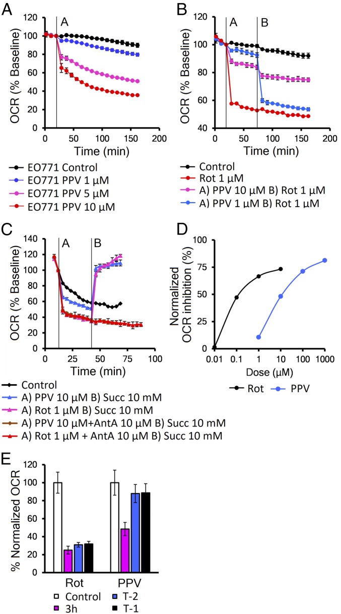
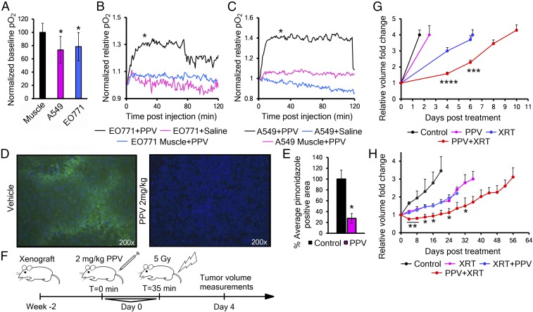
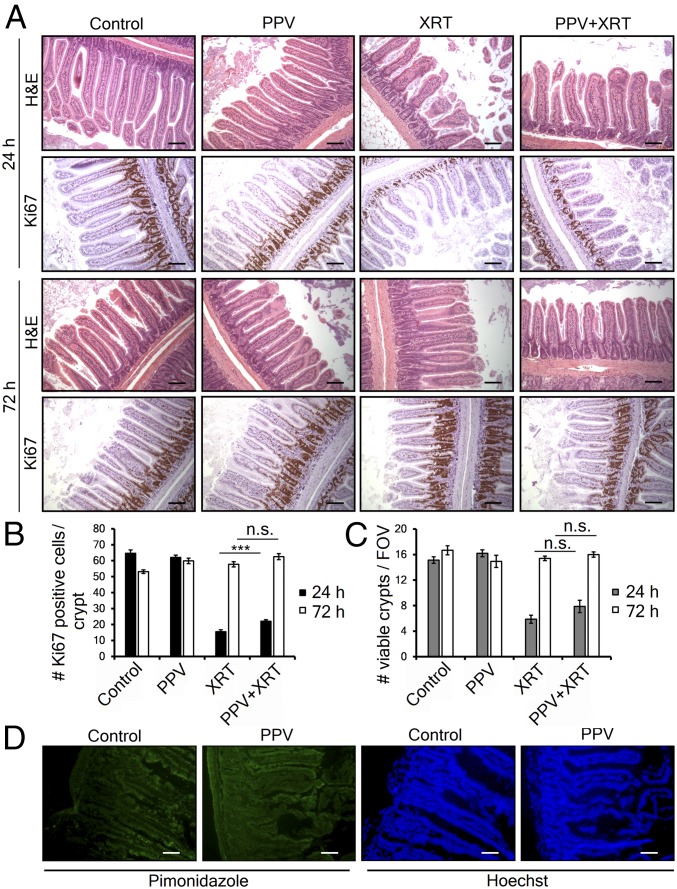
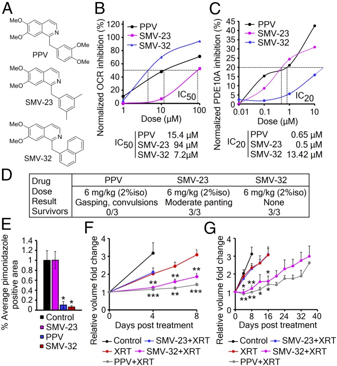
Tumor hypoxia reduces the effectiveness of radiation therapy by limiting the biologically effective dose. An acute increase in tumor oxygenation before radiation treatment should therefore significantly improve the tumor cell kill after radiation. Efforts to increase oxygen delivery to the tumor have not shown positive clinical results. Here we show that targeting mitochondrial respiration results in a significant reduction of the tumor cells' demand for oxygen, leading to increased tumor oxygenation and radiation response. We identified an activity of the FDA-approved drug papaverine as an inhibitor of mitochondrial complex I. We also provide genetic evidence that papaverine's complex I inhibition is directly responsible for increased oxygenation and enhanced radiation response. Furthermore, we describe derivatives of papaverine that have the potential to become clinical radiosensitizers with potentially fewer side effects. Importantly, this radiosensitizing strategy will not sensitize well-oxygenated normal tissue, thereby increasing the therapeutic index of radiotherapy.
Keywords: hypoxia; metabolism; mitochondria; radiosensitization.
Conflict of interest statement
The authors declare no conflict of interest.
Figures

Comment in
-
A potential solution for eliminating hypoxia as a cause for radioresistance.Proc Natl Acad Sci U S A. 2018 Oct 16;115(42):10548-10550. doi: 10.1073/pnas.1814212115. Epub 2018 Oct 9. Proc Natl Acad Sci U S A. 2018. PMID: 30301796 Free PMC article. No abstract available.
Similar articles
-
Evaluating Tumor Hypoxia Radiosensitization Via Electron Paramagnetic Resonance Oxygen Imaging (EPROI).Mol Imaging Biol. 2024 Jun;26(3):435-447. doi: 10.1007/s11307-023-01855-0. Epub 2023 Sep 18. Mol Imaging Biol. 2024. PMID: 37721686 Free PMC article.
-
Hypoxic tumor cell radiosensitization: role of the iNOS/NO pathway.Bull Cancer. 2008 Mar;95(3):282-91. doi: 10.1684/bdc.2008.0592. Bull Cancer. 2008. PMID: 18390408 Review.
-
The small molecule AU14022 promotes colorectal cancer cell death via p53-mediated G2/M-phase arrest and mitochondria-mediated apoptosis.J Cell Physiol. 2018 Jun;233(6):4666-4676. doi: 10.1002/jcp.26234. Epub 2018 Jan 15. J Cell Physiol. 2018. PMID: 29030986
-
Next-Generation Hypoxic Cell Radiosensitizers: Nitroimidazole Alkylsulfonamides.J Med Chem. 2018 Feb 8;61(3):1241-1254. doi: 10.1021/acs.jmedchem.7b01678. Epub 2018 Jan 11. J Med Chem. 2018. PMID: 29253343
-
Manipulation of tumor oxygenation and radiosensitivity through modification of cell respiration. A critical review of approaches and imaging biomarkers for therapeutic guidance.Biochim Biophys Acta Bioenerg. 2017 Aug;1858(8):700-711. doi: 10.1016/j.bbabio.2017.01.002. Epub 2017 Jan 12. Biochim Biophys Acta Bioenerg. 2017. PMID: 28088332 Review.
Cited by
-
Targeting Mitochondrial Metabolism to Reverse Radioresistance: An Alternative to Glucose Metabolism.Antioxidants (Basel). 2022 Nov 7;11(11):2202. doi: 10.3390/antiox11112202. Antioxidants (Basel). 2022. PMID: 36358574 Free PMC article. Review.
-
Targeting tumor hypoxia and mitochondrial metabolism with anti-parasitic drugs to improve radiation response in high-grade gliomas.J Exp Clin Cancer Res. 2020 Oct 7;39(1):208. doi: 10.1186/s13046-020-01724-6. J Exp Clin Cancer Res. 2020. PMID: 33028364 Free PMC article. Review.
-
Targeting hallmarks of cancer to enhance radiosensitivity in gastrointestinal cancers.Nat Rev Gastroenterol Hepatol. 2020 May;17(5):298-313. doi: 10.1038/s41575-019-0247-2. Epub 2020 Jan 31. Nat Rev Gastroenterol Hepatol. 2020. PMID: 32005946 Review.
-
Targeting Radiation Resistance in Oesophageal Adenocarcinoma with Pyrazinib-Functionalised Gold Nanoparticles.Cancers (Basel). 2024 Nov 29;16(23):4007. doi: 10.3390/cancers16234007. Cancers (Basel). 2024. PMID: 39682192 Free PMC article.
-
Applications of nanocomposites based on zeolitic imidazolate framework-8 in photodynamic and synergistic anti-tumor therapy.RSC Adv. 2022 Jun 9;12(26):16927-16941. doi: 10.1039/d2ra01102f. eCollection 2022 Jun 1. RSC Adv. 2022. PMID: 35754870 Free PMC article. Review.
References
-
- Brown JM, Giaccia AJ. The unique physiology of solid tumors: Opportunities (and problems) for cancer therapy. Cancer Res. 1998;58:1408–1416. - PubMed
-
- Harrison DK, Vaupel P. Heterogeneity in tissue oxygenation: From physiological variability in normal tissues to pathophysiological chaos in malignant tumours. Adv Exp Med Biol. 2014;812:25–31. - PubMed
Publication types
MeSH terms
Substances
Grants and funding
LinkOut - more resources
Full Text Sources
Other Literature Sources
Medical
