. 2018 Sep 25;115(39):E9075-E9084.
doi: 10.1073/pnas.1809842115. Epub 2018 Sep 10.

Structure-specific DNA replication-fork recognition directs helicase and replication restart activities of the PriA helicase

Affiliations

Structure-specific DNA replication-fork recognition directs helicase and replication restart activities of the PriA helicase

Tricia A Windgassen et al. Proc Natl Acad Sci U S A. .

Abstract

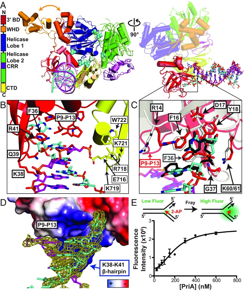
DNA replication restart, the essential process that reinitiates prematurely terminated genome replication reactions, relies on exquisitely specific recognition of abandoned DNA replication-fork structures. The PriA DNA helicase mediates this process in bacteria through mechanisms that remain poorly defined. We report the crystal structure of a PriA/replication-fork complex, which resolves leading-strand duplex DNA bound to the protein. Interaction with PriA unpairs one end of the DNA and sequesters the 3'-most nucleotide from the nascent leading strand into a conserved protein pocket. Cross-linking studies reveal a surface on the winged-helix domain of PriA that binds to parental duplex DNA. Deleting the winged-helix domain alters PriA's structure-specific DNA unwinding properties and impairs its activity in vivo. Our observations lead to a model in which coordinated parental-, leading-, and lagging-strand DNA binding provide PriA with the structural specificity needed to act on abandoned DNA replication forks.

Keywords: DNA repair; DNA replication restart; X-ray crystallography; cross-link mapping; protein–DNA complex.


Conflict of interest statement

The authors declare no conflict of interest.

Figures

Fig. 1.
Fig. 1.
Structure of PriA/DNA replication-fork complex resolves the dsDNA leading-arm interaction mechanism. (A, Left) Domain architecture and structure of K. pneumoniae PriA-dsDNA. (A, Middle) The two molecules from the asymmetric unit have been superimposed. Molecule A is in the bright colors used in the domain architecture diagram to the left and molecule B is in pale versions of those colors. Nascent leading strand (cyan) and parental leading strand (magenta) are shown as cartoons. Zinc atoms (gray) and sulfates (cyan) are shown as spheres. The orange arrow points to the two different locations of the WHD in molecule A and molecule B. (A, Right) Rotation for a side view of the 3′BD interaction with DNA from both PriA molecules in the asymmetric unit. The 3′BD and DNA from molecule A are in bright colors and from molecule B in pale colors, as in A, Middle. DNA is shown in sticks. The remaining PriA domains are shown in transparent cartoon and are those from molecule A. See also SI Appendix, Fig. S1 and Table S2. (B) Close-up view of the dsDNA interactions of the 3′BD and CTD of molecule A. K718, K719, and K721 side chains did not have sufficient electron density for modeling. (C) Close-up view of the 3′OH-terminal nucleotide binding pocket superimposed with the dinucleotide-bound EcPriA 3′BD structure [gray protein and black nucleotides (14)]. Residues involved in terminal nucleotide coordination are labeled in black. (D) Surface electrostatics view of the 3′BD with dsDNA Fo − Fc omit map contoured at 3.0σ. (E) Solution equilibrium DNA fraying analysis using 2-AP fluorescent base analog placed at the terminal base in the nascent leading strand. Data are mean ± SD (error bars) of three replicates.
Fig. 2.
Fig. 2.
PriA/DNA cross-linking maps a PriA WHD interface with the parental duplex. (A) PriA WHD (wheat) with dsDNA from the cocrystal structures of OhrR (gray, ref. 22) and HNF-3 (black, ref. 23), where the WH domains of the three proteins were structurally superimposed. The four positions where Bpa incorporation yielded EcPriA DNA fork cross-links are labeled (EcPriA residue/KpPriA residue) and shown in sticks from the KpPriA apo structure (9). (B) Diagram of PE assay using 32P-5′-labeled primers (red arrows) annealed to either the parental leading or lagging strand within the cross-linking DNA fork (SI Appendix, Table S1). Red dotted line depicts PE, with “X” representing a block to PE by a covalent protein/DNA cross-link. (C) Sequence of the cross-linking DNA fork, with significant PE blocked products marked by dotted lines from the cross-linked Bpa site. Number marks indicate the length of the PE product for either parental lagging or leading PE. (D) Urea PAGE of PE on cross-linked parental lagging strand samples with sequencing ladder (right: T, C, A, and G labels indicate ddA, ddG, ddT, or ddC, respectively, was added). Red lines highlight significant PE bands not present in the negative control (lane 2). (E) Primer 2 for the parental leading strand and sequencing ladder as in D. Representative gels of at least three replicates. See also SI Appendix, Fig. S2.
Fig. 3.
Fig. 3.
The PriA WHD is not required for PriA/DNA replication-fork interaction in vitro. Fluorescence anisotropy of a fluorescein-labeled DNA substrate [forked (Top), ds (Middle), or ss (Bottom) DNA (SI Appendix, Table S1)] measured with increasing PriA concentrations. Data are mean ± SD and fit to a single-site-specific model. Dissociation constants from fit are listed (right box). See also SI Appendix, Fig. S4.
Fig. 4.
Fig. 4.
The WHD is required for orienting PriA on DNA forks lacking a nascent leading strand in vitro. (A) PAGE of helicase products from increasing concentrations of PriA (0.1, 0.5, 1, 5, 10, and 50 nM) added to a three-stranded 32P-5′-labeled synthetic DNA fork type depicted between gel panels in black (1 nM; see SI Appendix, Table S1), where stars indicates the site of the 32P-5′ label. Helicase products are depicted in gray. Left gel panels: SSB (62.5 nM, tetramers) was preincubated with the DNA fork. Right gel panels: SSB was omitted. (B) Two-strand DNA fork type assessed as in A. (C) Four-strand DNA fork type assessed as in A and B. Gels are representative of at least three replicates. See also SI Appendix, Figs. S4 and S5.
Fig. 5.
Fig. 5.
EcPriA ∆WHD helicase specificity dysfunction selectively inactivates the PriA–PriC replication restart pathway in vivo. (A) Phase contrast (Left), GFP fluorescence (Middle; measure of SOS induction levels through sulA-gfp fusion), and mCherry fluorescence (Right; visualization of nucleoids through hupA::mcherry fusion) microscopy images of E. coli. (Scale bars, 10 μm.) (B) Quantification from images as in A for mutants used in this study (SI Appendix, Fig. S6 and Table S3). The Par phenotype was defined by cells containing multiple poorly condensed nucleoids as assessed by the visualization of nucleoids. GFP relative fluorescence intensity (RFI) was a measure for SOS induction and generated by taking at least three different images of cells on three different days.
Fig. 6.
Fig. 6.
priAWHD-containing strains exacerbate the UV sensitivity of recG-null strains. priA and priA mutant effects on UV sensitivity of recG-null strains (SI Appendix, Table S3). Surviving fraction after exposure to increasing levels of UV (0, 40, 60, and 80 J/m2). Data are means of four replicates ±SD. **P < 0.01 between indicated point and recG::kan alone.
Fig. 7.
Fig. 7.
Model of the full PriA/DNA replication-fork complex and roles of the 3′BD and WHD in PriA orientation on a DNA fork. (A) PriA/DNA fork model. The model was constructed from the KpPriA/DNA structure (Fig. 1, leading arm), dsDNA from structural superimposition with the WHD of OhrR (22) onto the PriA WHD (parental duplex), and structural superimposition of DNA from the RecQ/DNA complex (50) with the helicase core (lagging arm). The overall assembly was energy-minimized using the SYBYL-X 2.1 molecular modeling system. The PriA/DNA fork model fits PriABpa-DNA fork cross-linking constraints from this or a previous study (latter indicated by asterisk) (20), with dotted lines indicating the site of the mutated EcPriA residue in the KpPriA structure and the nucleotide site(s) of the cross-link (from PE disruption). (B) Scheme of PriA on a DNA fork that would result in the varied helicase products observed in Fig. 4.

References

    1. Cox MM, et al. The importance of repairing stalled replication forks. Nature. 2000;404:37–41. - PubMed
    1. Mangiameli SM, Merrikh CN, Wiggins PA, Merrikh H. Transcription leads to pervasive replisome instability in bacteria. eLife. 2017;6:e19848. - PMC - PubMed
    1. Windgassen TA, Wessel SR, Bhattacharyya B, Keck JL. Mechanisms of bacterial DNA replication restart. Nucleic Acids Res. 2018;46:504–519. - PMC - PubMed
    1. Jezewska MJ, Bujalowski W. Interactions of Escherichia coli replicative helicase PriA protein with single-stranded DNA. Biochemistry. 2000;39:10454–10467. - PubMed
    1. Szymanski MR, Jezewska MJ, Bujalowski W. The Escherichia coli PriA helicase-double-stranded DNA complex: Location of the strong DNA-binding subsite on the helicase domain of the protein and the affinity control by the two nucleotide-binding sites of the enzyme. J Mol Biol. 2010;402:344–362. - PMC - PubMed

Publication types