Comparative Study
. 2018 Dec;17(12):2412-2433.
doi: 10.1074/mcp.RA118.001036. Epub 2018 Sep 10.

Comparative Secretome Analyses of Human and Zoonotic Staphylococcus aureus Isolates CC8, CC22, and CC398

Affiliations
Comparative Study

Comparative Secretome Analyses of Human and Zoonotic Staphylococcus aureus Isolates CC8, CC22, and CC398

Tobias Busche et al. Mol Cell Proteomics. 2018 Dec.

Abstract

The spread of methicillin-resistant Staphylococcus aureus (MRSA) in the community, hospitals and in livestock is mediated by highly diverse virulence factors that include secreted toxins, superantigens, enzymes and surface-associated adhesins allowing host adaptation and colonization. Here, we combined proteogenomics, secretome and phenotype analyses to compare the secreted virulence factors in selected S. aureus isolates of the dominant human- and livestock-associated genetic lineages CC8, CC22, and CC398. The proteogenomic comparison revealed 2181 core genes and 1306 accessory genes in 18 S. aureus isolates reflecting the high genome diversity. Using secretome analysis, we identified 869 secreted proteins with 538 commons in eight isolates of CC8, CC22, and CC398. These include 64 predicted extracellular and 37 cell surface proteins that account for 82.4% of total secretome abundance. Among the top 10 most abundantly secreted virulence factors are the major autolysins (Atl, IsaA, Sle1, SAUPAN006375000), lipases and lipoteichoic acid hydrolases (Lip, Geh, LtaS), cytolytic toxins (Hla, Hlb, PSMβ1) and proteases (SspB). The CC398 isolates showed lower secretion of cell wall proteins, but higher secretion of α- and β-hemolysins (Hla, Hlb) which correlated with an increased Agr activity and strong hemolysis. CC398 strains were further characterized by lower biofilm formation and staphyloxanthin levels because of decreased SigB activity. Overall, comparative secretome analyses revealed CC8- or CC22-specific enterotoxin and Spl protease secretion as well as Agr- and SigB-controlled differences in exotoxin and surface protein secretion between human-specific and zoonotic lineages of S. aureus.

Keywords: MRSA; Microbiology; Pathogens; Proteogenomics; Secretome; Staphylococcus aureus; Virulence; clonal complexes; virulence factor secretion.


Figures

None
Graphical abstract
Fig. 1.
Fig. 1.
The core and accessory proteogenomes of 18 S. aureus CC8, CC22, CC398 isolates. The protein occurrence treemap (right) visualizes all 3487 unique proteins based on their presence and absence in the proteogenomes of the 18 S. aureus isolates. The 3487 unique proteins were sorted into functional categories based on TIGRfam and Aureowiki annotations (Table S3) (60). High diversity of the proteogenome across 18 S. aureus isolates is indicated in this protein occurrence treemap showing 2181 core and 1306 accessory proteins. The core proteins are colored in violet and the accessory proteins are color-coded based on their frequencies in <18 isolates. The protein frequency color code is shown below the treemap. Two legends of the treemap are presented on the left showing “top level functions” (TIGRfam level 1) and “sub level functions” (TIGRfam level 2) of the proteins as classified in Table S3. The colors in the legends (left) differentiate the functional classes of all proteins and are not related to the color code of the protein frequency treemap (right).
Fig. 2.
Fig. 2.
Phylogenomic clustering of the proteogenomes of 18 S. aureus CC8, CC22, CC398 isolates. Phylogenetic clustering of the proteogenomes of the 18 S. aureus isolates is based on protein frequencies, revealing lineage-specific branches. In total, 2181 and 1306 proteins belong to the core (violet) and accessory proteogenomes, respectively. Accessory proteins are present in <18 isolates and are color-coded based on their frequencies in the 18 isolates. The numbers of the accessory proteins that are present in 1–17 genomes are given as frequencies. The phylogenetic tree is based on the FASTA proteome data translated from the 18 genome sequences that are mapped to the PAN-IDs of the Aureowiki database. The presence and absence of all unique proteins in the 18 isolates is shown in supplemental Table S3 that was used for this phylogenomic clustering.
Fig. 3.
Fig. 3.
Overview and functional categories of the 869 secreted proteins that are quantified in the secretomes of eight S. aureus isolates of CC8, CC22 and CC398. A, The Venn diagram shows the numbers of proteins that were quantified in the secretomes of the eight S. aureus isolates of CC8 (38879, 38880), CC22 (17787, 24749) and CC398 (38885, 27969, SK41 and SK42) using label-free quantitative proteomics. Most identified virulence factors belong to the core genome while also unique proteins were identified that define lineage-specific secreted proteins. The Venn diagram was generated using the BioInfoRX Venn diagram plotter (http://bioinforx.com/free/bxarrays/venndiagram.php#). B, The 869 secreted proteins were classified according to TIGRfam and Aureowiki annotations including also core and accessory proteins (supplemental Tables S6, S8).
Fig. 4.
Fig. 4.
The subcellular localization of 869 secreted proteins (A, B) and the 50 most abundant secreted virulence factors in all secretomes of the eight isolates of CC8, CC22 and CC398 (C). A, B, The 869 secreted proteins of the eight S. aureus isolates were analyzed by PSORTb version 3.0.2 (73) for their subcellular localization into predicted extracellular, cell wall-associated, membrane and cytoplasmic proteins (supplemental Table S6). The diagrams show the distribution of proteins in the different compartments based on their percentages of protein numbers (A) and percentages of protein abundances (B) in relation to all proteins identified in the total secretome. This indicates that predicted 64 extracellular and 37 cell wall proteins represent with 82.4% the most abundant secreted proteins. Although 70% of proteins were identified as cytoplasmic proteins, these are low abundant and contribute only with 12.9% to the total secretome. C, The 50 most abundantly secreted extracellular proteins are listed according to their amounts that constitute 86.5% of the total secretome including the major autolysins (Atl, Sle1, IsaA, 6375), lipases and lipoteichoic acid hydrolase (Lip, Geh, LtaS), hemolytic toxins (Hla, Hlb, PSMβ1) among the top ten secreted virulence factors (supplemental Table S8). Thus, the majority of the secretome is highly conserved across different genetic lineages.
Fig. 5.
Fig. 5.
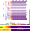
Voronoi secretome treemaps for comparison of the secretomes of the eight S. aureus isolates of CC8, CC22 and CC398. The secreted proteins that were quantified in the secretomes of the eight S. aureus isolates of CC8, CC22 and CC398 are visualized in Voronoi secretome treemaps. The secreted proteins were classified in functional categories according to TIGRfam and Aureowiki annotations as shown in supplemental Tables S6. The LFQ intensities of each protein were used for calculation of the normalized 99 percentile protein abundance in each of the eight secretomes (supplemental Table S8). The normalized 99 percentile of each protein across the 8 secretomes denotes the cell size of the treemap. The yellow-orange-magenta color code indicates the normalized 99 percentile abundance of the proteins in the secretomes of the different S. aureus isolates. The eight secretome profiles were also used for phylogenetic clustering indicating lineage-specific groups of secreted proteins. These CC8, CC22 and CC398 specific clusters of secretome profiles identified CC-specific secretome profiles because of genomic and regulatory differences. The higher secretion of toxins (Hla and Hlb) and lower secretion of many surface-associated proteins points to a higher Agr activity in CC398 strains. In addition, the secretome profiles revealed also an abundant core secretome of S. aureus virulence factors that is conserved across lineages.
Fig. 6.
Fig. 6.
Volcano Plots of differentially secreted proteins in the secretomes between CC8, CC22, CC398. The log2 fold changes of >+1.5 and <−1.5 (p value <0.05) are shown as significant differences in the average abundance of secreted proteins in the genetic lineages CC22 versus CC398 (A), CC8 versus CC398 (B) and CC8 versus CC22 (C) as calculated in supplemental Tables S7A–S7E. Only secreted proteins were considered that were identified in all 3 clonal complexes to reveal regulatory secretome differences. Although Agr-controlled surface-associated proteins were significantly down-regulated in CC398 isolates (green spots), exotoxins (Hla, Hlb) and extracellular enzymes (Aur, SspB, SspB2) were up-regulated in CC398 versus CC22 and CC8 (red spots). More cytoplasmic proteins are secreted in CC398 versus CC8 (black spots). Other significantly differentially secreted proteins were labeled with black symbols according to their sub-cellular localization as given in the legend. Non-significantly differentially secreted proteins between the CCs are gray spots.
Fig. 7.
Fig. 7.
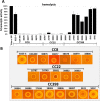
Hemolytic activities of S. aureus isolates of CC8, CC22 and CC398 indicate increased α- and β-hemolysis in CC398. Extracellular fractions were harvested from 18 S. aureus during the stationary phase after 16 h of growth. Total hemolytic activities of filtered culture supernatants were analyzed with 2% sheep blood erythrocyte solution. A, Lysis of erythrocytes was measured as absorbance at 405 nm. Hemolysis was normalized to the Triton-X100 positive control, which was considered as 100% hemolysis. Secreted proteins of the CC398 isolates displayed much higher hemolytic activities than CC8 and CC22 isolates. The increased hemolysis in most CC398 strains (except for 38884 and 38888) correlates with intact β-hemolysin expression due the absence of the β-hemolysin converting prophage ΦSa3. The phage ΦSa3 disrupts the hlb locus in the majority of CC8 and CC22 isolates (except for 38881). Strains 38881 and 38884 have intact hlb, but are agrC-defective. In addition, elevated α-hemolysin expression in CC398 contributes to strong hemolysis. The results are presented as average values of 5–10 independent biological experiments with measurements made in duplicates. Error bars represents S.E. B, The β-hemolysis can be visualized after spotting of 5 μl bacterial suspension on sheep blood agar plates as shown previously (77). Strong β-hemolytic rings can be visualized in most CC398 isolates except for strain 38888. The agr-defective strains 38881 and 38884 show only small β-hemolysis.
Fig. 8.
Fig. 8.
Biofilm formation, staphyloxanthin levels and structured macrocolony phenotypes of 18 S. aureus isolates of CC8, CC22 and CC398. A, Biofilm formation was analyzed from overnight cultures that were diluted to an OD580 of 0.5 in TSB with 1% glucose. 200 μl cells were transferred in triplicate to microtiter plate wells for attachment. The attached biofilm cells were stained using 0.1% crystal violet solution in the microtiter plate wells and resuspended with 0.1% SDS for quantitative measurements of the biofilm crystal violet solution at an OD595. For comparison, the laboratory CC8 strains S. aureus 8325–4 and USA300 were included in the biofilm assays. B, Staphyloxanthin levels were measured from cell pellets of 1 ml overnight cultures after methanol extraction as absorbance at 463 nm and normalized to the OD600 of the cell culture (463/600 nm ratio). C, Structured macrocolony formation and staphyloxanthin pigmentations of the strains was analyzed by spotting 2 μl cell suspension on TSB-agar with 100 mm MgCl2 for 5 days. S. aureus CC398 isolates with increased α- and β-hemolysin secretion showed lower ability for biofilm formation (A), lower staphyloxanthin levels (B) and white-yellow pigmented macrocolonies (C) because of reduced SigB activity. The results of the biofilm and staphyloxanthin measurements in (A, B) are shown as average values of 5 independent biological experiments. Error bars are S.E.
Fig. 9.
Fig. 9.
Transcriptional analyses indicate differential Agr and SigB activities in human and zoonotic S. aureus isolates. A, For Northern blotting analyses RNA was isolated of selected S. aureus CC8, CC22 and CC398 isolates grown in TSB medium and harvested during the exponential growth and stationary phase after 3 and 6 h of growth. Agr and SigB activities were analyzed using digoxygenin-labeled RNA probes specific for RNAIII, hla and spa (Agr), asp23 and sigB (SigB). The methylene-blue stained bands of the 16S and 23S rRNAs in the Northern blots are shown as loading controls at the bottom. The Northern blotting experiments were performed in 3 biological replicate experiments. B, Schematics of the Agr and SigB regulatory network for expression of secreted toxins, exoenzymes, surface factors that are involved in hemolysis, biofilm formation and staphyloxanthin production in S. aureus. CC398 isolates showed increased hla transcription and decreased asp23 and sigB operon transcription compared with CC8 or CC22 strains. This indicates lower SigB activity and increased Agr activity in CC398 strains resulting in higher α- and β-hemolysis as well as lower surface factors, biofilm production and staphyloxanthin amounts. Positive and negative regulations are indicated. Broken arrows show induction or repression of virulence factors and phenotypes in CC398 strains as revealed in this study.

Similar articles

Cited by

References

    1. Wertheim H. F. L., Melles D. C., Vos M. C., van Leeuwen W., van Belkum A., Verbrugh H. A., and Nouwen J. L. (2005) The role of nasal carriage in Staphylococcus aureus infections. Lancet Infect. Dis. 5, 751–762 - PubMed
    1. Weidenmaier C., Goerke C., and Wolz C. (2012) Staphylococcus aureus determinants for nasal colonization. Trends Microbiol. 20, 243–250 - PubMed
    1. Lowy F. D. (1998) Staphylococcus aureus infections. N. Engl. J. Med. 339, 520–532 - PubMed
    1. Boucher H. W., and Corey G. R. (2008) Epidemiology of methicillin-resistant Staphylococcus aureus. Clin. Infect. Dis. 46, S344–S349 - PubMed
    1. Archer G. L. (1998) Staphylococcus aureus: a well-armed pathogen. Clin. Infect. Dis. 26, 1179–1181 - PubMed

Publication types

MeSH terms