. 2018 Oct 17;38(42):9072-9090.
doi: 10.1523/JNEUROSCI.0637-18.2018. Epub 2018 Sep 10.

Hypothalamic CCL2/CCR2 Chemokine System: Role in Sexually Dimorphic Effects of Maternal Ethanol Exposure on Melanin-Concentrating Hormone and Behavior in Adolescent Offspring

Affiliations

Hypothalamic CCL2/CCR2 Chemokine System: Role in Sexually Dimorphic Effects of Maternal Ethanol Exposure on Melanin-Concentrating Hormone and Behavior in Adolescent Offspring

Guo-Qing Chang et al. J Neurosci. .

Abstract

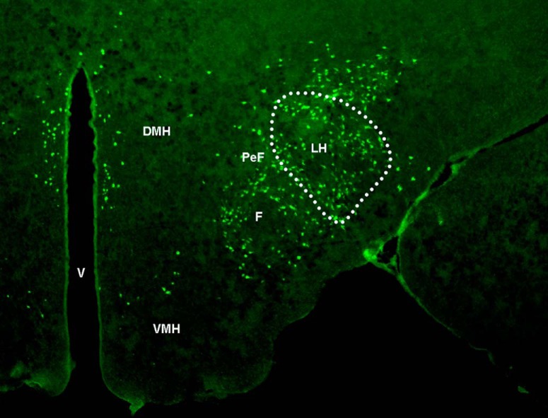
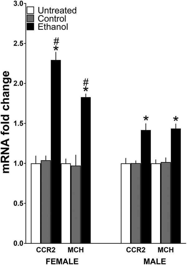
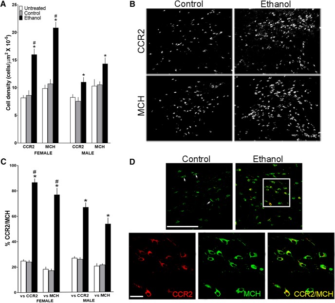
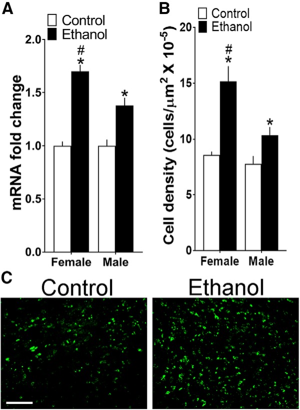
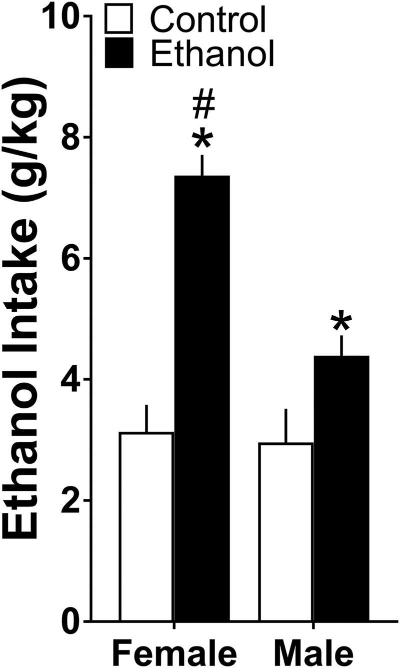
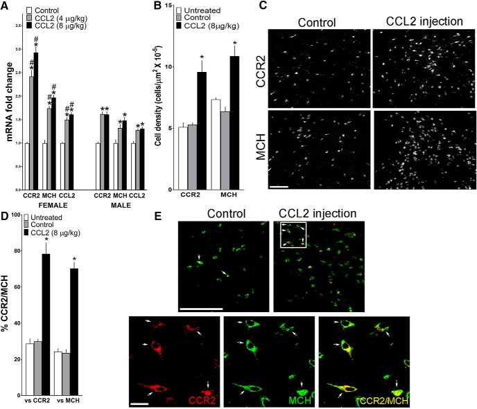
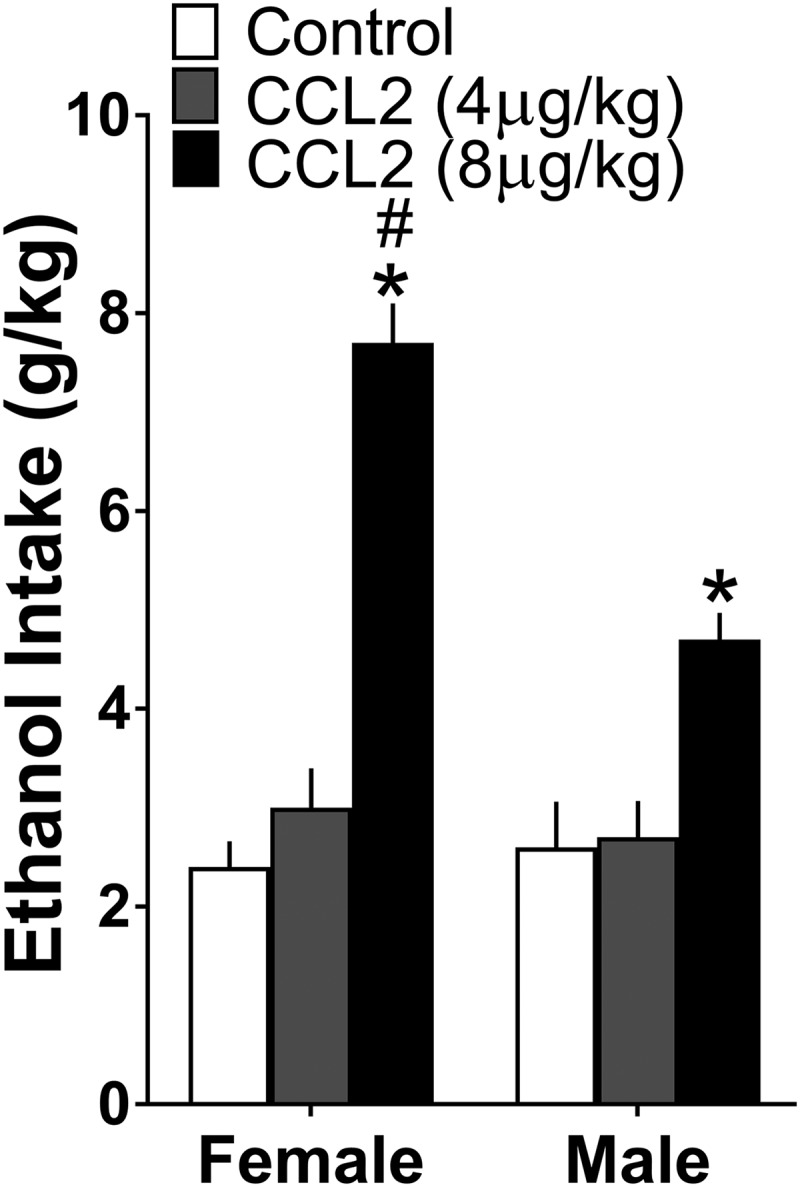
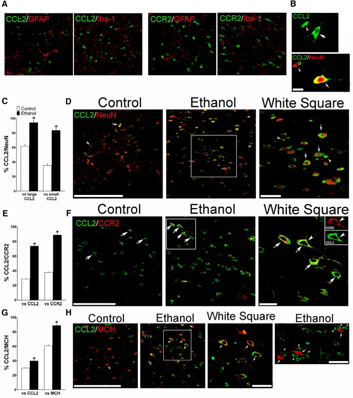
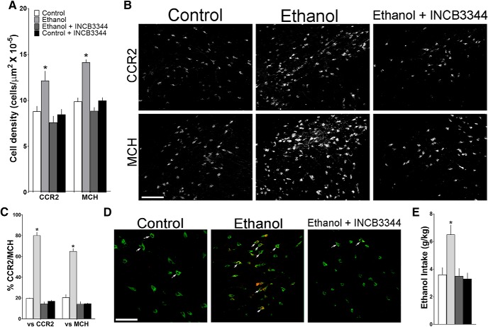
Clinical and animal studies show that ethanol exposure and inflammation during pregnancy cause similar behavioral disturbances in the offspring. While ethanol is shown to stimulate both neuroimmune and neurochemical systems in adults, little is known about their anatomical relationship in response to ethanol in utero and whether neuroimmune factors mediate ethanol's effects on neuronal development and behavior in offspring. Here we examined in female and male adolescent rats a specific population of neurons concentrated in lateral hypothalamus, which coexpress the inflammatory chemokine C-C motif ligand 2 (CCL2) or its receptor CCR2 with the orexigenic neuropeptide, melanin-concentrating hormone (MCH), that promotes ethanol drinking behavior. We demonstrate that maternal administration of ethanol (2 g/kg/d) from embryonic day 10 (E10) to E15, while having little impact on glia, stimulates expression of neuronal CCL2 and CCR2, increases density of both large CCL2 neurons colocalizing MCH and small CCL2 neurons surrounding MCH neurons, and stimulates ethanol drinking and anxiety in adolescent offspring. We show that these neuronal and behavioral changes are similarly produced by maternal administration of CCL2 (4 or 8 μg/kg/d, E10-E15) and blocked by maternal administration of a CCR2 antagonist INCB3344 (1 mg/kg/d, E10-E15), and these effects of ethanol and CCL2 are sexually dimorphic, consistently stronger in females. These results suggest that this neuronal CCL2/CCR2 system closely linked to MCH neurons has a role in mediating the effects of maternal ethanol exposure on adolescent offspring and contributes to the higher levels of adolescent risk factors for alcohol use disorders described in women.SIGNIFICANCE STATEMENT Ethanol consumption and inflammatory agents during pregnancy similarly increase alcohol intake and anxiety in adolescent offspring. To investigate how neurochemical and neuroimmune systems interact to mediate these disturbances, we examined a specific population of hypothalamic neurons coexpressing the inflammatory chemokine CCL2 and its receptor CCR2 with the neuropeptide, melanin-concentrating hormone. We demonstrate in adolescent offspring that maternal administration of CCL2, like ethanol, stimulates these neurons and increases ethanol drinking and anxiety, and these effects of ethanol are blocked by maternal CCR2 antagonist and consistently stronger in females. This suggests that neuronal chemokine signaling linked to neuropeptides mediates effects of maternal ethanol exposure on adolescent offspring and contributes to higher levels of adolescent risk factors for alcohol use disorders in women.

Keywords: CCR2 antagonist; anxiety; ethanol drinking; lateral hypothalamus; neuroimmune; prenatal.


Figures

Figure 1.
Figure 1.
Representative photomicrograph illustrates, at low magnification (2.5×) and anterior–posterior level of bregma −3.12 (Paxinos and Watson, 2005), the LH area and MCH-labeled neurons within it (outlined by dotted line) that were examined in this study. PeF, Perifornical hypothalamus; F, fornix; V, ventricle; DMH, dorsomedial hypothalamus; VMH, ventromedial hypothalamus.
Figure 2.
Figure 2.
Maternal administration of ethanol (2 g/kg/d, E10–E15) compared with isocaloric control solution and untreated control groups affects mRNA expression of CCR2 and MCH in the LH of adolescent offspring (n = 9/group/sex), measured using qRT-PCR and represented in the figure as mRNA fold change compared with the untreated control group (with average ratio scores given in parentheses). Ethanol compared with control groups (n = 9/group/sex) significantly increased in female offspring mRNA levels of MCH (untreated = 0.203 ± 0.012, control = 0.198 ± 0.027, ethanol = 0.372 ± 0.016) and CCR2 (untreated = 0.150 ± 0.0143, control = 0.157 ± 0.009, and ethanol = 0.344 ± 0.034) and in male offspring mRNA levels of MCH (untreated = 0.210 ± 0.010, control = 0.214 ± 0.012, and ethanol = 0.302 ± 0.018) and CCR2 (untreated = 0.151 ± 0.010, control = 0.155 ± 0.005, and ethanol = 0.215 ± 0.018), with the effect significantly greater in females, which exhibited a particularly large increase in CCR2. Data are mean ± SEM. *p < 0.05 versus control groups. #p < 0.05 versus male groups.
Figure 3.
Figure 3.
Maternal ethanol administration (2 g/kg/d, E10–E15) compared with isocaloric control and untreated control groups affects CCR2+ and MCH+ neurons in the LH of adolescent offspring (n = 7/group/sex), as measured using IF. A, Ethanol compared with control groups significantly increased the density of CCR2+ and MCH+ single-labeled neurons in both female and male offspring, with the effects in females significantly larger. B, This effect in female offspring is illustrated in representative immunostaining images of CCR2+ and MCH+ single-labeled neurons. C, Ethanol increased the density of CCR2+/MCH+ double-labeled neurons in both female and male offspring, again with this effect significantly stronger in females. D, This effect of ethanol on double-labeled neurons is illustrated in the confocal images from female offspring showing CCR2+ (red) and MCH+ (green) single-labeled neurons and CCR2+/MCH+ (yellow) double-labeled neurons in the LH. Top, A few CCR2+/MCH+ neurons (arrows) in control offspring and many in ethanol-exposed offspring, with those in the white square illustrated (bottom) at higher magnification for CCR2+ and MCH+ single-labeled and CCR2+/MCH+ double-labeled neurons. Scale bars, 200 μm. Data are mean ± SEM. *p < 0.05 versus control group. #p < 0.05 versus male groups.
Figure 4.
Figure 4.
Maternal ethanol administration (2 g/kg/d, E10–E15) compared with control solution also affects CCL2 in the LH of the adolescent offspring. A, Ethanol increased mRNA levels of CCL2, measured using qRT-PCR and represented in the figure as mRNA fold change compared with the control group (with average ratio scores given in parentheses), in female (control = 0.249 ± 0.015, ethanol = 0.425 ± 0.031) and male (control = 0.220 ± 0.013, ethanol = 0.305 ± 0.25) offspring (n = 9/group/sex), with this effect stronger in females. B, Ethanol increased the density of CCL2+ cells measured using IF in the LH of female and male offspring (n = 8/group/sex), again with this effect greater in females. C, This effect of ethanol versus control on CCL2+ cells is illustrated in the photomicrographs in female offspring. Scale bar, 200 μm. Data are mean ± SEM. *p < 0.05 versus control group. #p < 0.05 versus male group.
Figure 5.
Figure 5.
Maternal ethanol administration (2 g/kg/d, E10–E15) compared with isocaloric control solution stimulated ethanol drinking over 24 h in both female and male adolescent offspring (n = 10/group/sex), with this effect significantly stronger in females. Data are mean ± SEM. *p < 0.05 versus control group. #p < 0.05 versus male group.
Figure 6.
Figure 6.
Maternal injection of CCL2 (4 and 8 μg/kg/d, E10–E15) compared with vehicle control group affects in adolescent offspring the expression of CCR2, MCH, and CCL2, measured using qRT-PCR and represented in the figure as mRNA fold change compared with the control group (with average ratio scores given in parentheses), and the density of CCR2+ and MCH+ neurons in the LH measured using IF. A, CCL2 administration compared with control group (n = 9/group/sex) significantly increased in female offspring mRNA levels of MCH (control = 0.237 ± 0.0155, 4 μg/kg/d = 0.413 ± 0.023, and 8 μg/kg/d = 0.468 ± 0.028), CCR2 (control = 0.127 ± 0.034, 4 μg/kg/d = 0.308 ± 0.041, and 8 μg/kg/d = 0.373 ± 0.046), and CCL2 (control = 0.222 ± 0.019, 4 μg/kg/d = 0.333 ± 0.018, and 8 μg/kg/d = 0.358 ± 0.017) and in male offspring mRNA levels of MCH (control = 0.241 ± 0.018, 4 μg/kg/d = 0.319 ± 0.026, and 8 μg/kg/d = 0.356 ± 0.027), CCR2 (control = 0.123 ± 0.027, 4 μg/kg/d = 0.199 ± 0.012, and 8 μg/kg/d = 0.198 ± 0.013), and CCL2 (control = 0.225 ± 0.009, 4 μg/kg/d = 0.289 ± 0.010, and 8 μg/kg/d = 0.295 ± 0.013), with a significantly stronger effect in females as indicated in particular by an unusually large increase in CCR2 mRNA. B, CCL2 injection increased the density of CCR2+ and MCH+ neurons in female offspring. C, This effect of CCL2 versus control on cell density is illustrated in representative immunostaining images of single-labeled cells. D, CCL2 injection increased the density of CCR2+/MCH+ double-labeled neurons in the LH of the female offspring (n = 7/group). E, This effect of CCL2 versus control on double-labeled neurons is illustrated in the confocal images showing CCR2+ (red) and MCH+ (green) single-labeled neurons and CCR2+/MCH+ (yellow) double-labeled neurons in the LH. Top, A few CCR2+/MCH+ neurons in control offspring and many in CCL2-treated offspring (arrows), with those in the white square (bottom) at higher magnification for CCR2+ (left) and MCH+ (middle) single-labeled neurons and for CCR2+/MCH+ (right) double-labeled neurons (yellow or red/green). Scale bars, 200 μm. Data are mean ± SEM. *p < 0.05 versus control groups. #p < 0.05 versus male groups.
Figure 7.
Figure 7.
Maternal injection of CCL2 (4 or 8 μg/kg/d, E10–E15) compared with vehicle control group, like ethanol administration stimulated ethanol drinking over 24 h period in both female and male adolescent offspring (n = 10/group/sex), with this effect significantly stronger in females. Data are mean ± SEM. *p < 0.05 versus control group. #p < 0.05 versus male group.
Figure 8.
Figure 8.
Maternal ethanol administration (2 g/kg/d, E10–E15) compared with control solution, while having no effect on glia, alters the density of both large and small CCL2+ neurons in the LH, as assessed by IF in female adolescent offspring (n = 7/group). A, Confocal images show in ethanol-treated offspring no colocalization of CCL2 or CCR2 (green) with the markers of astrocytes (GFAP) and microglia (Iba-1) (red). B, Two confocal images provide typical examples of the large (arrow) and small (arrowhead) CCL2+ single-labeled cells (green) in the LH (top) and of large (arrow) and small (arrowhead) CCL2+ neurons that double-labeled the neuronal marker NeuN (bottom), with CCL2 (green) and NeuN (red) revealing CCL2+/NeuN+ double-labeled neurons that are large (yellow/red center) or small (green/red center). C, Maternal ethanol administration increased the density of both large and small CCL2+ neurons that double-labeled NeuN, as indicated by the increased percentage of both large and small CCL2+/NeuN+ double-labeled neurons relative to total large or total small CCL2+ single-labeled cells, respectively. D, Representative confocal images show few large (arrow) and few small (arrowhead) CCL2+ (green) neurons that colabeled NeuN (red) in control offspring (left) but many CCL2+/NeuN+ neurons (green/red center) in ethanol-exposed offspring (middle), with those in the white square illustrated at higher magnification (right). E, Maternal ethanol administration increased the density of CCL2+ neurons, mostly large, that colabeled CCR2, as indicated by the increased percentage of large CCL2+/CCR2+ double-labeled neurons relative to total large CCL2+ or CCR2+ single-labeled neurons. F, Representative confocal images, illustrating mostly large CCL2+ (green) and CCR2+ (red) neurons that were double-labeled (green/yellow), show some CCL2+/CCR2+ neurons (arrow) in control offspring (left) and many more CCL2+/CCR2+ neurons (arrow) in ethanol-treated offspring (middle), with those in the white square illustrated at higher magnification (right). G, Maternal ethanol administration increased the density of CCL2+ neurons, mostly large, that colabeled MCH, as indicated by the increased percentage of large CCL2+/MCH+ double-labeled neurons relative to total large CCL2+ or MCH+ single-labeled neurons. H, Representative confocal images, illustrating mostly large CCL2+ (green) and MCH+ (red) neurons that become yellow/red or green/red when double-labeled, show few CCL2+/MCH+ neurons (arrow) in control offspring (left) and many CCL2+/MCH+ neurons (arrow) in ethanol-treated offspring (middle), with those in the white square illustrated at higher magnification (right). While few small CCL2+ cells were found to double-label CCR2 or MCH, the small CCL2+ cells (arrowhead, green) were sometimes seen adjacent to or surrounding large MCH+ neurons (arrow, red) as shown in at far right. Scale bars: A, 100 μm; B–H, 200 μm. Data are mean ± SEM. *p < 0.05 versus control.
Figure 9.
Figure 9.
Maternal injection of a CCR2 antagonist INCB3344 (1 mg/kg/d) during the period of maternal ethanol administration (2 g/kg/d, E10–E15) prevents the stimulatory effect of ethanol on CCR2+ and MCH+ single-labeled neurons and CCR2+/MCH+ double-labeled neurons in the LH of female adolescent offspring as measured by IF. A, Ethanol treatment increased the density of CCR2+ and MCH+ neurons compared with control, whereas INCB3344 treatment blocks this effect of ethanol in the offspring (n = 7/group). B, This effect is illustrated in representative images of CCR2+ and MCH+ single-labeled neurons. C, Ethanol treatment increased the density of CCR2+/MCH+ double-labeled neurons compared with control, and this effect is abrogated in the offspring by maternal injection of the CCR2 antagonist INCB3344. D, This effect of ethanol is illustrated in representative confocal images of CCR2+/MCH+ neurons (arrows, yellow) that double-label CCR2+ (red) and MCH+ (green), showing few in the control group (left), many in the ethanol group (middle), and few in the ethanol + INCB3344 offspring (right). E, Ethanol treatment increased 24 h ethanol drinking behavior in the adolescent offspring (n = 10/group), and this effect is blocked by maternal administration of the CCR2 antagonist. Scale bar, 200 μm. Data are mean ± SEM. *p < 0.05 versus control and versus ethanol + INCB3344.

Similar articles

Cited by

References

    1. Abrahao KP, Salinas AG, Lovinger DM (2017) Alcohol and the brain: neuronal molecular targets, synapses, and circuits. Neuron 96:1223–1238. 10.1016/j.neuron.2017.10.032 - DOI - PMC - PubMed
    1. Adachi N, Suzuki S, Matsuoka H, Fushimi S, Ono J, Ohta KI, Hirai Y, Miki T, Koshimizu H (2018) Corticotropin-releasing hormone-binding protein is up-regulated by brain-derived neurotrophic factor and is secreted in an activity-dependent manner in rat cerebral cortical neurons. J Neurochem. Advance online publication. Retrieved Jan. 22, 2018. doi: 10.1111/jnc.14310. - DOI - PubMed
    1. Alati R, Al Mamun A, Williams GM, O'Callaghan M, Najman JM, Bor W (2006) In utero alcohol exposure and prediction of alcohol disorders in early adulthood: a birth cohort study. Arch Gen Psychiatry 63:1009–1016. 10.1001/archpsyc.63.9.1009 - DOI - PubMed
    1. Alfonso-Loeches S, Pascual M, Guerri C (2013) Gender differences in alcohol-induced neurotoxicity and brain damage. Toxicology 311:27–34. 10.1016/j.tox.2013.03.001 - DOI - PubMed
    1. Anthony JC, Petronis KR (1995) Early-onset drug use and risk of later drug problems. Drug Alcohol Depend 40:9–15. 10.1016/0376-8716(95)01194-3 - DOI - PubMed

Publication types

LinkOut - more resources