Review
. 2019 Aug 1;9(8):a034199.
doi: 10.1101/cshperspect.a034199.

Vagus Nerve Stimulation at the Interface of Brain-Gut Interactions

Affiliations
Review

Vagus Nerve Stimulation at the Interface of Brain-Gut Interactions

Bruno Bonaz et al. Cold Spring Harb Perspect Med. .

Abstract

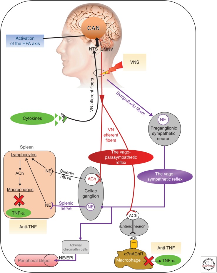
The vagus nerve, a key component of the cross-communication between the gut and the brain, is a major element of homeostasis sensing the "milieu intérieur" and boosting the nervous and endocrine responses to maintain the gastrointestinal health status. This nerve has anti-inflammatory properties regulating the gut through the activation of the hypothalamic-pituitary-adrenal axis and the release of cortisol and through a vagovagal reflex, which has an anti-tumor necrosis factor (TNF) effect called the cholinergic anti-inflammatory pathway. Stimulating this nerve is an interesting tool as a nondrug therapy for the treatment of gastrointestinal diseases in which brain-gut communication is dysfunctional, such as inflammatory bowel disorders and others. This review presents the rationale of vagal gastrointestinal physiology and diseases and the most recent advances in vagus nerve stimulation. It also highlights the main issues to be addressed in the future to improve this bioelectronic therapy for gastrointestinal disorders.


Figures

Figure 1.
Figure 1.
The brain–gut axis. The autonomic nervous system (ANS) is the bidirectional link between the central nervous system (CNS) and the gut via the enteric nervous system (ENS) and the immune, endocrine, and vascular systems. NTS, nucleus tractus solitarius; DMNV, dorsal motor nucleus of the vagus nerve.
Figure 2.
Figure 2.
Brain–gut integrative pathway. The gut afferents from the vagus and splanchnic nerves are transmitted to the nucleus tractus solitarius (NTS), in the medulla in close contact with the dorsal motor nucleus of the vagus nerve (DMNV), the origin of parasympathetic vagal efferents, thus creating an autonomic brainstem loop involved in the regulation of gastrointestinal motility, acid secretion, food intake, and satiety. This loop is modulated by an autonomic forebrain loop that includes nuclei in the pons, the hypothalamus, the hippocampus, the amygdala, the anterior cingulate, the insular, and the prefrontal cortices. This forebrain loop, also referred to as the central autonomic network, coordinates visceral information in an integrative dimension that includes neuroendocrine responses, emotions, cognition, and behavior. These two central loops explain how stress, feelings, and thoughts can influence gut functioning and vice versa. RVLM, rostral ventrolateral medulla; ILM, intermediolateralis nucleus.
Figure 3.
Figure 3.
Different pathways of the anti-inflammatory properties of the vagus nerve and how to target the vagus nerve for its anti-inflammatory properties. ACh, acetylcholine; CAN, central autonomic network; DMNV, dorsal motor nucleus of the vagus nerve; EPI, epinephrine; HPA, hypothalamic–pituitary–adrenal; NE, norepinephrine; NTS, nucleus tractus solitarius; TNF-α, tumor necrosis factor α; VNS, vagus nerve stimulation; α7nAChR, α7 nicotinic acetylcholine receptor (Bonaz et al. 2017).
Figure 4.
Figure 4.
Extrinsic (autonomic nervous system [ANS]) and enteric nervous system (ENS) innervation of the gut. The extrinsic innervation includes the vagal parasympathetic and sympathetic systems, with afferent sensory and efferent secretomotor fibers. The ENS is a complex neural network controlling various cell populations, including smooth muscle, mucosal secretory, endocrine, and immune/inflammatory cells, as well as microvasculature (for secretion, absorption, and motility). The ENS network is organized in several plexuses, located in various levels of the gut wall, which provides a partial autonomous control of gastrointestinal functions. The ANS provides the extrinsic innervation of the gut with (1) sensory neurons linked to vagal, thoracolumbar, and lumbosacral pathways that are located at the mucosal, myenteric, muscular, and vascular levels, and (2) vagal, sacral, and sympathetic axons for motor activity.
Figure 5.
Figure 5.
Effect of vagus nerve stimulation (VNS) during 6 months of follow-up in the seven patients presenting with an active Crohn’s disease at inclusion on (A) the Crohn’s disease activity index (CDAI); (B) C-reactive protein (CRP); (C) the Crohn’s disease endoscopic index of severity (CDEIS); (D) the vagal tone (high frequency in normalized units [HFnu]) and the sympathovagal balance (low frequency [LF]/high frequency [HF]). CDAI < 150, clinical remission. Cut-off level for CRP, 5 mg/L. CDEIS < 6, endoscopic remission. HFnu, homeostatic range. Balanced ratio of LF/HF (sympathovagal balance) ∼1. (From Bonaz et al. 2016a; modified, with permission, from the author.)

Similar articles

Cited by

References

    1. Abe C, Inoue T, Inglis MA, Viar KE, Huang L, Ye H, Rosin DL, Stornetta RL, Okusa MD, Guyenet PG. 2017. C1 neurons mediate a stress-induced anti-inflammatory reflex in mice. Nat Neurosci 20: 700–707. - PMC - PubMed
    1. Agostini A, Ballotta D, Righi S, Moretti M, Bertani A, Scarcelli A, Sartini A, Ercolani M, Nichelli P, Campieri M, et al. 2017. Stress and brain functional changes in patients with Crohn’s disease: A functional magnetic resonance imaging study. Neurogastroenterol Motil 29: 1–10. - PubMed
    1. Baciu MV, Bonaz BL, Papillon E, Bost RA, Le Bas JF, Fournet J, Segebarth CM. 1999. Central processing of rectal pain: A functional MR imaging study. AJNR Am J Neuroradiol 20: 1920–1924. - PMC - PubMed
    1. Barquist E, Bonaz B, Martinez V, Rivier J, Zinner MJ, Tache Y. 1996. Neuronal pathways involved in abdominal surgery-induced gastric ileus in rats. Am J Physiol 270: R888–R894. - PubMed
    1. Barreau F, Ferrier L, Fioramonti J, Bueno L. 2004. Neonatal maternal deprivation triggers long term alterations in colonic epithelial barrier and mucosal immunity in rats. Gut 53: 501–506. - PMC - PubMed

Publication types

LinkOut - more resources