. 2018 Oct;30(10):2383-2401.
doi: 10.1105/tpc.17.00586. Epub 2018 Sep 10.

CIK Receptor Kinases Determine Cell Fate Specification during Early Anther Development in Arabidopsis

Affiliations

CIK Receptor Kinases Determine Cell Fate Specification during Early Anther Development in Arabidopsis

Yanwei Cui et al. Plant Cell. 2018 Oct.

Abstract

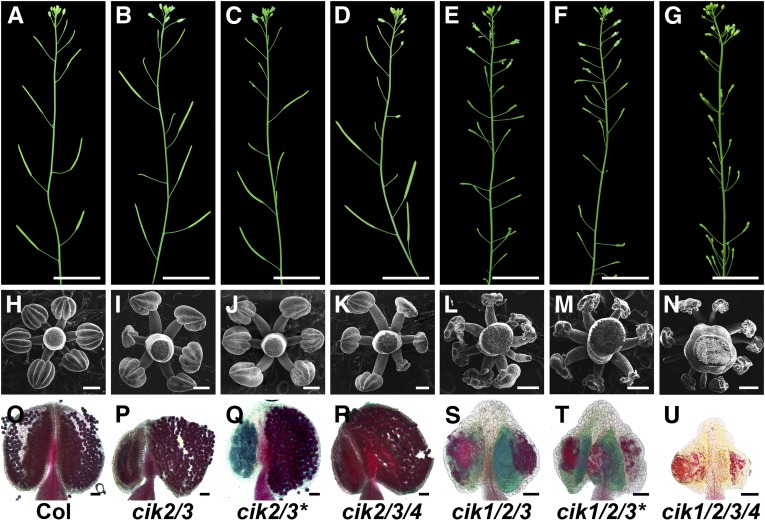
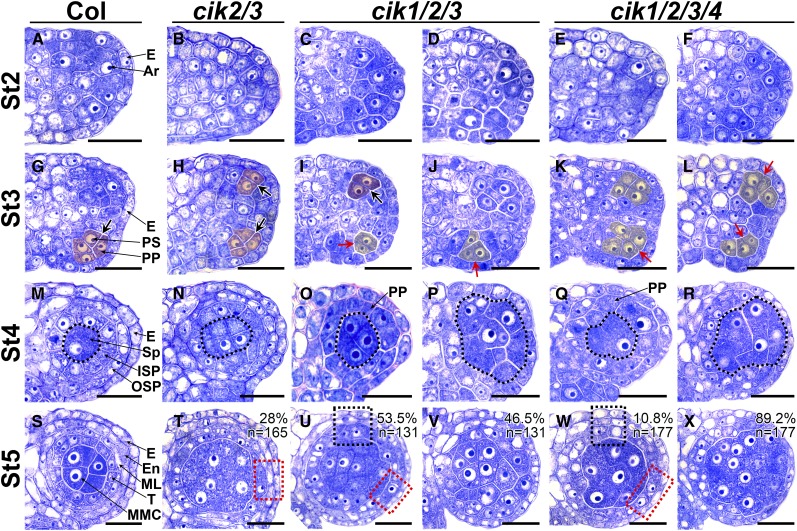
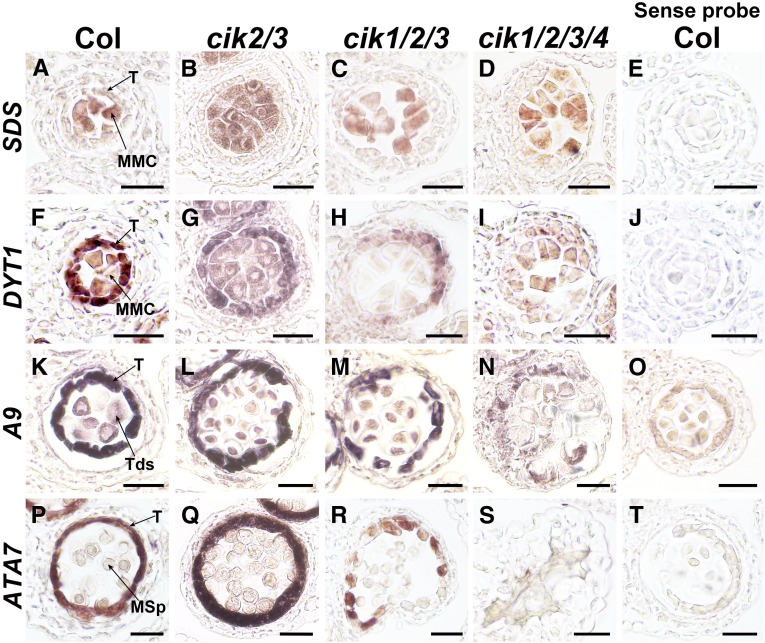
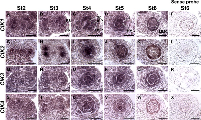
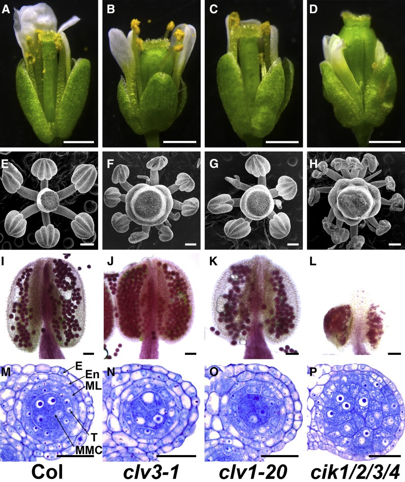
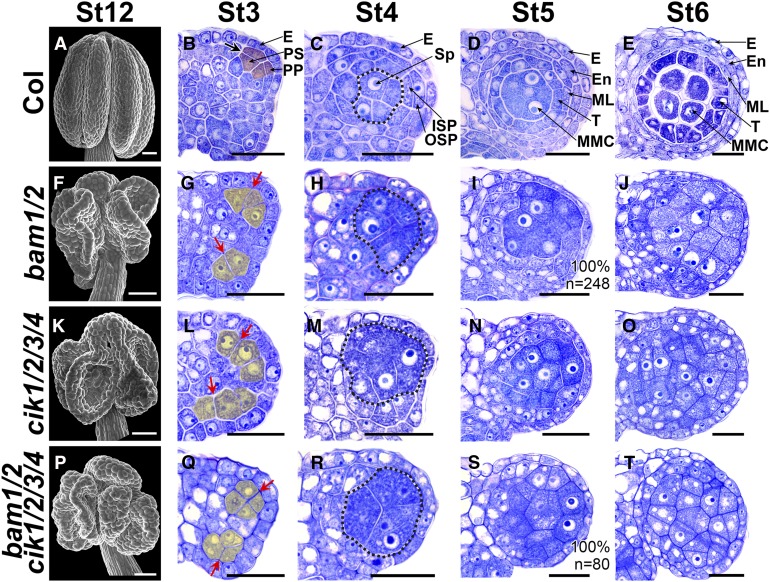
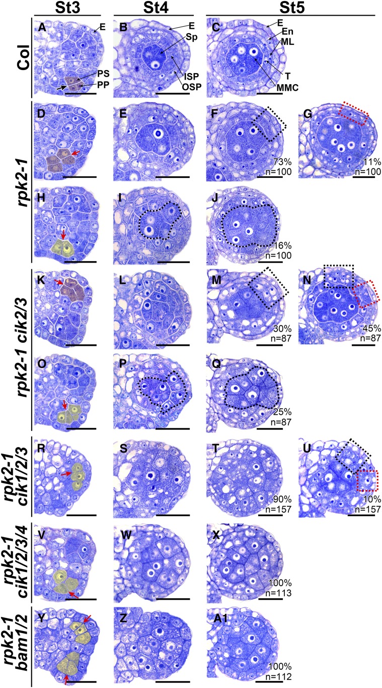
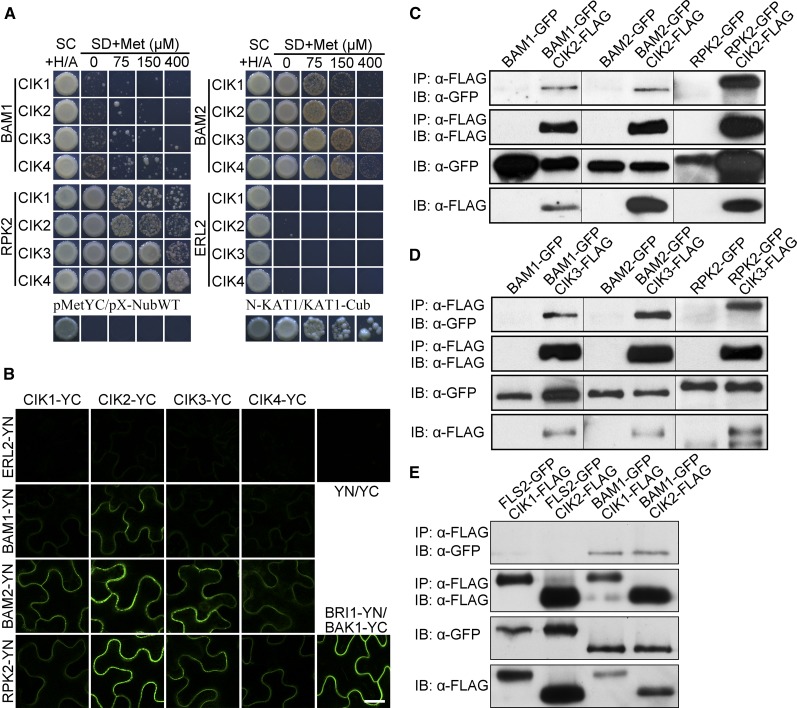
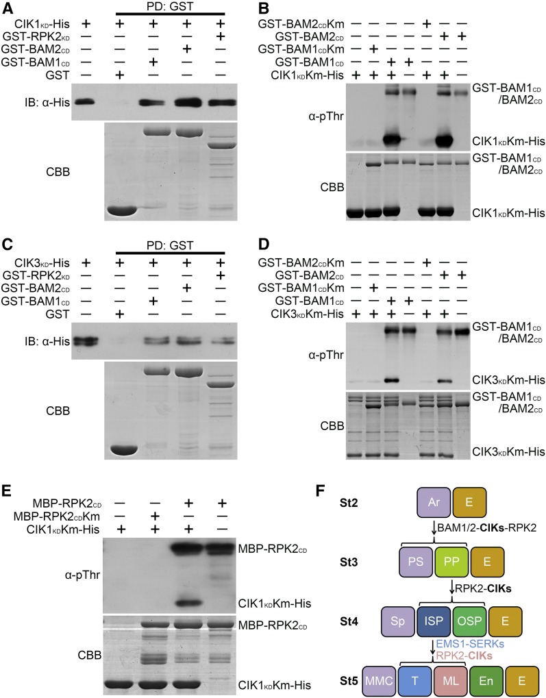
Appropriate cell division and differentiation ensure normal anther development in angiosperms. BARELY ANY MERISTEM 1/2 (BAM1/2) and RECEPTOR-LIKE PROTEIN KINASE2 (RPK2), two groups of leucine-rich repeat receptor-like protein kinases, are required for early anther cell specification. However, little is known about the molecular mechanisms underlying these two RLK-mediated signaling pathways. Here, we show that CLAVATA3 INSENSITIVE RECEPTOR KINASEs (CIKs), a group of novel coreceptor protein kinase-controlling stem cell homeostasis, play essential roles in BAM1/2- and RPK2-regulated early anther development in Arabidopsis thaliana The archesporial cells of cik1/2/3 triple and cik1/2/3/4 quadruple mutant anthers perform anticlinal division instead of periclinal division. Defective cell division and specification of the primary and inner secondary parietal cells occur in these mutant anthers. The disordered divisions and specifications of anther wall cells finally result in excess microsporocytes and a lack of one to three parietal cell layers in mutant anthers, resembling rpk2 or bam1/2 mutant anthers. Genetic and biochemical analyses indicate that CIKs function as coreceptors of BAM1/2 and RPK2 to regulate archesporial cell division and determine the specification of anther parietal cells.


Figures

Figure 1.
Figure 1.
Loss of Function of CIKs Leads to Reduced Fertility. (A) to (G) Inflorescence stems of 6-week-old plants. (H) to (N) Scanning electron microscopy images showing stamens at flower stage 12. (O) to (U) Alexander’s staining of mature anthers to show pollen viability. (A), (H), and (O), wild type (Col); (B), (I), and (P), cik2/3; (C), (J), and (Q), cik2/3* (the second cik2/3 mutant); (D), (K), and (R), cik2/3/4; (E), (L), and (S), cik1/2/3; (F), (M), and (T), cik1/2/3* (the second cik1/2/3 mutant); (G), (N), and (U), cik1/2/3/4. Bars = 2 cm in (A) to (G), 200 µm in (H) to (N), and 50 µm in (O) to (U).
Figure 2.
Figure 2.
Cell Division and Differentiation Are Abnormal in Anthers of cik Mutants. (A) to (F) Semithin sections of stage 2 (St2) anthers. The archesporial cells (Ar) are produced under the epidermis (E). No difference was observed between the wild type (A) and cik mutants ([B] to [F]). (G) to (L) Semithin sections of St3 anthers. The archesporial cell periclinally divides once to produce an outer primary parietal cell (PP) and an inner primary sporogenous cell (PS) in the wild type (G). No obvious defects can be observed in cik2/3 (H). Anticlinal cell divisions occur in cik1/2/3 ([I] and [J]) and cik1/2/3/4 ([K] and [L]). Thick black arrows indicate normal periclinal division of cells shaded in red; red arrows indicate anticlinal division of cells shaded in yellow. (M) to (R) Semithin sections of St4 anthers. The primary parietal cell divides once to produce an outer secondary parietal cell (OSP) and an inner secondary parietal cell (ISP) in the wild type (M) and cik2/3 (N). No OSP and ISP cells can be correctly specified in cik1/2/3 and cik1/2/3/4 ([O] to [R]). Sporogenous cells (Sp) and Sp-like cells are indicated with dotted loops. (S) to (X) Semithin sections of St5 anthers. The inner secondary parietal cell divides once to produce an outer middle layer cell (ML) and an inner tapetal cell (T), forming a locule with four cell layers that enclose the microspore mother cells (MMC) in the wild type (S). In cik2/3, 28% of locules display a discontinuous middle layer (dotted red rectangle) (T). In cik1/2/3, 53.5% of locules display one or two parietal cell layers (dotted red and black rectangles) (U), and 46.5% of locules produce MMC-like cells enclosed only by the epidermis (V). In cik1/2/3/4, 10.8% of locules have one or two parietal cell layers (dotted red and black rectangles) (W), and 89.2% of locules produce MMC-like cells enclosed only by the epidermis (X). All cik mutants produce extra MMC-like cells. Half anther is shown in (A) to (L); one locule is shown in (M) to (X). n indicates the total number of examined locules. Bars = 20 µm.
Figure 3.
Figure 3.
RNA in Situ Hybridization Analyses of MMC and Tapetum Markers in Wild-Type and cik Anthers. (A) to (D) SDS was detected in MMCs of wild-type (A), cik2/3 (B), cik1/2/3 (C), and cik1/2/3/4 (D) anthers at stage 6. (F) to (I) DYT1 was detected in the tapetum and MMCs of wild-type (F) and cik2/3 (G) anthers at stage 6. Higher expression in the tapetum than MMCs is observed. Weak DYT1 signal was detected in MMCs and the tapetum of the cik1/2/3 mutant (H) and only in MMC-like cells of the cik1/2/3/4 mutant (I). (K) to (N) A9 was detected in the tapetum and tetrads of wild-type (K), cik2/3 (L), and cik1/2/3 (M) anthers at stage 7. No typical A9 expression was detected in cik1/2/3/4 (N). (P) to (S) ATA7 was detected in the tapetum of wild-type (P), cik2/3 (Q), and cik1/2/3 (R) anthers at stage 8. No signal was detected in cik1/2/3/4 (S). Sense probes of SDS (E), DYT1 (J), A9 (O), and ATA7 (T) were used as negative controls. These experiments were repeated three times independently with similar results. MSp, microspore; T, tapetum; Tds, tetrads. Bars = 20 µm.
Figure 4.
Figure 4.
RNA in Situ Hybridization Showing the Expression Patterns of CIKs in Early Wild-Type Anthers. (A) to (E) CIK1 is expressed in the whole anther at stage 2 (St2) (A) and St3 (B), with higher level in L2-derived cells. The expression is concentrated in four locules with higher expression in the sporogenous cells and two layers of secondary parietal cells at St4 (C), MMCs, the tapetum and the middle layer at St5 (D), and MMCs and the tapetum at St6 (E). (G) to (K) CIK2 shows expression patterns similar to CIK1 except that the signal is more obvious in the primary sporogenous cells at St3 (H). (M) to (Q) CIK3 shows expression patterns similar to CIK1. (S) to (W) CIK4 shows expression patterns similar to CIK1. Sense probes of CIK1 (F), CIK2 (L), CIK3 (R), and CIK4 (X) were used as negative controls. One locule is shown for anthers at St4, St5, and St6. These experiments were repeated at least three times independently with similar results. ISP, inner secondary parietal cell; OSP, outer secondary parietal cell; PP, primary parietal cell; PS, primary sporogenous cell; Sp, sporogenous cell; T, tapetum. Bars = 20 µm.
Figure 5.
Figure 5.
clv Mutants Produce Functional Anthers. (A) to (D) Flowers of the wild type (A), clv3-1 (B), clv1-20 (C), and cik1/2/3/4 (D) at stage 13. Bars = 1 mm. (E) to (H) Scanning electron microscopy images showing anthers of the wild type (E), clv3-1 (F), clv1-20 (G), and cik1/2/3/4 (H) at flower stage 12. Bars = 200 µm. (I) to (L) Alexander’s staining of stage 13 anthers to show pollen viability of wild type (I), clv3-1 (J), clv1-20 (K), and cik1/2/3/4 (L). Bars = 50 µm. (M) to (P) Semithin sections showing one locule of stage 5 anthers of wild type (M), clv3-1 (N), clv1-20 (O), and cik1/2/3/4 (P). Bars = 20 µm. E, epidermis; En, endothecium; ML, middle layer; T, tapetum.
Figure 6.
Figure 6.
CIKs Genetically Interact with BAM1/2 to Regulate Archesporial Cell Division. (A) to (E) Wild-type anthers. Scanning electron microscopy image showing a mature anther with four plump lobes at flower stage 12 (St12) (A). Semithin sections showing normal anther development at stage 3 (St3) (B), St4 (C), St5 (D), and St6 (E). (F) to (J) bam1/2 anthers. Scanning electron microscopy image showing a mature anther with collapsed lobes at flower St12 (F). At St3, the archesporial cell divides anticlinally, and no primary parietal cell and primary sporogenous cell can be properly specified (G). At St4, no outer secondary parietal cell and inner secondary parietal cell are produced (H). MMC-like cells are enclosed only by the epidermis in St5 (I) and St6 (J) anthers. (K) to (O) cik1/2/3/4 anthers showing defects similar to bam1/2 mutants. (P) to (T) bam1/2 cik1/2/3/4 anthers showing defects similar to bam1/2 and cik1/2/3/4 mutants. Thick black arrow indicates normal periclinal division of cells shaded in red (B). Red arrows indicate abnormal anticlinal division of cells shaded in yellow ([G], [L], and [Q]). Sp-like cells are indicated with dotted loops. n indicates the total number of examined locules. E, epidermis; En, endothecium; ISP, inner secondary parietal cell; ML, middle layer; OSP, outer secondary parietal cell; PP, primary parietal cell; PS, primary sporogenous cell; Sp, sporogenous cell; T, tapetum. Bars = 50 µm in (A), (F), (K), and (P) and 20 µm in (B) to (E), (G) to (J), (L) to (O), and (Q) to (T).
Figure 7.
Figure 7.
CIKs Genetically Interact with RPK2 to Coordinate Cell Specification during Early Anther Development. (A) to (C) Wild-type anthers at stage 3 (St3) (A), St4 (B), and St5 (C). (D) to (J) Anthers of rpk2-1. At St3, some archesporial cells periclinally and symmetrically divide once to produce primary parietal cell- and primary sporogenous cell-like cells (shaded in red) (D). The primary parietal cell-like cells can divide once to produce two layers of secondary parietal cell-like cells at St4 (E). At St5, the MMC-like cells are enclosed by the tapetum-like cells and the endothecium-like cells (dotted black rectangle) (F) or by only one parietal cell layer (dotted red rectangle) (G). The other archesporial cells divide anticlinally (shaded in yellow), and no primary parietal cells can be produced (H), resulting in sporogenous cell-like cells (I) and MMC-like cells (J) enclosed only by the epidermis. (K) to (Q) Anthers of rpk2-1 cik2/3 showing phenotypes similar to rpk2-1. In 75% of locules, one or two parietal cell layers can be produced at St5 (dotted red or black rectangle) ([M] and [N]). In 25% of locules, the MMC-like cells (dotted loops) are enclosed only by the epidermis (Q). (R) to (U) Anthers of rpk2-1 cik1/2/3. Ninety percent of rpk2-1 cik1/2/3 locules produce MMC-like cells enclosed only by the epidermis (T). The other locules are enclosed by one or two parietal cell layers (dotted red or black rectangle) (U). (V) to (X) rpk2-1 cik1/2/3/4 anthers showing phenotypes similar to bam1/2. (Y) to (A1) rpk2-1 bam1/2 anthers showing phenotypes similar to bam1/2. Thick black arrow indicates normal periclinal division of cells shaded in red (A). Red arrows indicate abnormal periclinal division of cells shaded in red ([D] and [K]) or anticlinal division of cells shaded in yellow ([H], [O], [R], [V], and [Y]). Sp-like cells ([I] and [P]) and MMC-like cells ([J] and [Q]) are indicated with dotted loops. Percentages of each phenotype are indicated. n indicates the total number of examined locules. E, epidermis; En, endothecium; ISP, inner secondary parietal cell; ML, middle layer; OSP, outer secondary parietal cell; PP, primary parietal cell; PS, primary sporogenous cell; Sp, sporogenous cell; T, tapetum. Bars = 20 µm.
Figure 8.
Figure 8.
CIKs Physically Interact with BAM1/2 and RPK2. (A) mbSUS yeast two-hybrid results showing that CIKs interact with BAM1/2 and RPK2. SC, synthetic complete medium; SD, synthetic minimal medium. pMetYC/pX-NubWT, negative control; N-KAT1/KAT1-Cub, positive control. ERL2 was used as a negative control. (B) BiFC results showing that CIKs interact with BAM1/2 and RPK2 in epidermal cells of tobacco leaves. BRI1-YN/BAK1-YC, positive control; YN/YC, negative control. ERL2 was used as a negative control. Bar = 20 µm. (C) to (E) Coimmunoprecipitation analyses showing that CIKs interact with BAM1/2 and RPK2 in vivo. Total proteins were extracted from leaves of 3-week-old double transgenic plants expressing GFP-tagged BAMs, RPK2, or FLS2 and CIKs-FLAG. Proteins immunoprecipitated with an α-Flag antibody were analyzed with an α-GFP antibody (IP, α-Flag; IB, α-GFP) or an α-Flag antibody (IP, α-Flag; IB, α-Flag). Protein extracts before immunoprecipitation were analyzed with an α-GFP antibody (IB, α-GFP) or an α-Flag antibody (IB, α-Flag) as input controls. FLS2-GFP CIKs-FLAG double transgenic plants and BAM1-GFP, BAM2-GFP, or RPK2-GFP single transgenic plants were used as negative controls. These experiments were repeated at least three times independently with similar results.
Figure 9.
Figure 9.
CIKs Can Be Phosphorylated by BAM1/2 and RPK2. (A) and (C) CIK1KD and CIK3KD interact with BAM1CD, BAM2CD, and RPK2KD in vitro. CIK1KD-His (A) and CIK3KD-His (C) fusion proteins were incubated with glutathione beads bound with GST, GST-BAM1CD, GST-BAM2CD, or GST-RPK2KD. The pulled-down (PD) proteins were immunoblotted with an α-His antibody (IB, α-His). (B) and (D) In vitro kinase assays showing phosphorylation of CIK1KDKm (B) or CIK3KDKm (D) by BAM1CD and BAM2CD. BAM1CDKm and BAM2CDKm cannot phosphorylate CIK1KDKm and CIK3KDKm. (E) In vitro kinase assays showing phosphorylation of CIK1KDKm by RPK2CD. RPK2CDKm cannot phosphorylate CIK1KDKm. An α-pThr antibody was used to detect the phosphorylation levels. The input proteins were stained with Coomassie Brilliant Blue (CBB). These experiments were repeated three times independently with similar results. (F) CIKs function as coreceptors of BAM1/2 and RPK2 to regulate early anther development. CIKs physically interact with BAM1/2 and RPK2 to ensure specification and maintenance of the parietal cell fate. In the quadruple cik mutant, the primary parietal cell cannot be properly specified because of the altered division of the archesporial cell, which produces MMC-like cells enclosed only by the epidermis. The inner secondary parietal cell and the outer secondary parietal cell cannot be properly specified in some areas of cik anther wall either, resulting in MMC-like cells surrounded by one or two parietal cell layers and the epidermis. RPK2-CIKs and EMS1-SERKs complexes coordinate to determine the inner secondary parietal cell fate. The inner secondary parietal cell only forms the tapetal cell without the function of CIKs or RPK2, whereas it only forms the middle layer cell in ems1 or serk1/2 mutants. Anther stages (St) 2 to 5 are represented. Ar, archesporial cell; E, epidermis; En, endothecium; ML, middle layer; PP, primary parietal cell; PS, primary sporogenous cell; ISP, inner secondary parietal cell; OSP, outer secondary parietal cell; Sp, sporogenous cell; T, tapetum.
None

Similar articles

Cited by

References

    1. Albrecht C., Russinova E., Hecht V., Baaijens E., de Vries S. (2005). The Arabidopsis thaliana SOMATIC EMBRYOGENESIS RECEPTOR-LIKE KINASES1 and 2 control male sporogenesis. Plant Cell 17: 3337–3349. - PMC - PubMed
    1. Alexander M.P. (1969). Differential staining of aborted and nonaborted pollen. Stain Technol. 44: 117–122. - PubMed
    1. Anne P., Amiguet-Vercher A., Brandt B., Kalmbach L., Geldner N., Hothorn M., Hardtke C.S. (2018). CLERK is a novel receptor kinase required for sensing of root-active CLE peptides in Arabidopsis. Development 145: dev162354. - PMC - PubMed
    1. Azumi Y., Liu D., Zhao D., Li W., Wang G., Hu Y., Ma H. (2002). Homolog interaction during meiotic prophase I in Arabidopsis requires the SOLO DANCERS gene encoding a novel cyclin-like protein. EMBO J. 21: 3081–3095. - PMC - PubMed
    1. Brand U., Fletcher J.C., Hobe M., Meyerowitz E.M., Simon R. (2000). Dependence of stem cell fate in Arabidopsis on a feedback loop regulated by CLV3 activity. Science 289: 617–619. - PubMed

Publication types

MeSH terms

Substances

LinkOut - more resources