. 2018 Oct;30(10):2447-2462.
doi: 10.1105/tpc.18.00266. Epub 2018 Sep 10.

Maize Oxalyl-CoA Decarboxylase1 Degrades Oxalate and Affects the Seed Metabolome and Nutritional Quality

Affiliations

Maize Oxalyl-CoA Decarboxylase1 Degrades Oxalate and Affects the Seed Metabolome and Nutritional Quality

Jun Yang et al. Plant Cell. 2018 Oct.

Abstract

The organic acid oxalate occurs in microbes, animals, and plants; however, excessive oxalate accumulation in vivo is toxic to cell growth and decreases the nutritional quality of certain vegetables. However, the enzymes and functions required for oxalate degradation in plants remain largely unknown. Here, we report the cloning of a maize (Zea mays) opaque endosperm mutant that encodes oxalyl-CoA decarboxylase1 (EC4.1.1.8; OCD1). Ocd1 is generally expressed and is specifically induced by oxalate. The ocd1 mutant seeds contain a significantly higher level of oxalate than the wild type, indicating that the ocd1 mutants have a defect in oxalate catabolism. The maize classic mutant opaque7 (o7) was initially cloned for its high lysine trait, although the gene function was not understood until its homolog in Arabidopsis thaliana was found to encode an oxalyl-CoA synthetase (EC 6.2.1.8), which ligates oxalate and CoA to form oxalyl-CoA. Our enzymatic analysis showed that ZmOCD1 catalyzes oxalyl-CoA, the product of O7, into formyl-CoA and CO2 for degradation. Mutations in ocd1 caused dramatic alterations in the metabolome in the endosperm. Our findings demonstrate that ZmOCD1 acts downstream of O7 in oxalate degradation and affects endosperm development, the metabolome, and nutritional quality in maize seeds.


Figures

Figure 1.
Figure 1.
Kernel Phenotypes of the Wild Type and ocd1-1 Mutant. (A) A self-pollinated ear segregating opaque mutants. (B) and (C) Mature kernels of the wild type (B) and ocd1-1 (C) observed in a light box. (D) and (E) Cross sections of mature wild-type (D) and ocd1-1 (E) kernels. (F) and (G) Scanning electron micrographs of the peripheral area of the mature wild-type (F) and ocd1-1 (G) endosperm. (H) Quantification of the starch content in the wild-type and ocd1-1 mature seeds. (I) 100-kernel weight of wild type and ocd1-1. Bars = 1 cm in (A), 0.5 cm in (B) to (E), and 20 µm in (F) and (G). Error bars show sd from three biological replicates. ***P < 0.001 as determined by Student’s t tests.
Figure 2.
Figure 2.
Protein Contents and PBs in the Wild-Type and ocd1-1 Endosperm. (A) and (B) SDS-PAGE analysis of zein (A) and nonzein (B) proteins in the wild type and ocd1-1. Total protein loaded in each lane was equal to 200 μg of maize flour. The size of each zein protein band is indicated beside it. M, protein markers in (A) from top to bottom correspond to 37, 25, 20, 15, and 10 kD and in (B) correspond to 250, 150, 100, 75, 50, 37, 25, and 20 kD. (C) and (D) Quantification of zein, nonzein, total protein (C), and total lysine (D) contents in wild-type and ocd1-1 mature seeds. (E) and (F) Ultrastructure of the 20-DAP endosperms of the wild type (E) and ocd1-1 (F). Bars = 10 μm. (G) and (H) Enlarged images of boxed areas in (E) and (F). Bars = 1 μm. (I) and (J) PB size (I) and number (J) in the 20-DAP endosperms of the wild type and ocd1-1. Error bars in (C) and (D) show the sd calculated from at least three biological replicates. For the PB size and number analysis, more than 20 endosperm cells in the wild type and ocd1-1 were calculated. *P < 0.05 and ***P < 0.001. SG, starch granular.
Figure 3.
Figure 3.
Map-Based Cloning and Genetic Confirmation of ocd1. (A) Fine mapping of ocd1 using the F2 population of ocd1 and B73. The numbers under each bar indicate the number of recombinants identified by the corresponding molecular marker. (B) Schematic representation of the Ocd1 gene structure with mutant alleles indicated. The black bold line and gray line indicate exons and introns, respectively. The triangles indicate Mu insertions in the three alleles. The indicated primers were used to characterize the mutant as shown in (C) to (E). (C) Identification of the Mu insertion in ocd1-1. The primers were used as indicated in (B). (D) Identification of the Mu insertion in ocd1-2 and ocd1-3. The suggested TIR6 was used as the insertion-specific primer. (E) RT-PCR analysis of the Ocd1 expression. The Ubi gene was used as an internal control. (F) Self-pollinated ears of ocd1-2/+ (left panel) and ocd1-3/+ (right panel). Both ears segregate opaque seeds. Bars = 1 cm. (G) Ears of a heterozygous ocd1-1/+ plant pollinated by ocd1-2/+ (left panel) and ocd1-3/+ (right panel) pollen. Bars = 1 cm.
Figure 4.
Figure 4.
The Transcript and Protein Expression Pattern of Ocd1 during Seed Development. (A) RT-qPCR analysis of Ocd1 expression during seed development. z1C was used as an endosperm-specific marker. All expression levels are normalized to that of Ubi. Three replicates for each RNA sample were made and error bars represent ±sd. (B) Immunofluorescent assay of the OCD1 distribution during seed development. E, endosperm; Em, embryo; P, pericarp, S, seed. Bars = 500 μm.
Figure 5.
Figure 5.
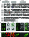
Domain, Sequence, and Subcellular Localization of ZmOCD1. (A) Schematic of the ZmOCD1 protein with conserved domains indicated. aa, amino acids. (B) Alignment of the ZmOCD1 protein and its homologs from Arabidopsis and O. formigenes. The key amino acids for enzyme functions are indicated by different colored shapes. Red squares, ThDP binding sites; orange circles, ADP binding sites; blue diamonds, Mg binding sites; and purple triangles, active-site residues. (C) Transient expression of the free 35S:GFP and 35S:ZmOCD1-GFP fusion (left panel) in Arabidopsis mesophyll protoplast cells and in tobacco leaf epidermal cells (right panel). Bars = 25 µm. (D) Subcellular localization of ZmOCD1 in 35S:ZmOCD1-GFP transgenic plants. The GFP fluorescence in cotyledon (left panel) and root tip (right panel) cells from the T2 plants was observed.
Figure 6.
Figure 6.
Biochemical Function of Recombinant ZmOCD1. (A) Partial pathway for oxalate degradation. The first two steps of oxalate degradation are proposed to be catalyzed by O7 and ZmOCD1. (B) and (C) In vitro enzyme activity analysis. Purified recombinant ZmOCD1 (B) or the empty vector control (C) was incubated with oxalyl-CoA and the cofactors. (D) and (E) In vivo enzyme activity analysis. 20-DAP endosperm extracts of the wild type (D) or ocd1-1 (E) were incubated with oxalyl-CoA and the cofactors. Product formation was analyzed by LC-MS/MS. The relative intensities of the LC-MS/MS transitions of oxalyl-CoA (m/z+ = 840.1, red line) and formyl-CoA (m/z+ = 796.1, blue line) are shown. The LC-MS/MS analyses were performed under the positive mode (M/H)+.
Figure 7.
Figure 7.
Oxalate-Induced Expression of Ocd1 in Roots. (A) Ocd1 expression is induced by different oxalate concentrations. (B) Ocd1 expression is induced by different times. (C) Ocd1 expression is induced by different organic acids. The wild-type roots were treated with different conditions as indicated. All data were normalized relative to the expression of Ubi. The values are indicated as the means ± sd of three independent treatments.
Figure 8.
Figure 8.
Oxalate Contents and Metabolome Analysis in the Wild Type and ocd1. (A) Measurement of oxalate contents in mature wild-type, W22, o7, and ocd1-1 seeds. Error bars represent ±sd from three biological replicates. (B) Significant fold changes of metabolites in wild-type and ocd1-1 20-DAP endosperms revealed by targeted metabolomics analyses. (C) Principal component analysis of nontargeted metabolomes. Metabolites were extracted from six individual endosperms at 20 DAP and examined under positive and negative mode. (D) Relative contents of variant saccharides with significant fold changes in ocd1-1 compared to the wild type. (E) Relative contents of variant amino acids with significant fold changes in ocd1-1 compared to the wild type. (F) Relative contents of taurine and hypotaurine in the wild type and ocd1-1. (G) Concentrations of ATP in the wild type and ocd1-1. Levels of ATP were quantified enzymatically. (H) Relative content of indole acetic acid in the wild type and ocd1-1. Levels of metabolites (except for ATP) in wild type were designated 1. The error bars indicate ±sd from six biological replicates.
Figure 9.
Figure 9.
Proposed Model for ZmOCD1 Function and the Opaque Phenotype. Due to the loss of function of oxalyl-CoA decarboxylase in ocd1, the reactions downstream of oxalyl-CoA are blocked (in gray). The dashed line indicates putative a feedback inhibition, which results in an excessive accumulation of oxalate in the ocd1 endosperm. The altered metabolites in the ocd1 endosperm may cause reduced synthesis of zeins and starch. The former directly affects the vitreous endosperm formation and the latter leads to the kernel weight loss. Dramatic metabolome perturbations, as well as other unidentified pathways, may contribute to the opaque phenotype in ocd1 kernels in a way that is independent of the zein protein decrease.

Comment in

  • Nutritious corn.
    Lei L. Lei L. Nat Plants. 2018 Oct;4(10):741. doi: 10.1038/s41477-018-0281-z. Nat Plants. 2018. PMID: 30287956 No abstract available.

Similar articles

Cited by

References

    1. Bateman D.F., Beer S.V. (1965). Simultaneous production and synergistic action of oxalic acid and polygalacturonase during pathogenesis by Sclerotium rolfsii. Phytopathology 55: 204–211. - PubMed
    1. Berthold C.L., Moussatche P., Richards N.G., Lindqvist Y. (2005). Structural basis for activation of the thiamin diphosphate-dependent enzyme oxalyl-CoA decarboxylase by adenosine diphosphate. J. Biol. Chem. 280: 41645–41654. - PubMed
    1. Brzezicha-Cirocka J., Grembecka M., Szefer P. (2016). Oxalate, magnesium and calcium content in selected kinds of tea: impact on human health. Eur. Food Res. Technol. 242: 383–389.
    1. Casteels M., Sniekers M., Fraccascia P., Mannaerts G.P., Van Veldhoven P.P. (2007). The role of 2-hydroxyacyl-CoA lyase, a thiamin pyrophosphate-dependent enzyme, in the peroxisomal metabolism of 3-methyl-branched fatty acids and 2-hydroxy straight-chain fatty acids. Biochem. Soc. Trans. 35: 876–880. - PubMed
    1. Castellaro A.M., Tonda A., Cejas H.H., Ferreyra H., Caputto B.L., Pucci O.A., Gil G.A. (2015). Oxalate induces breast cancer. BMC Cancer 15: 761–773. - PMC - PubMed

Publication types

MeSH terms