. 2018 Sep 10;19(9):2683.
doi: 10.3390/ijms19092683.

The Protective Effect of a Long-Acting and Multi-Target HM-3-Fc Fusion Protein in Rheumatoid Arthritis

Affiliations

The Protective Effect of a Long-Acting and Multi-Target HM-3-Fc Fusion Protein in Rheumatoid Arthritis

Ruijing Huang et al. Int J Mol Sci. .

Abstract

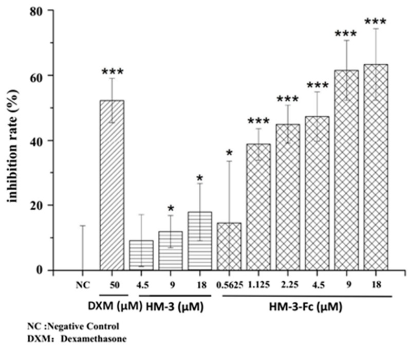
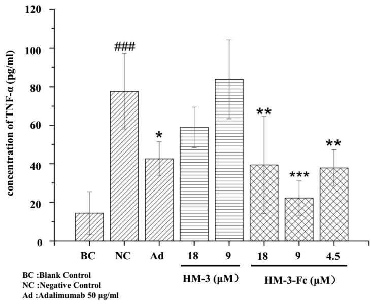
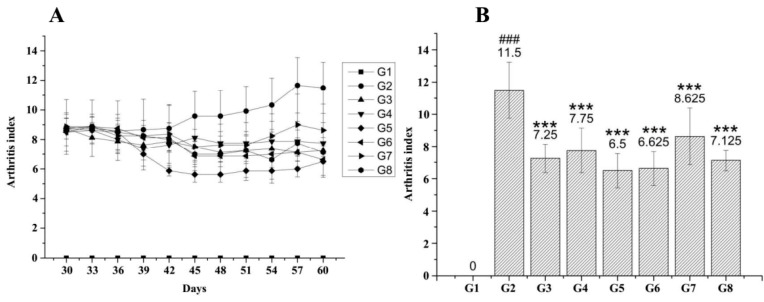
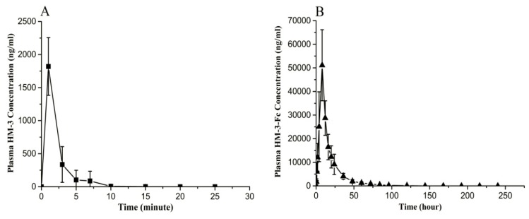
Current treatment of rheumatoid arthritis (RA) is limited by relative shortage of treatment targets. HM-3 is a novel anti-RA polypeptide consisting of 18 amino acids with integrin αVβ3 and α5β1 as targets. Previous studies confirmed that HM-3 effectively inhibited the synovial angiogenesis and the inflammatory response. However, due to its short half-life, the anti-RA activity was achieved by frequent administration. To extend the half-life of HM-3, we designed a fusion protein with name HM-3-Fc, by combination of modified Fc segment of immunoglobulin 4 (IgG4) with HM-3 polypeptide. In vitro cell experiments demonstrated that HM-3-Fc inhibited the proliferation of splenic lymphocytes and reduced the release of TNF-α from macrophages. The pharmacodynamics studies on mice paw in Collagen-Induced Arthritis (CIA) model demonstrated that HM-3-Fc administered once in 5 days in the 50 and 25 mg/kg groups, or once in 7 days in the 25 mg/kg group showed a better protective effect within two weeks than the positive control adalimumab and HM-3 group. Preliminary pharmacokinetic studies in cynomolgus confirmed that the in vivo half-life of HM-3-Fc was 15.24 h in comparison with 1.32 min that of HM-3, which demonstrated that an Fc fusion can effectively increase the half-life of HM-3 and make it possible for further reduction of subcutaneous injection frequency. Fc-HM-3 is a long-acting active molecule for RA treatment.

Keywords: Fc-domain of immunoglobulin G4; HM-3; TNF-α; half-life; inflammatory response; pharmacodynamics; rheumatoid arthritis; synovial angiogenesis.


Conflict of interest statement

The authors declare no conflict of interest.

Figures

Figure 1
Figure 1
The viability (VIA%), viable cell density (VCD) and yield of HM-3-Fc cells cultured for 15 days. Sterilely taken cells from bioreactor were stained by 0.4% trypan blue and then counted with a Countstar IC-1000 cell counter. The results of viable cells are summarized; VIA% = number of viable cells/total cell number × 100%. The protein expression amount was detected by size exclusion chromatography-high pressure liquid chromatography (SEC-HPLC) method.
Figure 2
Figure 2
Physiochemical characteristics of HM-3-Fc. (1) Purity analysis of HM-3-Fc (A). Columns: TOSOH, TSKgel G3000SWxL PN: 008543, Mobile Phase: 0.4 M NaCl 0.03 M Na2HPO4 0.02 M NaH2PO4 Solution with 10% Acetonitrile. Isocratic elution, Flow: 0.5 mL/min, Temperature: 25 °C, Detection fluorescence (FL): 214 nm, Injected volume: 10 μL. (2) CE-SDS analysis of HM-3-Fc. Non-reduced capillary electrophoresis (CE) analysis (B) and reduced CE-SDS analysis (C). Sample concentration: 1 mg/mL heated in water bath at 70 °C for 10 min. The sample was injected to the anode with reverse polarity using −5 kV for 20 s. Separation voltage was −15 kV with reverse polarity during the 30 min separation. Detection wavelength was 214 nm. (3) cIEF analysis of HM-3-Fc. Holograph of cIEF profile (D) and amplified part of cIEF graph (E). B1 = basic peak 1, A1–A4 = acidic peaks 1–5. The samples were detected by Beckman MDQ capillary electrophoresis system using a neutral coated capillary. Inject sample for 99.0 s at 20 psi. Focusing: 15 min at 25 kV under normal polarity. Mobilization: 30 min at 30 kV under normal polarity. Detection wavelength was 280 nm.
Figure 3
Figure 3
Specific and high affinity binding of HM-3-Fc with integrin αvβ3 and α5β1. A flow cytometry method was setup with FITC-protein A (FITC-A) as a probe that binds the Fc part of HM-3-Fc. The dark line indicated the fluorescence signal for untreated synovial cells and it was used to define the threshold of positive fluorescence signal. The light line indicated the fluorescence signal for HM-3-Fc and FITC-A treated synovial cells. (AC) The binding rates of synovial cells after incubation with 1, 10 or 75 μM HM-3-Fc before incubated with FITC-A. (DF) The binding rates of synovial cells that were blocked with anti-αvβ3, anti-α5β1, or their combination.
Figure 4
Figure 4
The effects of HM-3-Fc on splenic lymphocytes viability. Cells were stimulated with ConA, based on which 50 μmol/L Dexamethasone (DXM) was used as a positive control drug. In the other groups, cells were incubated with different concentrations of HM-3 or HM-3-Fc as indicated in the Figure. In this study, proliferation inhibition rate (PI) was used to express the inhibitory effect of HM-3-Fc on the proliferation of modeled splenic lymphocytes. The formula is as follows: PI = 1 − Atest/Acontrol100%. Atest is the absorbance of the drug group. Acontrol is the absorbance of the negative control group. Results were presented as mean ± SD (n = 6). * p < 0.05 and *** p < 0.001.
Figure 5
Figure 5
The effects of HM-3 and HM-3-Fc on the expression of TNF-α by U937 macrophages. Cells without LPS induction were used as a normal control. The other cells were inducted with LPS and the cells without drug treatment were used as a negative control. The other cells were incubated with HM-3 or HM-3-Fc at indicated concentrations, with 50 μg/mL Adalimumab as a positive control drug. Results were presented as mean ± SD (n = 3). ### p < 0.001 vs. Blank Control. * p < 0.05, ** p < 0.01 and *** p < 0.001 vs. Negative Control.
Figure 6
Figure 6
Inhibitory effect of HM-3-Fc on angiogenesis in zebrafish. Zebrafish were subcutaneously injected with HM-3 or HM-3-Fc and also with Avastin as a positive control. After 24 h, intestinal vascular area was visualized and measured and then angiogenesis inhibition rates were calculated based on the vascular area of each group. (A) Typical photos of zebrafish intestinal vascular area in different groups (150×). (B) The intestinal vascular areas of zebrafish in each group were visualized and measured by Nikon NIS-Elements D 3.10. And the areas of intestinal blood vessels in the experiment were calculated (S). (C) Angiogenesis inhibition rate = (1 − S(Normal Group)/S (Experiment Group)) × 100%. Results were presented as mean ± SD (n = 12). ** p < 0.01, *** p < 0.001.
Figure 7
Figure 7
HM-3-Fc treatments decreased arthritis grading of CIA mice. Mice were immunized with an emulsion of bull collagen type II and Complete Freund’s adjuvant (CFA) on day 0 and subsequently with an injection of incomplete Freund’s adjuvant (IFA) on day 21. On the 30th day of the experiments, mice were randomized in different groups for treatments with different drugs. Arthritis grading (A) of mice in each group was measured every three days until the 60th day. Comparison of arthritis index of the mice in each group was shown in panel (B). For G2, n = 12; for G3–G8, n = 8. ### p < 0.001 vs. G1. *** p < 0.001 vs. G2.
Figure 8
Figure 8
Serum drug concentration versus time curves of HM-3 and HM-3-Fc after subcutaneous injection in cynomolgus. (A) Serum drug concentration versus time curve of HM-3 after s.c. administration of 0.5 mg/kg HM-3 in cynomolgus. (B) Serum drug concentration versus time curve of HM-3-Fc after s.c. administration of 5.77 mg/kg HM-3-Fc in cynomolgus. Data were shown as mean ± SEM (standard error of mean).

Similar articles

Cited by

References

    1. Paleolog E.M., Miotla J.M. Angiogenesis in arthritis: Role in disease pathogenesis and as a potential therapeutic target. Angiogenesis. 1998;2:295–307. doi: 10.1023/A:1009229508096. - DOI - PubMed
    1. Szekanecz Z., Koch A.E. Vascular involvement in rheumatic diseases: ‘Vascular rheumatology’. Arthritis Res. Ther. 2008;10:224. - PMC - PubMed
    1. Muz B., Khan M.N., Kiriakidis S., Paleolog E.M. The role of hypoxia and HIF-dependent signalling events in rheumatoid arthritis. Arthritis Res. Ther. 2009;11:201. doi: 10.1186/ar2568. - DOI - PMC - PubMed
    1. Jackson J.R. The codependence of angiogenesis and chronic inflammation. FASEB J. 1997;11:457–465. doi: 10.1096/fasebj.11.6.9194526. - DOI - PubMed
    1. Marrelli A. Angiogenesis in rheumatoid arthritis: A disease specific process or a common response to chronic inflammation? Autoimmun. Rev. 2011;10:595–598. doi: 10.1016/j.autrev.2011.04.020. - DOI - PubMed

MeSH terms

LinkOut - more resources