

Gold Rod-Polyethylene Glycol-Carbon Dot Nanohybrids as Phototheranostic Probes
- PMID: 30201913
- PMCID: PMC6165167
- DOI: 10.3390/nano8090706
Gold Rod-Polyethylene Glycol-Carbon Dot Nanohybrids as Phototheranostic Probes
Abstract
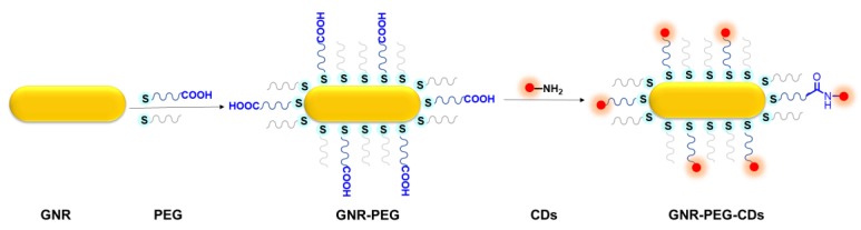
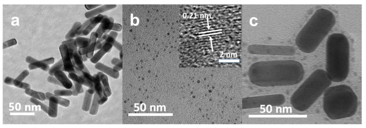
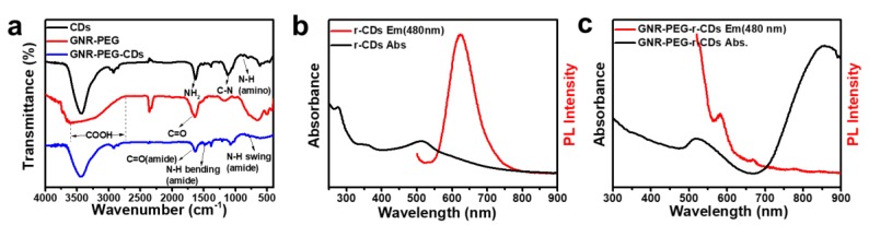
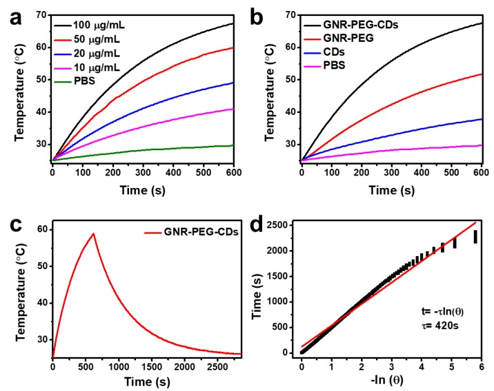
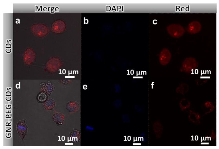
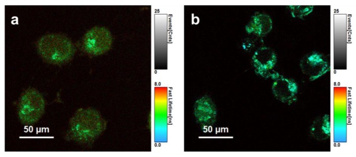
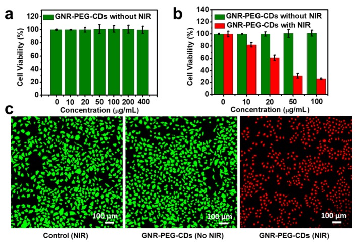
Emphasis using phototheranostics has been placed on the construction of multifunctional nanoplatforms for simultaneous tumor diagnosis and therapy. Herein, we put forth a novel nanosized luminescent material using the incorporation of red emissive carbon dots on gold nanorods through polyethylene glycol as a covalent linkage for dual-modal imaging and photothermal therapy. The novel nanohybrids, not only retain the optical properties of the gold nanorod and carbon dots, but also possess superior imaging performance in both confocal laser scanning microscopy and fluorescence lifetime imaging microscopy. The nanohybrids also exhibit excellent photothermal performance as phototheranostic nanohybrid probes for in vitro assays. This study promises a new multifunctional nanoplatform for cancer diagnostics and therapeutics.
Keywords: bioimaging; carbon dots; gold nanorod; nanohybrids; photothermal therapy.
Conflict of interest statement
There are no conflicts to declare.
Figures
Similar articles
-
Gold nanorod@silica-carbon dots as multifunctional phototheranostics for fluorescence and photoacoustic imaging-guided synergistic photodynamic/photothermal therapy.Nanoscale. 2016 Jul 14;8(26):13067-77. doi: 10.1039/c6nr03459d. Epub 2016 Jun 21. Nanoscale. 2016. PMID: 27326673
-
Preparation of photothermal-chemotherapy nanohybrids by complexation of gold nanorods with polyamidoamine dendrimers having poly(ethylene glycol) and hydrophobic chains.J Mater Chem B. 2020 Apr 14;8(14):2826-2833. doi: 10.1039/c9tb02163a. Epub 2020 Mar 13. J Mater Chem B. 2020. PMID: 32166301
-
Dual-responsive nanohybrid based on degradable silica-coated gold nanorods for triple-combination therapy for breast cancer.Acta Biomater. 2021 Jul 1;128:435-446. doi: 10.1016/j.actbio.2021.04.006. Epub 2021 Apr 20. Acta Biomater. 2021. PMID: 33862284
-
Phototheranostics for multifunctional treatment of cancer with fluorescence imaging.Adv Drug Deliv Rev. 2022 Oct;189:114483. doi: 10.1016/j.addr.2022.114483. Epub 2022 Aug 6. Adv Drug Deliv Rev. 2022. PMID: 35944585 Free PMC article. Review.
-
Gold Nanorods as Nanodevices for Bioimaging, Photothermal Therapeutics, and Drug Delivery.Chem Pharm Bull (Tokyo). 2017;65(7):625-628. doi: 10.1248/cpb.c17-00102. Chem Pharm Bull (Tokyo). 2017. PMID: 28674334 Review.
Cited by
-
Carbon Dots-Based Logic Gates.Nanomaterials (Basel). 2021 Jan 17;11(1):232. doi: 10.3390/nano11010232. Nanomaterials (Basel). 2021. PMID: 33477327 Free PMC article. Review.
-
Metal-Enhanced Fluorescent Carbon Quantum Dots via One-Pot Solid State Synthesis for Cell Imaging.ACS Appl Bio Mater. 2023 May 15;6(5):1798-1805. doi: 10.1021/acsabm.3c00040. Epub 2023 Apr 26. ACS Appl Bio Mater. 2023. PMID: 37125840 Free PMC article.
References
Grants and funding
LinkOut - more resources
Full Text Sources
Other Literature Sources
