. 2018 Oct;19(10):1093-1099.
doi: 10.1038/s41590-018-0201-4. Epub 2018 Sep 10.

Tissue signals imprint ILC2 identity with anticipatory function

Affiliations

Tissue signals imprint ILC2 identity with anticipatory function

Roberto R Ricardo-Gonzalez et al. Nat Immunol. 2018 Oct.

Abstract

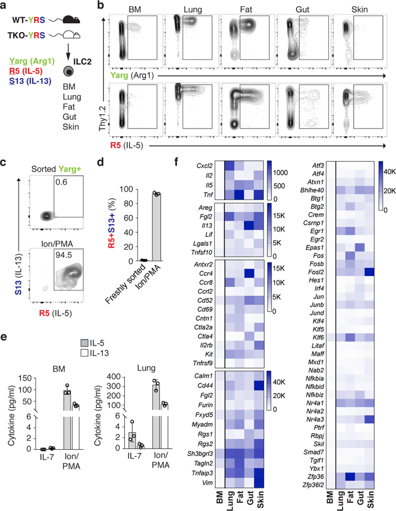
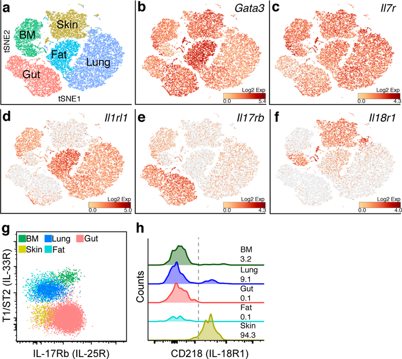
Group 2 innate lymphoid cells (ILC2s) are distributed systemically and produce type 2 cytokines in response to a variety of stimuli, including the epithelial cytokines interleukin (IL)-25, IL-33, and thymic stromal lymphopoietin (TSLP). Transcriptional profiling of ILC2s from different tissues, however, grouped ILC2s according to their tissue of origin, even in the setting of combined IL-25-, IL-33-receptor-, and TSLP-receptor-deficiency. Single-cell profiling confirmed a tissue-organizing transcriptome and identified ILC2 subsets expressing distinct activating receptors, including the major subset of skin ILC2s, which were activated preferentially by IL-18. Tissue ILC2 subsets were unaltered in number and expression in germ-free mice, suggesting that endogenous, tissue-derived signals drive the maturation of ILC2 subsets by controlling expression of distinct patterns of activating receptors, thus anticipating tissue-specific perturbations occurring later in life.


Conflict of interest statement

Competing interests

The authors declare no competing interests.

Competing interests: None declared.

Figures

Figure 1:
Figure 1:. Steady-state ILC2 activation in multiple peripheral tissues.
a, ILC2s were analyzed in multiple tissues from wild-type (WT) or Crlf2–/–IL25–/–Il1rl1–/– triple-deficient (TKO) mice on Arg1 (Yarg); R5 (IL-5); S13 (IL-13) combined triple-reporter (YRS) background. b, Representative flow cytometry of CD45+Lin- cells from indicated WT-YRS mouse tissues. c, d, Representative flow cytometry (c) and percentage (d) of sorted Yarg+ bone marrow (BM) ILC2 expressing R5 and S13 reporter alleles before and after stimulation with ionomycin (Ion) and phorbol 12-myristate 13-acetate (PMA). e, IL-5 and IL-13 in supernatants of Yarg+ ILC2 sorted from WT-YRS BM or lung tissue cultured in IL-7 or Ion/PMA for 24 h. f, RNA-seq analysis of select transcripts significantly enriched (FDR<0.01) in R5+ ILC2s from peripheral tissues (lung, gut, fat, skin) versus Yarg+ BM ILC2s. b, c, Data representative of 3 independent experiments and in (d-f) represent biological replicates (n=3; mean±SD). f, Data represent mean normalized read counts (fragments per million mapped reads) from biological replicates (n=3, BM; n=5, lung; n=6, fat, gut, skin).
Figure 2:
Figure 2:. ILC2 distribution and homeostatic function is independent of the microbiota.
a, b, Number of ILC2s (Lin-CD45+Thy1.2+CD25+) in lungs (a), and skin (b). c, c, Percentage of gut ILC2s (Lin-CD45+IL17rb+KLRG1+) out of total CD45+ cells. d,Number of spleen eosinophils in C57BL/6 mice housed in specific pathogen-free (SPF) or germ-free (GF) conditions. e, Quantitative RT-PCR (qPCR) analysis of Il5 transcript abundance among ILC2s sorted from SPF or GF mouse tissues. a-d, Data pooled from 2 or more independent experiments with n≥4 from each tissue and expressed as mean±SD. e, Data represent biological replicates (n=3; mean⩲SD) and are normalized to Rps17.
Figure 3:
Figure 3:. Tissue-resident ILC2s depend on distinct tissue signals.
a-b, Number of ILC2s (Lin-CD45+Thy1.2+CD25+) in lungs (a) and fat (b). c, percentage of ILC2s (Lin-CD45+KLRG1+) out of total CD45+ cells in gut among wild-type (WT) or Crlf2/IL25/Il1rl1/ triple-deficient (TKO) mice on Arg1 (Yarg); R5 (IL-5); S13 (IL-13) triple-reporter (YRS) backgrounds. d-f, Number of R5+ ILC2s (Lin-CD45+) in lungs (d), fat (e), and gut (f) tissues of WT-YRS and TKO-YRS mice. g, Numbers of eosinophils in spleen. h, Number of R5+ ILC2s in skin (Lin-CD45+) of WT-YRS and TKO-YRS mice. Data pooled from 2 or more independent experiments with n≥10 mice per group, represented as mean⩲SD; **, p<0.001; ***, p<0.0001.
Figure 4:
Figure 4:. Transcriptional heterogeneity of tissue-resident ILC2s.
a, t-distributed stochastic neighbor embedding (tSNE) plot representing ILC2 samples. ILC2s were sorted from lung, fat, gut, skin (Lin-CD45+Red5+), and bone marrow (BM, Lin-CD45+Yarg+) as outlined in the gating strategy shown in Supplementary Figure 1. b, RNA-seq analysis of differentially expressed transcripts (FDR<0.01) among ILC2s from each tissue versus all other tissues; select upregulated transcripts in each tissue highlighted. Data pooled from 3 independent experiments with n≥3 biological replicates per group.
Figure 5:
Figure 5:. Tissue map of ILC2 signals revealed by single-cell RNA sequencing.
a, tSNE plot representing 35,396 ILC2s sorted from BM (Lin-CD45+Yarg+), lung, fat, gut and skin (Lin-CD45+Red5+) analyzed by single-cell RNA sequencing (scRNA-seq). b-f, normalized relative expression of Gata3 (b), Il7r (c), Il1rl1 (d), Il17rb (e), and Il18r1 (f) transcripts. g-h, Representative flow cytometric staining for T1/ST2 (IL-33R), IL17Rb (IL-25R) (g), and CD218 (IL-18R1) (h) among ILC2s from indicated WT-YRS mouse tissues. g, h, Data are representative of 3 independent experiments. h, numbers represent the percentage of ILC2s positive for CD218 (right of dotted line).
Figure 6:
Figure 6:. IL-18 independently mediates ILC2 subset activation.
a, IL-13 in supernatants of ILC2s (Lin-CD45+Thy1.2+Red5+) sorted from skin or lung tissue and cultured in TSLP alone or TSLP and IL-18. b, Percentage of skin and lung ILC2s (Lin-CD45+Thy1.2+Red5+) expressing S13 reporter allele after intradermal injection of PBS or TSLP and IL-18. c, d, Percentage of skin ILC2s expressing S13 reporter allele (c) and R5 reporter median fluorescence intensity (MFI) (d) among skin ILC2s from WT and IL18-deficient (KO) mice on a R5; S13 dual-reporter background. e, Number of IL-18R1+R5+ ILC2s in lungs of WT-YRS and TKO-YRS mice 24 h after intranasal administration of PBS or recombinant IL-18. f, Percentage of IL-18R1+Yarg+ ILC2s sorted from BM of WT or ST2 KO mice expressing S13 reporter allele 24 h after culture in IL-7+IL-33 or IL-7+IL-18. g, Representative flow cytometry of R5 reporter expression among skin and lung ILC2s (Lin-CD45+Thy1.2+) from WT and IL-18–/–ST2–/–deficient (DKO) mice on a R5; S13 dual-reporter background. Numbers indicate percent R5 positive in each quadrant. Data in (a-f) pooled from 2 or more independent experiments for a total of at least 3 mice per group, represented as mean⩲SD; *, p<0.01; **, p<0.001; ***, p<0.0001.
Figure 7:
Figure 7:. Blunted type 2 skin inflammation in the absence of IL-18.
a-d, Total skin ILC2s (Lin-CD45+Thy1.2+CD25+) (a), R5+ ILC2s (Lin-CD45+Thy1.2+Red5+) (b), percentage of R5+ ILC2s expressing S13 reporter allele (c), and total eosinophils (d) in ears of wild type or IL-18KO mice treated with ethanol (EtOH) or MC903. Data pooled from 2–3 individual experiments with n≥6 for each experimental group and are represented as mean±SEM; *, p<0.05; **(p<0.005); ***, p<0.0005, ****, p<0.00005.

Comment in

References

    1. Moltke von, J. & Locksley RM I-L-C-2 it: type 2 immunity and group 2 innate lymphoid cells in homeostasis. Curr. Opin. Immunol 31, 58–65 (2014). - PMC - PubMed
    1. Klose CSN & Artis D Innate lymphoid cells as regulators of immunity, inflammation and tissue homeostasis. Nat. Immunol 17, 765–774 (2016). - PubMed
    1. Neill DR et al. Nuocytes represent a new innate effector leukocyte that mediates type-2 immunity. Nature 464, 1367–1370 (2010). - PMC - PubMed
    1. Klein Wolterink RGJ et al. Pulmonary innate lymphoid cells are major producers of IL-5 and IL-13 in murine models of allergic asthma. Eur. J. Immunol 42, 1106–1116 (2012). - PubMed
    1. Salimi M et al. A role for IL-25 and IL-33-driven type-2 innate lymphoid cells in atopic dermatitis. J. Exp. Med 210, 2939–2950 (2013). - PMC - PubMed

Publication types