. 2018 Sep 10;8(1):13525.
doi: 10.1038/s41598-018-31766-y.

Exploring the elusive composition of corpora amylacea of human brain

Affiliations

Exploring the elusive composition of corpora amylacea of human brain

Elisabet Augé et al. Sci Rep. .

Abstract

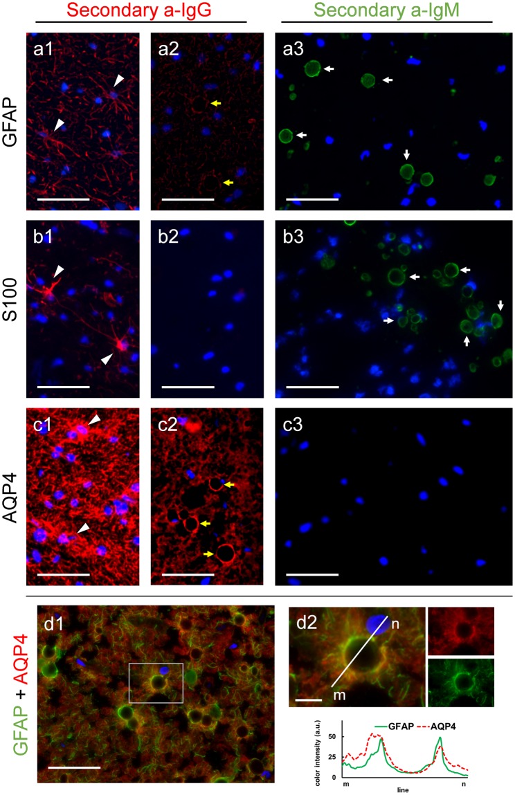
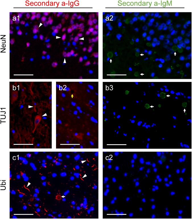
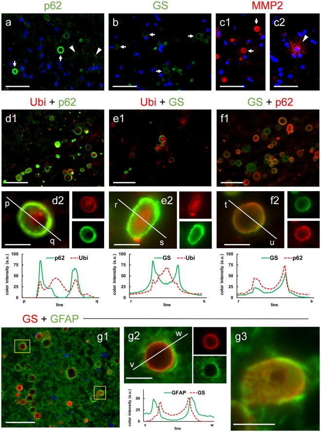
Corpora amylacea (CA) are polyglucosan bodies that accumulate in the human brain during ageing and are also present in large numbers in neurodegenerative conditions. Theories regarding the function of CA are regularly updated as new components are described. In previous work, we revealed the presence of some neo-epitopes in CA and the existence of some natural IgM antibodies directed against these neo-epitopes. We also noted that these neo-epitopes and IgMs were the cause of false staining in CA immunohistochemical studies, and disproved the proposed presence of β-amyloid peptides and tau protein in them. Here we extend the list of components erroneously attributed to CA. We show that, contrary to previous descriptions, CA do not contain GFAP, S100, AQP4, NeuN or class III β-tubulin, and we question the presence of other components. Nonetheless, we observe that CA contains ubiquitin and p62, both of them associated with processes of elimination of waste substances, and also glycogen synthase, an indispensable enzyme for polyglucosan formation. In summary, this study shows that it is imperative to continue reviewing previous studies about CA but, more importantly, it shows that the vision of CA as structures involved in protective or cleaning mechanisms remains the most consistent theory.


Conflict of interest statement

The authors declare no competing interests.

Figures

Figure 1
Figure 1
Absence of GFAP, S100 and AQP4 in CA. Representative images of human brain hippocampus sections immunostained with GFAP (a), S100 (b) and AQP4 (c). When using isotype-specific anti-IgG antibodies (a1, a2, b1 and b2) or anti-IgG(H&L) antibodies (c1c2) conjugated to a red fluorochrome in the second incubation, the positive labeling of astrocytes can be observed (arrowheads in a1, b1 and c1), but CA are not labelled and are visible just as a dark hole when astrocyte processes encircled them (yellow arrows in a2 and c2). By using an isotype-specific anti-IgM antibody conjugated to a green fluorochrome in the second incubation, labeling of CA can be observed as shown in a3 and b3 (white arrows) but not in c3, indicating that GFAP and S100, but not AQP4, are IgM contaminated. Double immunostaining with GFAP and AQP4 show these two markers surrounding the CA (d1). d2 shows an inset of d1, where a CA is magnified. The different color channels are shown in small images next to the corresponding ones, and the histogram profiles of green and red intensities on the plotted line show the similar distribution of both markers around the CA. Hoechst (blue) was used for nuclear staining. Scale bar in d2: 10 μm, other scale bars: 50 μm.
Figure 2
Figure 2
Absence of NeuN and class III β-tubulin and presence of ubiquitin in CA. Representative images of human brain hippocampus sections immunostained with NeuN (a), anti-class III β-tubulin (TUJ1) (b) and anti-ubiquitin (Ubi) (c). As expected, when using isotype-specific anti-IgG antibodies conjugated to a red fluorochrome in the second incubation, NeuN stained the neuronal soma (arrowheads in a1), TUJ1 stains both neuronal soma and neurites (arrowheads in b1), and Ubi stained some dystrophic neurites (arrowheads in c1). CA were not labeled with NeuN (a1) and TUJ1 (yellow arrow in b2), but became labeled with Ubi (white arrow in c1). When using isotype-specific anti-IgM antibodies conjugated to a green fluorochrome in the second incubation, labeling of CA was observed in a2 and b3 (white arrows), but not in c2, indicating the presence of contaminant IgMs in the vials of NeuN and TUJ1, but not in the Ubi vial. Hoechst (blue) was used for nuclear staining. Scale bar: 50 μm.
Figure 3
Figure 3
Immunolabeling of CA with the markers p62, GS and MMP2. Representative images of human brain hippocampus sections immunostained with p62 (a), GS (b) and MMP2 (c1 and c2). The three markers positively stained the CA (arrows). p62 also stained some dystrophic neurites (arrowheads in a), while MMP2 stained some thin filaments (arrowheads in c2). Hoechst (blue) was used for nuclear staining. d1d2: double immunostaining with anti-ubiquitin (Ubi) (red) and p62 (green); e1e2: double immunostaining with Ubi (red) and GS (green); f1f2: double immunostaining with GS (green) and p62 (red). In some CA, p62 and GS are clearly visible in the peripheral region (d2, e2 and f2), while ubiquitin is concentrated in the central zone (d2 and e2). The different color channels from d2, e2 and f2 are shown in small images next to the corresponding ones. The histogram profiles of green and red intensities on the plotted lines illustrate the peripheral location of p62 and GS, and show that ubiquitin is concentrated in the central zone but is also present at the periphery. Double staining with GS and GFAP showed that GFAP staining is surrounding that of GS, which indicated that the astrocytic processes are surrounding CA (g1). This is clearly shown when CA is magnified (g2). When the histogram profiles are traced, it can be observed that the peaks of intensity of GFAP staining appear externally to those of GS staining. In some cases, CA become sliced in a tangential plane, and then the GS staining appear throughout the surface of the CA, and the astrocytic processes stained with GFAP are surrounding this surface (g3). Scale bars on d2, e2, f2, g2 and g3: 10 μm; other scale bars: 50 μm.

References

    1. Cavanagh JB. Corpora-amylacea and the family of polyglucosan diseases. Brain Res. Rev. 1999;29:265–295. doi: 10.1016/S0165-0173(99)00003-X. - DOI - PubMed
    1. Cissé S, Perry G, Lacoste-Royal G, Cabana T, Gauvreau D. Immunochemical identification of ubiquitin and heat-shock proteins in corpora amylacea from normal aged and Alzheimer’s disease brains. Acta Neuropathol. 1993;85:233–240. doi: 10.1007/BF00227716. - DOI - PubMed
    1. Keller JN. Age-related neuropathology, cognitive decline, and Alzheimer’s disease. Ageing Res. Rev. 2006;5:1–13. doi: 10.1016/j.arr.2005.06.002. - DOI - PubMed
    1. Mizutani T, Satoh J, Morimatsu Y. Axonal polyglucosan body in the ventral posterolateral nucleus of the human thalamus in relation to aging. Acta Neuropathol. 1987;74:9–12. doi: 10.1007/BF00688332. - DOI - PubMed
    1. Mrak RE, Griffin ST, Graham DI. Aging-associated changes in human brain. J. Neuropathol. Exp. Neurol. 1997;56:1269–1275. doi: 10.1097/00005072-199712000-00001. - DOI - PubMed

Publication types