

Role of CT and MRI in the design and development of orthopaedic model using additive manufacturing
- PMID: 30202151
- PMCID: PMC6128794
- DOI: 10.1016/j.jcot.2018.07.002
Role of CT and MRI in the design and development of orthopaedic model using additive manufacturing
Erratum in
-
Erratum regarding previously published articles.J Clin Orthop Trauma. 2020 Nov-Dec;11(6):1169-1171. doi: 10.1016/j.jcot.2020.09.032. Epub 2020 Sep 26. J Clin Orthop Trauma. 2020. PMID: 33013141 Free PMC article.
-
Erratum regarding missing Declaration of Competing Interest statements in previously published articles.J Clin Orthop Trauma. 2020 Nov-Dec;11(6):1178. doi: 10.1016/j.jcot.2020.10.026. Epub 2020 Oct 15. J Clin Orthop Trauma. 2020. PMID: 33078052 Free PMC article.
-
Erratum regarding previously published articles.J Clin Orthop Trauma. 2020 Nov-Dec;11(6):1172-1174. doi: 10.1016/j.jcot.2020.10.044. Epub 2020 Oct 23. J Clin Orthop Trauma. 2020. PMID: 33192025 Free PMC article.
Abstract
Objective: To study the role of Computed tomography (CT) and Magnetic resonance imaging (MRI) for design and development of orthopaedic model using additive manufacturing (AM) technologies.
Methods: A significant number of research papers in this area are studied to provide the direction of development along with the future scope.
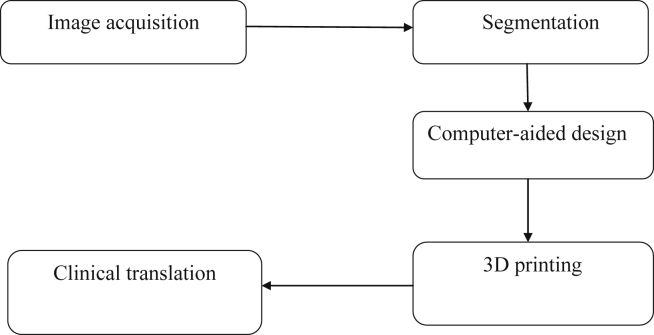
Results: Briefly discussed various steps used to create a 3D model by Additive Manufacturing using CT and MRI scan. These scanning technologies are used to produce medical as well as orthopaedic implants by using AM technologies. The images so produced are exported in different software like OsiriX Imaging Software, 3D slicer, Mimics, Magics, 3D doctor and InVesalius to produce a 3D digital model. Various criteria's achieved by CT and MRI scan for design and development of orthopaedic implant using additive manufacturing are also discussed briefly. AM model created by this process show exact shape, size, dimensions, textures, colour and features.
Conclusion: AM technologies help to convert the digital model into a 3D physical object, thereby improving the understanding of patient anatomy for treatment as well as for educational purpose. These scanning technologies have various applications to enhance the AM in the field of orthopaedic. In orthopaedic every patient model is a customised unit, sourced from the individual patient. 3D CAD data captured by these scanning technologies are directly exported in standard triangulate language (STL) format for printing by AM technologies. Crossestion of the physical model fabricated by this process shows a patient's anatomy if the model prepared by using the bone-like material.
Keywords: Additive manufacturing (AM); Computed tomography (CT); Magnetic resonance imaging (MRI); Orthopaedic.
Figures
References
-
- Werner H., dos Santos J.R., Fontes R. Additive manufacturing models of fetuses built from three-dimensional ultrasound, magnetic resonance imaging and computed tomography scan data. Ultrasound Obstet Gynecol. 2010;36(3):355–361. - PubMed
-
- Herrmann K.H., Gartner C., Gullmar D., Kramer M., Reichenbach J.R. 3D printing of MRI compatible components: why every MRI research group should have a low-budget 3D printer. Med Eng Phys. 2014;36(10):1373–1380. - PubMed
LinkOut - more resources
Full Text Sources
Other Literature Sources
Miscellaneous
