

Cervical pedicle screw guiding jig, an innovative solution
- PMID: 30202153
- PMCID: PMC6128793
- DOI: 10.1016/j.jcot.2018.07.010
Cervical pedicle screw guiding jig, an innovative solution
Erratum in
-
Erratum regarding missing declaration of competing interest statements in previously published articles.J Clin Orthop Trauma. 2020 Nov-Dec;11(6):1175. doi: 10.1016/j.jcot.2020.10.023. Epub 2020 Oct 15. J Clin Orthop Trauma. 2020. PMID: 33192026 Free PMC article.
Abstract
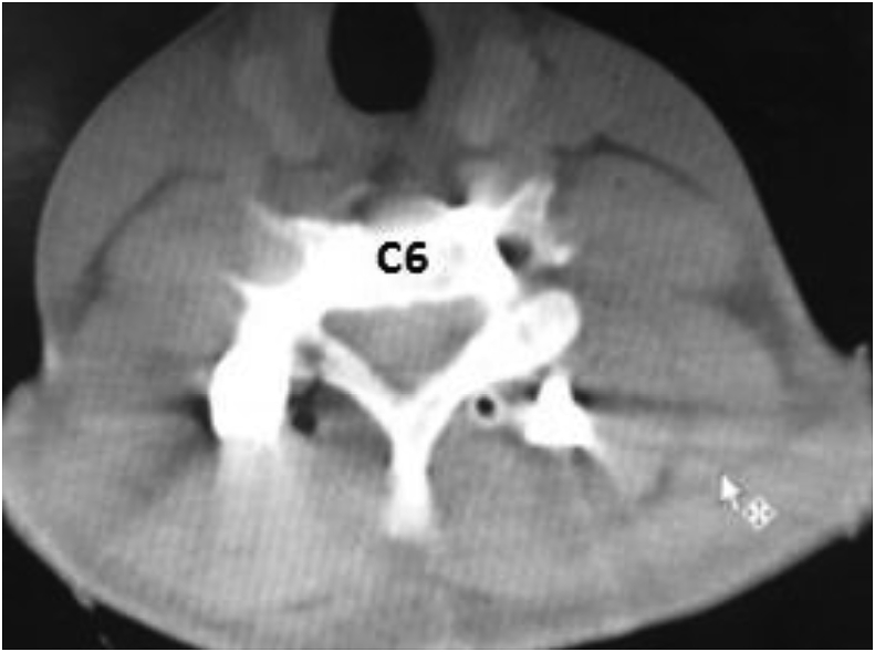
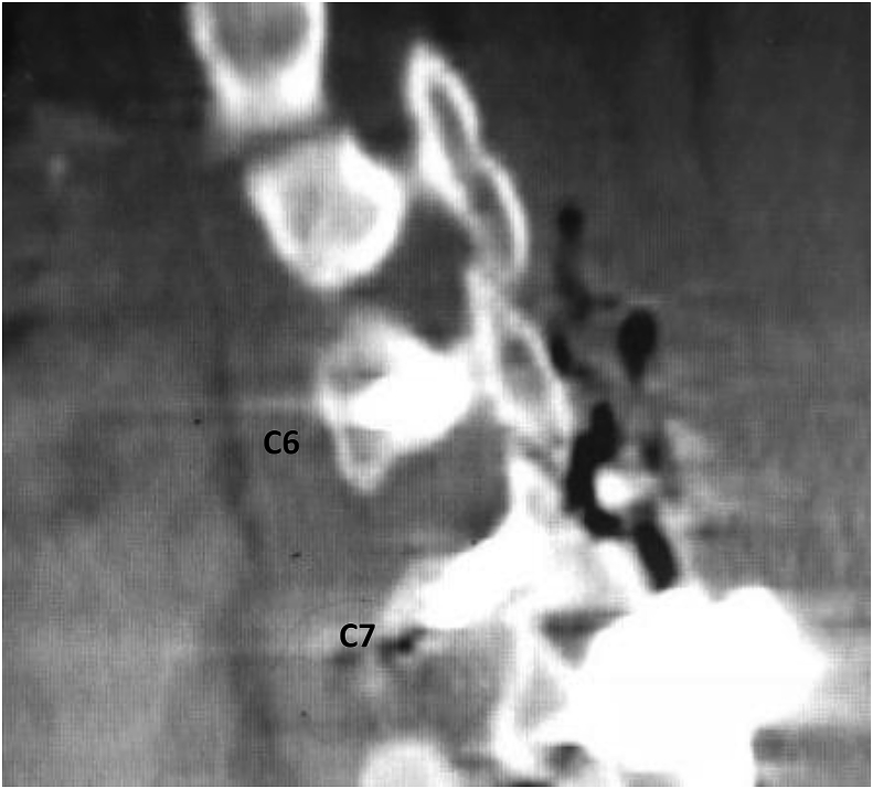
Pedicle screws are one the commonest used modality in spinal instrumentation. However, the method of pedicle screw fixation in cervical spine as compared to thoracic and lumbar spine is still technically demanding because it carries the risk of catastrophic damage to the surrounding neurovascular structures We have utilized virtual planning and 3D (3-dimension) printing to develop a patient specific jig to guide the accurate placement of pedicle screws. A patient with bifacetal dislocation C7 over D1 classified as flexion-distraction injury type 3 who was planned for decompression and fusion by posterior instrumentation at C6, C7, D1 and D2 was selected. A CT scan with 1 mm cuts was used to produce DICOM images of the same. Using these DICOM images virtual planning was done on MIMICS and 3 MATICS software to create patient specific jigs. These jigs were then 3D printed using a 3D printer and used for accurate placement of pedicle screws intra-operatively after adequate sterilization. Our procedure is low cost but high technology based. It is simple, accurate, and very cost effective. The technology transfer is very easy and can be adopted easily.
Keywords: 3d printing; Cervical spine; Jig; Pedicle screw.
Figures


References
-
- Panjabi M.M., Shin E.K., Chen N.C., Wang J.L. Internal morphology of human cervical pedicles. Spine. 2000 May 15;25(10):1197–1205. - PubMed
-
- Ebraheim N.A., Xu R., Knight T., Yeasting R.A. Morphometric evaluation of lower cervical pedicle and its projection. Spine. 1997 Jan 1;22(1):1–5. - PubMed
-
- Anon . American Spine Injury Association; Atlanta, GA: 2008. International Standards for Neurological Classification of Spinal Injury.
-
- Hoppenfield S., Boer P.D., Buckley R. fifth ed. Lippincott Williams and Wilkins; New York, NY: 2017. Surgical Exposures in Orthopaedics: the Anatomic Approach; pp. 715–738. (chapter 6)
-
- Raley D.A., Mobbs R.J. Retrospective computed tomography scan analysis of percutaneously inserted pedicle screws for posterior transpedicular stabilization of the thoracic and lumbar spine: accuracy and complication rates. Spine. 2012 May 20;37(12):1092–1100. - PubMed
Publication types
LinkOut - more resources
Full Text Sources
Other Literature Sources
Miscellaneous
