. 2018 Sep 6:155:27.
doi: 10.1186/s41065-018-0066-4. eCollection 2018.

Overexpression of the Drosophila ATR homologous checkpoint kinase Mei-41 induces a G2/M checkpoint in Drosophila imaginal tissue

Affiliations

Overexpression of the Drosophila ATR homologous checkpoint kinase Mei-41 induces a G2/M checkpoint in Drosophila imaginal tissue

Fabienne E Bayer et al. Hereditas. .

Abstract

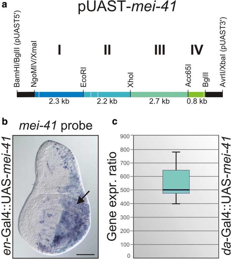
Background: DNA damage generally results in the activation of ATM/ATR kinases and the downstream checkpoint kinases Chk1/Chk2. In Drosophila melanogaster, the ATR homologue meiotic 41 (mei-41) is pivotal to DNA damage repair and cell cycle checkpoint signalling. Although various mei-41 mutant alleles have been analyzed in the past, no gain-of-function allele is yet available. To fill this gap, we have generated transgenic flies allowing temporal and tissue-specific induction of mei-41.

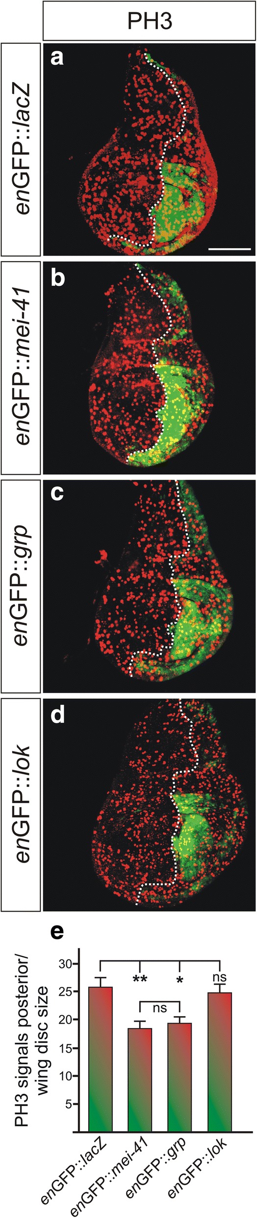
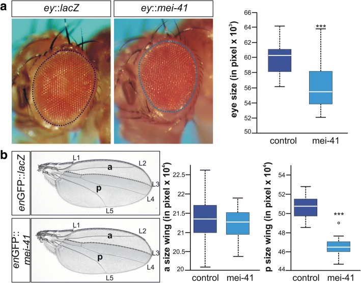
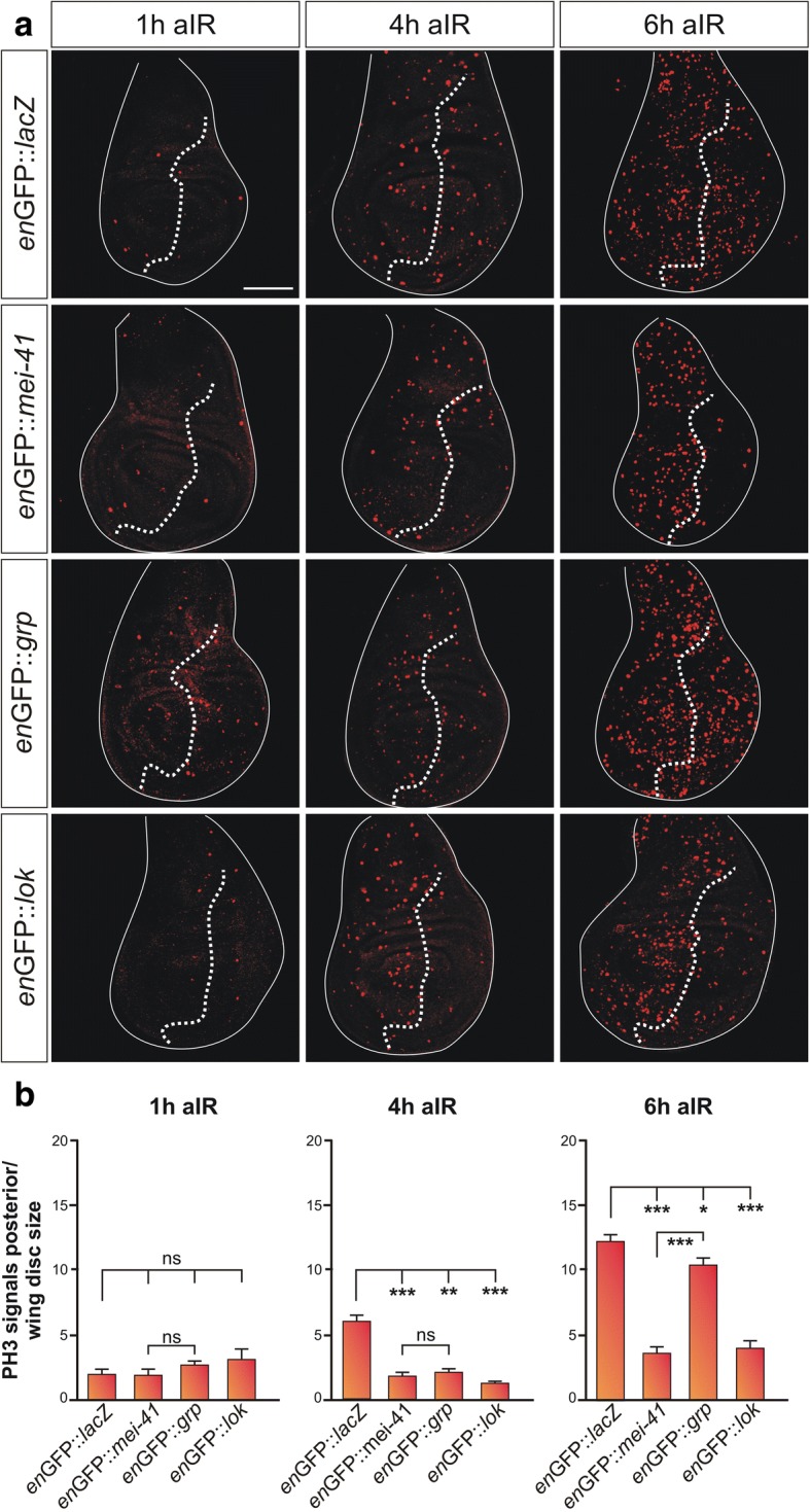
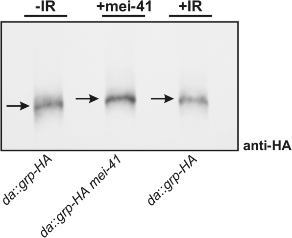
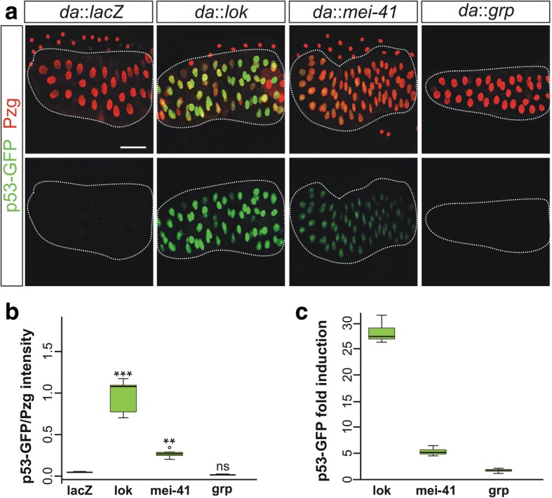
Results: Overexpression of mei-41 in wing and eye anlagen affects proliferation and a G2/M checkpoint even in the absence of genomic stress. Similar consequences were observed following the overexpression of the downstream kinase Grapes (Grp) but not of Loki (Lok), encoding the respective Drosophila Chk1 and Chk2 homologues, in agreement with their previously reported activities. Moreover, we show that irradiation induced cell cycle arrest was prolonged in the presence of ectopic mei-41 expression. Similar to irradiation stress, mei-41 triggered the occurrence of a slower migrating form of Grp, implying specific phosphorylation of Grp in response to either signal. Using a p53R-GFP biosensor, we further show that overexpression of mei-41 was sufficient to elicit a robust p53 activation in vivo.

Conclusion: We conclude that overexpression of the Drosophila ATR homologue mei-41 elicits an effectual DNA damage response irrespective of irradiation.

Keywords: ATR; DNA damage checkpoint; Mei-41; Overexpression; p53 activation.


Conflict of interest statement

Not applicable.Not applicable.The authors declare that there are no competing financial, personal, or professional interests.Springer Nature remains neutral with regard to jurisdictional claims in published maps and institutional affiliations.

Figures

Fig. 1
Fig. 1
Generation of a UAS-mei-41 overexpression line. (a) Cloning scheme of the pUAST-mei-41 construct. Coloured line corresponds to the mei-41 gene; intron positions are depicted as pale dashes. Segments derived from plasmid vectors are depicted in black and are not to scale. Genomic mei-41 DNA was PCR-amplified in fragments I-IV, which were eventually merged and shuttled into pUAST transformation vector in the correct 5′-3′ orientation. (b) UAS-mei-41 flies were crossed to en-Gal4-GFP, and induction of mei-41 expression was visualized in the posterior compartment of a wing imaginal disc by in situ hybridization (arrow). Size bar represents 100 μm. (c) Overexpression of mei-41 was quantified by qRT-PCR. To this end, UAS-mei-41 was ubiquitously induced with da-Gal4, and mRNA isolated from third instar larvae. Compared to control (da::lacZ), mei-41 is about 500-fold overexpressed. Data were assembled from three biological and two technical replicates. Mini-max depicts 95% confidence, median corresponds to expression ratio. Gapdh and beta-Tubulin56D served as reference genes. Efficiencies for mei-41 (0.92), for gapdh (0.94) and for beta-Tubulin56D (0.95) were accounted for determining relative quantities [44]
Fig. 2
Fig. 2
Overexpression of mei-41 induces G2/M cell cycle arrest. (a-d) The checkpoint kinases mei-41, grp and lok (chk2) as indicated were overexpressed in the posterior compartment of wing imaginal discs using en-Gal4-GFP (green) (b-d). UAS-lacZ served as control (a). To image M phase, discs were stained with anti-Phospho-Histone H3 (PH3) antibodies (red) indicating the number of mitotic cells. GFP labelling was used to determine the antero-posterior boundary (dotted line). (e) Quantification of PH3 signals showed significant downregulation upon overexpression of mei-41 and grp but not of lok. Significance was tested by ANOVA two-tailed Tukey-Kramer approach (** p < 0.01; * p < 0.05; ns: not significant). Bars represent standard error (SEM) from 13 to 17 analyzed discs per genotype. Size bar represents 100 μm in all panels
Fig. 3
Fig. 3
Overexpression of mei-41 results in smaller adult tissue size. (a) UAS-mei-41 was overexpressed during eye development using ey-Gal4; likewise was UAS-lacZ serving as control. Eye size of adult female flies was measured as indicated. Compared to the control, mei-41 overexpression caused a significant decrease in eye size (n ≥ 26). (b) Effects on tissue size of the overexpression of UAS-mei-41 with en-Gal4-GFP in the posterior compartment of the developing wing were compared to those of the control UAS-lacZ (n ≥ 20). Size of anterior and posterior compartments was measured as indicated. Whereas no difference was detected in the anterior (a), the posterior compartment (p, shaded grey) was significantly smaller when mei-41 was overexpressed. Longitudinal veins L1-L5 are labelled. Data are depicted as boxplots with center lines showing the medians, box limits indicate the 25th and 75th percentiles as determined by BoxPlotR software; whiskers extend 1.5 times the interquartile range. Statistics were done using ANOVA, and significance determined with Dunnett’s approach (*** p < 0.001)
Fig. 4
Fig. 4
Cell cycle re-entry is delayed in cells overexpressing mei-41. (a) The indicated kinase or the lacZ control was overexpressed in the posterior compartment of wing imaginal discs using en-Gal4-GFP. Third instar larvae of the given genotypes were irradiated with 40 Gy and allowed to recover for one, four and six hours (1 h, 4 h, 6 h aIR) respectively, before dissection of the wing imaginal discs. Mitotic cells are highlighted by Phospho-Histone H3 antibody staining (red). GFP labelling was used to determine the antero-posterior boundary (indicated by the dotted line). IR-stress caused a cell cycle arrest, only few cells appear in M phase one hour after irradiation. Entry into mitosis reappears with time in the control (enGFP::lacZ), as well as in the anterior compartment of all genotypes as DNA damage response proceeds. Not so in the posterior compartment, though, where either mei-41 or lok is overexpressed. However, overexpression of grp allowed the cell cycle to resume 6 h post-IR. Size bar represents 100 μm in all panels. (b) Quantification of PH3 signals in the posterior compartment reveals no significant difference amongst the different genotypes 1 h after irradiation (aIR). Re-entry into mitosis is observed in the wild type but not the other genotypes at 4 h aIR. Even at 6 h aIR, significantly less mitotic cells are observed in the posterior compartments overexpressing either mei-41 or lok, whereas grp overexpression had only minor lasting effects. *** p < 0.001; ** p < 0.01; * p < 0.5; ns, not significant) according to ANOVA two-tailed Tukey-Kramer approach for multiple comparisons. Bars represent standard of the mean (SEM). At least 16 wing discs were analyzed for each genotype and time point
Fig. 5
Fig. 5
Overexpression of mei-41 results in an electrophoretic mobility shift of Grp. Mobility of HA-tagged Grp protein was analyzed in response to either irradiation (1 h after 40 Gy IR) or the overexpression of mei-41. Western analysis combined with Phos-Tag™-PAGE was performed using anti-HA antibodies and proteins extracts from imaginal discs of 25 larvae from each genotype. Note similar mobility shift in response to IR or mei-41 overexpression indicative of a phosphorylation of Grp protein. (Genotypes are: da-Gal4/+; da-Gal4/UAS-grp-HA and da-Gal4/+; da-Gal4/UAS-grp-HA UAS-mei-41)
Fig. 6
Fig. 6
Response of the p53R-GFP reporter on mei-41 overexpression. (a) In response to p53 activation, nuclear GFP is expressed from the p53R-GFP reporter [36]. This system was used to assay p53 activation in consequence of the overexpression of either lok, mei-41 or grp; lacZ served as control. Salivary glands were analyzed; their nuclei visualized with the nuclear marker Pzg [39]. In contrast to lacZ and grp, overexpression of lok and to a lesser degree mei-41 resulted in a robust induction of the p53R-GFP reporter. Size bar represents 100 μm in all panels. (b) Quantification of nuclear p53R-GFP intensity was determined relative to the mean intensity of the nuclear marker protein Pzg (n = 84). Induction of lok strongly induced p53R-GFP nuclear accumulation. Also mei-41 caused a significant nuclear accumulation of p53R-GFP, whereas grp did not. *** p < 0.001; ** p < 0.01; ns. not significant according to ANOVA two tailed Dunnet’s approach. (c) Expression levels of GFP were quantified by qRT-PCR in third instar larvae. Overexpression of lok and mei-41 with da-Gal4 considerably increased p53R-GFP reporter gene activity in relation to the lacZ-control: lok 27.5-fold, mei-41 4.9-fold and grp 1.6-fold. Data were assembled from four biological and two technical replicates. Mini-max depicts 95% confidence, median corresponds to expression ratio. As reference genes, cyp33 and Tbp were used. Efficiencies for GFP (0.91), for cyp33 (0.96) and Tbp (0.95) were accounted for in determining relative quantities [44]

Similar articles

Cited by

References

    1. Zhou BB, Elledge SJ. The DNA damage response: putting checkpoints in perspective. Nature. 2000;408:433–439. doi: 10.1038/35044005. - DOI - PubMed
    1. Melo J, Toczyski D. A unified view of the DNA-damage checkpoint. Curr Opin Cell Biol. 2002;14:237–245. doi: 10.1016/S0955-0674(02)00312-5. - DOI - PubMed
    1. Song YH. Drosophila melanogaster: a model for the study of DNA damage checkpoint response. Mol Cells. 2005;19:167–179. - PubMed
    1. Shaltiel IA, Krenning L, Bruinsma W, Medema RH. The same, only different - DNA damage checkpoints and their reversal throughout the cell cycle. J Cell Sci. 2015;128:607–620. doi: 10.1242/jcs.163766. - DOI - PubMed
    1. Abraham RT. Cell cycle checkpoint signaling through the ATM and ATR kinases. Genes Dev. 2001;15:2177–2196. doi: 10.1101/gad.914401. - DOI - PubMed

Substances

LinkOut - more resources