Review
. 2015 Sep 10;1(2):CNC8.
doi: 10.2217/cnc.15.8. eCollection 2016 Mar.

The King-Devick test of rapid number naming for concussion detection: meta-analysis and systematic review of the literature

Affiliations
Review

The King-Devick test of rapid number naming for concussion detection: meta-analysis and systematic review of the literature

Kristin M Galetta et al. Concussion. .

Abstract

Background: Vision encompasses a large component of the brain's pathways, yet is not represented in current sideline testing.

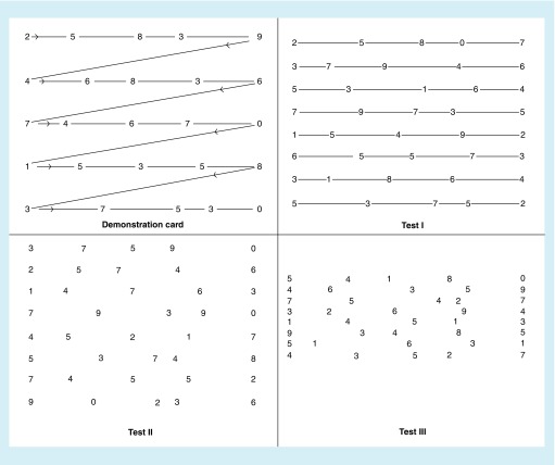
Objectives: We performed a meta-analysis of published data for a vision-based test of rapid number naming (King-Devick [K-D] test).

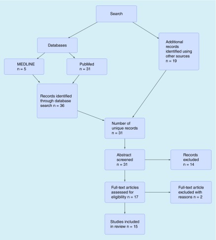
Studies & methods: Pooled and meta-analysis of 15 studies estimated preseason baseline K-D scores and sensitivity/specificity for identifying concussed versus nonconcussed control athletes.

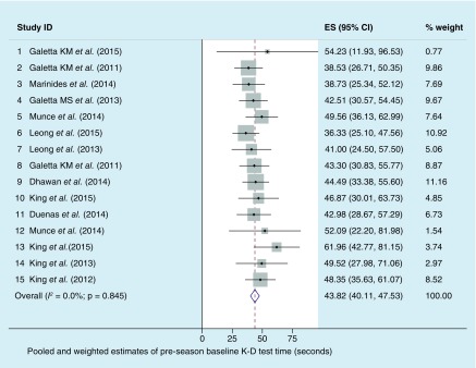
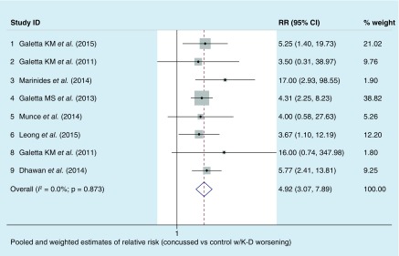
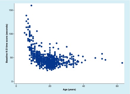
Result: Baseline K-D (n = 1419) showed a weighted estimate of 43.8 s (95% CI: 40.2, 47.5; I2 = 0.0%; p=0.85 - indicating very little heterogeneity). Sensitivity was 86% (96/112 concussed athletes had K-D worsening; 95% CI: 78%, 92%); specificity was 90% (181/202 controls had no worsening; 95% CI: 85%, 93%).

Conclusion: Rapid number naming adds to sideline assessment and contributes a critical dimension of vision to sports-related concussion testing.

Keywords: King-Devick test; concussion; meta-analysis; rapid number naming; saccades; sports; vision.


Conflict of interest statement

Financial & competing interests disclosure S Galetta has received consulting honoraria from Biogen and Genzyme. LJ Balcer has received consulting honoraria from Biogen and Genzyme, and has served on a clinical trial advisory board for Biogen. DF Leong is an employee of King-Devick Test, Inc. The authors have no other relevant affiliations or financial involvement with any organization or entity with a financial interest in or financial conflict with the subject matter or materials discussed in the manuscript apart from those disclosed. No writing assistance was utilized in the production of this manuscript.

Figures

<b>Figure 1.</b>
Figure 1.. Major cortical areas involved in control of eye movements and visual processing, with projections illustrating saccade generation in black.
Major cortical areas involved in control of eye movements and visual processing, with projections illustrating saccade generation in black. Saccades are initiated by signals sent from the frontal, parietal or supplementary eye fields to the superior colliculus, which then projects to the brainstem gaze centers. In parallel, the FEF also initiates saccades via direct connections to the BGC. In the indirect pathway, the substantia nigra pars reticulata inhibits the superior colliculus, preventing saccade generation. To turn off this inhibition, the FEFs are activated prior to a saccade, which then inhibits the substantia nigra pars reticulata via the caudate. The saccade pathways are a multidistributed network, but the FEF primarily generates voluntary- or memory-guided saccades, the parietal eye field – reflexive saccades, the SEF – saccades in coordination with body movements as well as successive saccades and the DLPC – antisaccades, the inhibition of reflexive saccades and the advanced planning of saccades. Cerebellar projections (shown in blue) fine-tune the saccades, given that cerebellar lesions can lead to saccadic dysmetria. The nucleus reticularis tegmenti pontis receives projections from the FEF and the superior colliculus (projection not shown) and in turn projects to the cerebellar ocular V. The Vinhibits the ipsilateral caudal fastigial nucleus, which then projects to the BGC to enhance saccades moving to the contralateral side and tamp down saccades moving to the ipsilateral side, likely via both inhibitory and excitatory connections [33,70,71]. BGC: Brainstem gaze centers; CN: Caudate; DLPC: Dorsolateral prefrontal cortex; FEF: Frontal eye field; FN: Fastigial nucleus; NRTP: Nucleus reticularis tegmenti pontis; PEF: Parietal eye fields; SC: Superior colliculus; SEF: Supplementary eye field; SNPR: Substantia nigra pars reticulata; V: Vermis.
<b>Figure 2.</b>
Figure 2.. Demonstration and test cards for the King-Devick test, a candidate rapid sideline screening for concussion based on speed of rapid number naming.
To perform the King-Devick test, participants are asked to read the numbers on each card from left to right as quickly as possible, but without making any errors. Following completion of the demonstration card (upper left), subjects are then asked to read each of the three test cards in the same manner. The times required to complete each card are recorded in seconds using a stopwatch. The sum of the three test card time scores constitutes the summary score for the entire test, the King-Devick time score. Numbers of errors made in reading the test cards are also recorded; misspeaks on numbers are recorded as errors only if the subject does not immediately correct the mistake before going on to the next number.
<b>Figure 3.</b>
Figure 3.. Study selection process for pooled and meta-analyses.
<b>Figure 4.</b>
Figure 4.. Distribution of preseason baseline time scores for the King-Devick test.
Dots represent point estimates of each study mean (or ES); sizes of the gray boxes reflect the weights of the studies in the meta-analysis. Bars are 95% CI. The diamond shows the weighted estimate for the mean preseason K-D baseline score; this is determined from fixed-effects models account for study N and precision (narrowness of 95% CI). I2 statistic values were 0.0%; p = 0.85; indicating very little heterogeneity between studies in calculation of the weighted estimate. Stated differently, the nonsignificance of the I2 test for heterogeneity suggests that the differences between the studies are explicable by random variation rather than systematic factors. ES: Effect size; K-D: King-Devick. Data taken from [29,30,47,48,49,50,51,52,53,54,55,56,72,73,74].
<b>Figure 5.</b>
Figure 5.. Distribution of relative risk of concussed versus control athlete status in the setting of any worsening of time score from preseason baseline for the King-Devick test.
Dots represent point estimates of each study's relative risk; size of the gray box reflects the weight of the study in meta-analysis. Bars are 95% CI. The diamond shows the weighted estimate for the relative risk; this is determined from fixed-effects models account for study N and precision (narrowness of 95% CI). I2 statistic values were 0.0%; p = 0.87, indicating very little heterogeneity between studies in calculation of the weighted estimate. Stated differently, the nonsignificance of the I2 test for heterogeneity suggests that the differences between the studies are explicable by random variation. Study ID numbers are not consecutive since some studies did not have postinjury data. RR: Relative risk; K-D: King-Devick. Data taken from [29,30,47,48,50,53,54,72].
<b>Figure 6.</b>
Figure 6.. Relation of rapid number naming (King-Devick) scores to athlete age.
The scores improve (are faster) with age among youth (age 18 years and younger), then appear stable with perhaps some increase by the end of the fourth decade. K-D: King-Devick.
<b>Figure 7.</b>
Figure 7.. Comparisons of receiver operating characteristic curve areas for the three sideline tests for distinguishing concussed versus nonconcussed control athletes on the sideline among athletes who underwent all measures (n = 23).
King-Devick = red line versus timed tandem gait = blue line versus Standardized Assessment of Concussion = green line. Receiver operating characteristic curve areas represent the probability that a test or combination can correctly distinguish between two categories (concussed vs nonconcussed control).

References

    1. McCrory P, Meeuwisse WH, Aubry M, et al. Consensus statement on concussion in sport: the 4th International Conference on Concussion in Sport held in Zurich, November 2012. Br. J. Sports Med. 2013;47(5):250–258. - PubMed
    1. Plassman BL, Havlik RJ, Steffens DC, et al. Documented head injury in early adulthood and risk of Alzheimer's disease and other dementias. Neurology. 2000;55(8):1158–1166. - PubMed
    1. Eibner C, Schell TL, Jaycox LH. Care of war veterans with mild traumatic brain injury. N. Engl. J. Med. 2009;361(5):537. author reply 537–538. - PubMed
    1. Xydakis MS, Robbins AS, Grant GA. Mild traumatic brain injury in U.S. soldiers returning from Iraq. N. Engl. J. Med. 2008;358(20):2177. author reply 2179. - PubMed
    1. Walsh DV, Capó-Aponte JE, Jorgensen-Wagers K, et al. Visual field dysfunctions in warfighters during different stages following blast and nonblast mTBI. Mil. Med. 2015;180(2):178–185. - PubMed