. 2018 Sep 5:6:e5580.
doi: 10.7717/peerj.5580. eCollection 2018.

Cell-wall-degrading enzymes produced in vitro and in vivo by Rhizoctonia solani, the causative fungus of peanut sheath blight

Affiliations

Cell-wall-degrading enzymes produced in vitro and in vivo by Rhizoctonia solani, the causative fungus of peanut sheath blight

Cai Yun Xue et al. PeerJ. .

Abstract

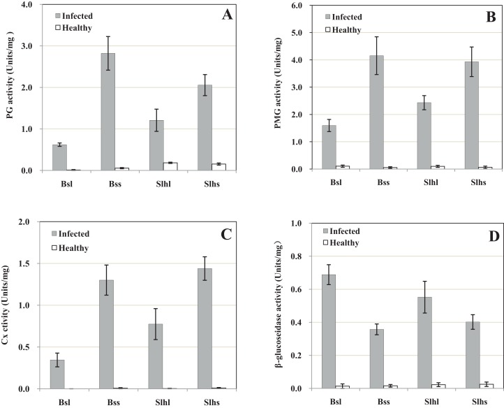
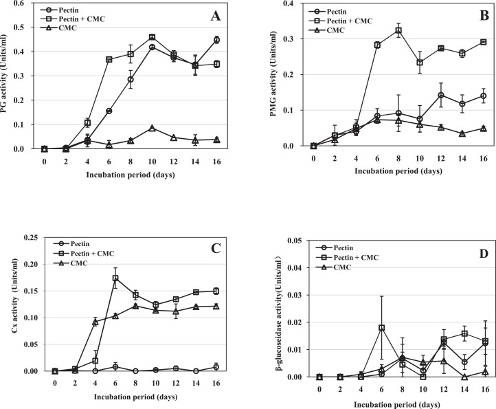
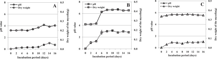
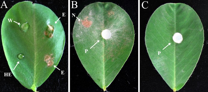
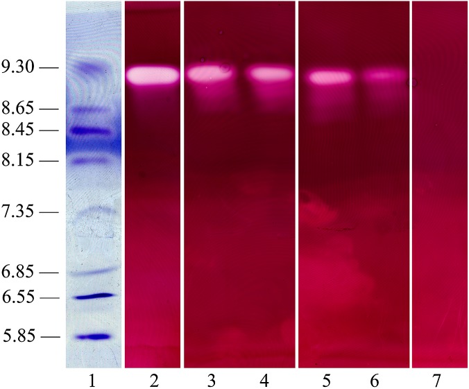
Rhizoctonia solani causes the disease peanut sheath blight, involving symptoms of maceration and necrosis of infected tissue, mainly caused by cell-wall-degrading enzymes (CWDEs). This study investigated the production of CWDEs including polygalacturonase (PG), polymethyl-galacturonase (PMG), cellulase (Cx) and β-glucosidase by R. solani in vitro (in liquid culture) and in vivo (in peanut plants). Significant PG, PMG, Cx and β-glucosidase activities were detected in infected tissues including stalk and leaves of Baisha and Silihong peanut cultivars. Extracts of healthy tissue showed little or no such activities. In shaken liquid cultures of R. solani in medium containing pectin or pectin plus carboxymethyl cellulose (CMC) as the carbon source(s), PG and PMG were notably active. Significant Cx activity was detected in cultures with CMC or pectin plus CMC as the carbon source(s). However, only a very low level of β-glucosidase activity was observed in cultures with any of the tested carbon sources. An increase of pH was recorded in decayed peanut tissues and liquid culture filtrates; the filtrate pH and fungal growth positively correlated. The fungal growth and/or pH were important factors for the production of PG, PMG and Cx in culture with pectin plus CMC as the carbon source. A single active PG isozyme with isoelectric point around 9.2 was detected in culture filtrates and in infected peanut tissues by the method of isoelectric focusing electrophoresis. The crude enzymes extracted from liquid culture of R. solani induced decay of healthy peanut leaves.

Keywords: Cell-wall-degrading enzymes; IEF; Peanut; Peanut sheath blight disease; Rhizoctonia solani.


Conflict of interest statement

The authors declare that they have no competing interests.

Figures

Figure 1
Figure 1. Activity of cell-wall-degrading enzymes including polygalacturonase (PG), polymethyl-galacturonase (PMG), cellulase (Cx) and β-glucosidase in peanut tissue infected by R. solani and healthy tissue.
Healthy tissues were inoculated with sterile corn kernels. Samples were collected 10 days post-infection (dpi). (A) PG activity. (B) PMG activity. (C) Cx activity. (D) β-glucosidase activity. Bsl, Baisha cultivar leaf; Bss, Baisha cultivar stalk; Slhl, Silihong cultivar leaf; Slhs, Silihong cultivar stalk. Standard errors (vertical bars) were calculated from three replicates.
Figure 2
Figure 2. Activity of R. solani cell-wall-degrading enzymes including PG, PMG, Cx and β-glucosidase in liquid shaken cultures containing various carbon sources.
Liquid medium with various carbon sources including 1% pectin, 0.5% pectin + 0.5% CMC or 1% CMC were used in the experiment, grown at 25 °C with shaking at 100 rpm. (A) PG activity. (B) PMG activity. (C) Cx activity. (D) β-glucosidase activity. Standard errors (vertical bars) were calculated from three replicates.
Figure 3
Figure 3. pH value of the culture filtrate and mycelial dry weight of R. solani in liquid shaken cultures containing various carbon sources.
Liquid medium with various carbon sources including 1% pectin, 0.5% pectin + 0.5% CMC or 1% CMC were used in the experiment, grown at 25 °C with shaking at 100 rpm. (A) Pectin as the sole carbon source. (B) Pectin plus CMC as the carbon source. (C) CMC as the sole carbon source. Standard errors (vertical bars) were calculated from three replicates.
Figure 4
Figure 4. Effects of crude enzyme extract from R. solani culture and R. solani mycelium inoculated onto peanut leaf for 2 days.
Leaves of Bs cultivar were treated with a five μl drop of R. solani crude enzyme extract or mycelial plugs five mm in diameter. The crude enzyme extracts were collected 10 dpi from medium containing pectin plus CMC as carbon sources incubated with R. solani. All treated leaves were incubated at 27 °C. (A) Crude enzyme extract inoculation. (B) Mycelial plug inoculation. (C) Sterile potato dextrose agar plug inoculation. W, distilled water (control); E, crude enzyme; HE, heated crude enzyme (control); N, necrotic lesion; P, mycelial plug. Treatment with “E” resulted in symptoms of necrotic lesions, leaves treated with “W” or “HE” showed no necrotic lesions.
Figure 5
Figure 5. Polygalacturonase isozyme produced in shaken liquid cultures and in infected tissue inoculated with R. solani (both for 10 days).
The crude enzyme extracts were collected from liquid medium containing pectin plus CMC as carbon sources incubated with R. solani. Samples were separated on an ultrathin layer polyacrylamide isoelectric focusing gel (pH 3.5–10.0), followed by agarose overlay activity staining. The PG band is white, visualized using a light box, with isoelectric point around 9.2. Lane 1, pI makers; 2, extract from culture filtrate; 3, extract from infected stalk of Baisha (Bs) cultivar; 4, extract from infected leaf of Bs cultivar; 5, extract from infected stalk of Silihong (Slh) cultivar; 6, extract from infected leaf of Slh cultivar; 7, control (heat-treated extract).

References

    1. Amit KC, Shubha C, Sushmita C, Shridha C. In vitro and in vivo production of polygalacturonase, polymethylgalacturonase and cellulase enzymes by Alternaria solani at different incubation periods. African Journal of Plant Science. 2014;8(5):248–253. doi: 10.5897/ajps2013.1114. - DOI
    1. Anderson JR, Bentley S, Irwin JAG, Mackie JM, Neate S, Pattemore JA. Characterisation of Rhizoctonia solani isolates causing root canker of lucerne in Australia. Australasian Plant Pathology. 2004;33(2):241–247. doi: 10.1071/ap04012. - DOI
    1. Balali GR, Kowsari M. Pectic zymogram variation and pathogenicity of Rhizoctonia solani AG-4 to bean (Phaseolus vulgaris) isolates in Isfahn, Iran. Mycopathologia. 2004;158(3):377–384. doi: 10.1007/s11046-004-2227-4. - DOI - PubMed
    1. Bartz FE, Cubeta MA, Toda T, Naito S, Ivors KL. An in planta method for assessing the role of basidiospores in Rhizoctonia foliar disease of tomato. Plant Disease. 2010;94(5):515–520. doi: 10.1094/pdis-94-5-0515. - DOI - PubMed
    1. Brien PAO, Zamani M. Production of pectic enzymes by barepatch isolates of Rhizoctonia solani AG 8. Australasian Plant Pathology. 2003;32(1):65–72. doi: 10.1071/ap02073. - DOI

LinkOut - more resources