Review
. 2019:251:3-36.
doi: 10.1007/164_2018_168.

Brown Adipose Tissue Development and Metabolism

Affiliations
Review

Brown Adipose Tissue Development and Metabolism

Su Myung Jung et al. Handb Exp Pharmacol. 2019.

Abstract

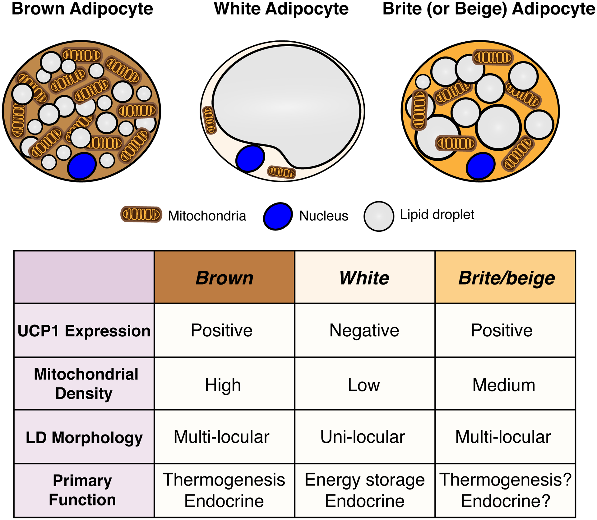
Brown adipose tissue is well known to be a thermoregulatory organ particularly important in small rodents and human infants, but it was only recently that its existence and significance to metabolic fitness in adult humans have been widely realized. The ability of active brown fat to expend high amounts of energy has raised interest in stimulating thermogenesis therapeutically to treat metabolic diseases related to obesity and type 2 diabetes. In parallel, there has been a surge of research aimed at understanding the biology of rodent and human brown fat development, its remarkable metabolic properties, and the phenomenon of white fat browning, in which white adipocytes can be converted into brown like adipocytes with similar thermogenic properties. Here, we review the current understanding of the developmental and metabolic pathways involved in forming thermogenic adipocytes, and highlight some of the many unknown functions of brown fat that make its study a rich and exciting area for future research.

Keywords: Adipogenesis; Beige adipocyte; Brite adipocyte; Brown adipose tissue; Development; Glucose and lipid metabolism; Lineage tracing; Progenitor cells; Thermogenesis; Ucp1.


Figures

Figure 1.
Figure 1.. General characteristics of brown, white and brite/beige adipocytes.
A stimulated brown adipocyte (left) contains numerous small lipid droplets, many mitochondria, and expresses high levels of uncoupling protein 1 (UCP1), which is embedded in the inner mitochondrial membrane and required for thermogenesis. The color of brown fat reflects the high iron content of mitochondria. A white adipocyte (middle) in contrast contains a single large lipid droplet, fewer mitochondria, and does not express UCP1. A Brite/beige adipocyte (right) is characteristically intermediate between brown and white adipocyte, having multiple lipid droplets (though often larger than those seen in a brown adipocyte), more mitochondria than a white adipocyte, and it expresses UCP1.
Figure 2.
Figure 2.. Adipose tissue anatomy and plasticity
(A) Cartoons showing brown and white fat depots in mice that are acclimated to thermoneutrality (30°C ~ 32°C), mild cold (20°C ~ 22°C), and severe cold (6°C ~ 10°C). The color and size of each depot is modeled such that it reflects the observed differences in mice acclimated to each temperature. A key showing the gradient of “browning” or “britening/beiging” is provided below each model. (B) Hematoxylin and Eosin staining of the indicated brown and white fat depots at each temperature. Note that at thermoneutrality, brown adipocytes contain larger single lipid droplets. At 20–22°C, the standard mouse facility temperature, brown adipocytes exhibit their stimulated morphology of being multi-locular (see Figure 1) while white adipocytes remain unilocular though SWAT adipocyte size is reduced likely reflecting in part a higher level of lipolysis that is necessary to fuel the active brown fat depots. At severe cold temperatures, (6–10°C), additional morphological changes can been see in BAT (i.e. lipid droplets become more uniform), and under these conditions, brite/beige adipocytes also from in the subcutaneous WAT. Of note, the browning capacity of WAT depots is not dependent on a depot being subcutaneous or visceral because, for example, the retroperitoneal visceral WAT depot has high britening/beiging capacity (not shown) while the perigonadal visceral WAT (shown) does not. [Abbreviations] iBAT, interscapular BAT; sBAT, subscapular BAT; cBAT, cervical BAT; paBAT, peri-aortic BAT; prBAT, peri-renal BAT; asWAT, anterior subcutaneous WAT; psWAT, posterior subcutaneous WAT; mWAT, mesenteric WAT; rWAT, retroperitoneal WAT; pgWAT, perigonadal WAT. The images in this figure are based primarily on experiments with C57Bl/6 mice.
Figure 3.
Figure 3.. Brown fat locations in humans
(A) Newborn infants have large interscapular and peri-renal BAT depots. (B) In adults, smaller BAT depots are located in the cervical, supraclavicular, axillary, peri-aortic, paravertebral and suprarenal regions. The mapping of these depots in adults is largely based on glucose uptake measurements by 18F-FDG-PET/CT imaging, which shows increased glucose flux at colder temperatures (shown in figure) and on post-mortem resections. The molecular and functional nature of individual (putative) BAT depots remains unclear in humans. Also note that the amount of BAT is highly variable between individuals, but when active BAT is present, it has been shown to correlate with improved metabolism (not shown, discussed in text). Emerging advances in BAT imaging will likely confirm additional depots.
Figure 4.
Figure 4.. Model of the heterogeneity and complexity in brown and brite/beige adipocyte development.
Several multi-potent cell populations that are mainly mesodermal and express specific transcription factors (e.g. En1, Myf5, Pax3, Prx1) appear to give rise heterogeneously to thermogenic adipocytes in different depots. Note that there is overlap shared with some markers but not with others. For example, Pax3 and Myf5 together may mark a pool of early precursors that give rise to iBAT, but only Pax3 marks a precursor pool that gives rise to some visceral pgWAT adipocytes (discussed in text). The significance of this heterogeneity is not understood. Additionally, there are several populations of brown and brite/beige adipocytes for which potential lineage markers remain unidentified. Also note that the brown and brite/beige adipocytes shown in this figure are depicted in their active state (i.e. upon β-adrenergic stimulation), but in vivo brown and brite/beige adipocytes are not necessarily present at the same time, such as in mild cold conditions (see Figure 2).

References

    1. ABU-REMAILEH M, WYANT GA, KIM C, LAQTOM NN, ABBASI M, CHAN SH, FREINKMAN E & SABATINI DM 2017. Lysosomal metabolomics reveals V-ATPase- and mTOR-dependent regulation of amino acid efflux from lysosomes. Science, 358, 807–813. - PMC - PubMed
    1. ACOSTA JR, JOOST S, KARLSSON K, EHRLUND A, LI X, AOUADI M, KASPER M, ARNER P, RYDEN M & LAURENCIKIENE J 2017. Single cell transcriptomics suggest that human adipocyte progenitor cells constitute a homogeneous cell population. Stem Cell Res Ther, 8, 250. - PMC - PubMed
    1. AHFELDT T, SCHINZEL RT, LEE Y-K, HENDRICKSON D, KAPLAN A, LUM DH, CAMAHORT R, XIA F, SHAY J, RHEE EP, CLISH CB, DEO RC, SHEN T, LAU FH, COWLEY A, MOWRER G, AL-SIDDIQI H, NAHRENDORF M, MUSUNURU K, GERSZTEN RE, RINN JL & COWAN CA 2012. Programming human pluripotent stem cells into white and brown adipocytes. Nature Cell Biology, 14, 209–219. - PMC - PubMed
    1. AMBROSI TH, SCIALDONE A, GRAJA A, GOHLKE S, JANK AM, BOCIAN C, WOELK L, FAN H, LOGAN DW, SCHURMANN A, SARAIVA LR & SCHULZ TJ 2017. Adipocyte Accumulation in the Bone Marrow during Obesity and Aging Impairs Stem Cell-Based Hematopoietic and Bone Regeneration. Cell Stem Cell, 20, 771–784.e6. - PMC - PubMed
    1. ATIT R, SGAIER SK, MOHAMED OA, TAKETO MM, DUFORT D, JOYNER AL, NISWANDER L & CONLON RA 2006. beta-catenin activation is necessary and sufficient to specify the dorsal dermal fate in the mouse. Developmental Biology, 296, 164–176. - PubMed