. 2018 Oct;30(43):e1803618.
doi: 10.1002/adma.201803618. Epub 2018 Sep 11.

Bioresponsive Nanoparticles Targeted to Infectious Microenvironments for Sepsis Management

Affiliations

Bioresponsive Nanoparticles Targeted to Infectious Microenvironments for Sepsis Management

Can Yang Zhang et al. Adv Mater. 2018 Oct.

Abstract

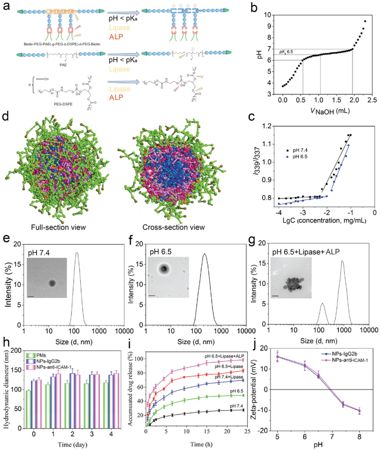
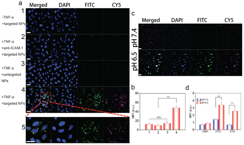
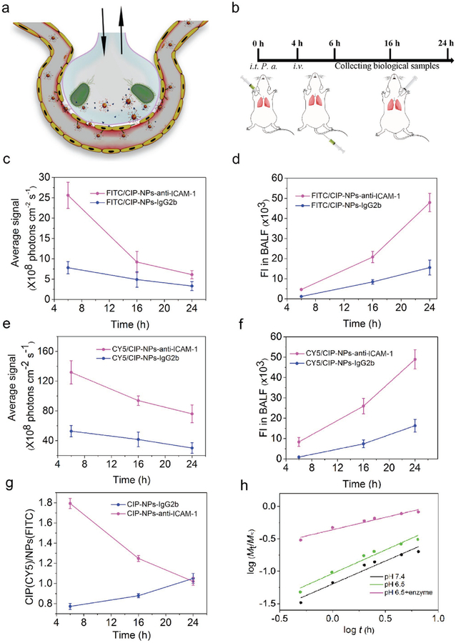
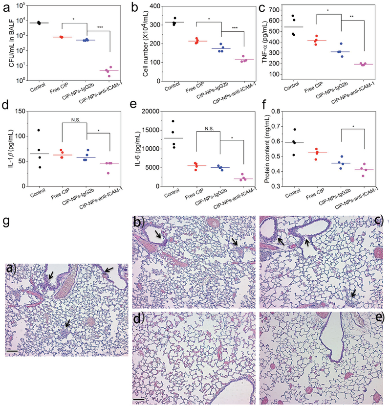
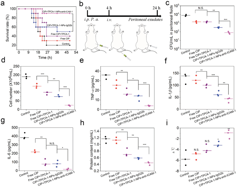
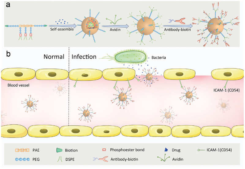
Sepsis is a life-threatening disease resulted from a dysregulated host immune response to bacterial infections, continuing to cause high morbidity and mortality worldwide. Despite discoveries of many potential therapeutic targets, effective treatments of sepsis are lacking. Here, a strategy is reported to target infectious microenvironments (IMEs) via bioresponsive nanoparticles that simultaneously eliminate bacteria and alleviate the host inflammation response, thus managing sepsis in mice. The nanoparticle is made of copolymers sensitive to pH and bacterial enzymes to self-assemble into a micelle loaded with both an antibiotic (ciprofloxacin) and an anti-inflammatory agent ((2-[(aminocarbonyl)amino]-5-(4-fluorophenyl)-3-thiophenecarboxamide). In addition, the nanoparticle is conjugated with intercellular adhesion molecule-1 antibodies to target IMEs. Nanoparticle targeting to IMEs and local cues as triggers to deliver therapeutics in on-demand manners is demonstrated using an acute lung bacterial infection mouse model. In the sepsis mouse model induced by peritonitis at a lethal dose of bacterial invasion, it is shown that concurrently targeting pathogens and excessive inflammation pathways is valuable to manage the sepsis. The study illustrates not only the development of a new delivery system but also the mechanism-based therapy of nanomedicine for infectious diseases.

Keywords: infectious microenvironments; nanoparticle; sepsis; targeted drug delivery.


Conflict of interest statement

Conflict of Interest

The authors declare no conflict of interest.

Figures

Figure 1.
Figure 1.
Characterization of NPs and drug release profile of CIP-NPs-Abs. a) Structure and pH/enzyme-sensitive amphiphilic block copolymer. PAE moieties are pH/bacterium lipase-responsive segments, resulting from tertiary amine residues and ester bonds of a polymer chain. Under acid/lipase conditions, tertiary amine residues are protonated and become positive accompanied by cleavage of ester bonds. PEG-DSPE moieties are lipase and ALP-responsive blocks. Green trapezoids represent biotin groups; Blue balls represent PEG units; Yellow shackles represent PAE segments; Red shackles represent phosphoester bonds. b) pKa of a copolymer was measured by acid–base titration. c) Copolymer CMC at pH 7.4 or 6.5 was determined by pyrene fluorescence spectroscopy. d) Coarse-grained simulations of self-assembly of CIP-PMs using a DPD method. A copolymer chain is divided into five types of beads: peach, HDD (1,6-hexanediol diacrylate) units in PAE and DSPE; PEG (light green); biotin (dark green); AP (3-amino-1-propanol) units in PAE (pink); CIP molecule (blue). Figure S10 in the Supporting Information shows the detail about the simulation process. e–g) Representative DLS size and TEM images of NPs-anti-ICAM-1 incubated in PBS at pH 7.4 (e), pH 6.5 (f) or pH 6.5 with enzymes (g) (Pseudomonas lipase, 1 mg mL−1 and ALP, 500 unit mL−1) for 2 h. Scale bars: 100 nm. h) Serum stability of PMs and NPs-anti-ICAM-1/IgG2b incubated in PBS with 20% FBS at pH 7.4 at 37°C. i) In vitro drug release of CIP-NPs-anti-ICAM-1 in different buffers (Pseudomonas lipase, 1 mg mL−1 and ALP, 500 unit mL−1). j) Zeta-potential of CIP-NPs-anti-ICAM-1/IgG2b in PBS as the function of pH. The data are shown as mean ± s.d. (n = 3 independent experiments).
Figure 2.
Figure 2.
Abs-coated NPs target inflamed endothelium and bind to bacteria in vitro. a) Confocal laser scanning microscope (CLSM) images of HUVECs treated with Abs-coated NPs under various conditions. (1, unactivated HUVECs treated with CY5/FITC-labeled CIP-NPs-anti-ICAM-1; 2, activated HUVECs coated with anti-ICAM-1 and treated with CY5/FITC-labeled CIP-NPs-anti-ICAM-1; 3, activated HUVECs treated with CY5/FITC-labeled CIPNPs-IgG2b; 4, activated HUVECs treated with CY5/FITC-labeled CIP-NPs-anti-ICAM-1; 5, enlarged image of the box in image 4.) Scale bars, 20 μm. b) Flow cytometry mean fluorescence intensity of HUVECs treated with Abs-coated NPs under various conditions. c) pH-dependent interactions between CY5/FITC-labeled CIP-NPs-anti-ICAM-1 and P. aeruginosa at pH 7.4 or 6.5 imaged by CLSM. Scale bars, 10 μm. d) Fluorescence intensities of FITC and CY5 in bacteria were quantified based on confocal images. Data are shown as mean ± s.d. (n = 6 independent experiments). Statistical analysis was conducted using Student’s t-test of Origin 8.5, p values: ***p < 0.001, N.S. (no significant difference) p > 0.05.
Figure 3.
Figure 3.
NPs-anti-ICAM-1 targeted to IMEs in vivo. a) Schematic shows an alveolus that is a major component of the lung. The alveolus contains endothelial/epithelial monolayers forming the interface of airway and bloodstream. Bacteria (P. aeruginosa) invade in the lung via airway, and subsequently activate inflammation responses to highly express ICAM-1 on the lumen of blood vessels and enhance lung permeability. NPs-anti-ICAM-1 binds to inflamed vasculature and local infection environments trigger drug release from pH/enzyme-responsive NPs. b) Experimental protocol of the acute lung inflammation model after intratracheal administration of P. aeruginosa (106 CFU per animal). c,d) Fluorescence intensity of FITC (c) and CY5 (e) of lung tissues after BALF removal from mice. d,f) Fluorescence intensity of FITC (d) and CY5 (f) in BALF. g) Fluorescence intensity ratios of CY5 to FITC in BALF. h) Drug release dynamics of CIP from Abs-coated NPs based on in vitro drug release experiments of Figure 2i. Data are shown as mean ± s.d. (n = 3 independent experiments).
Figure 4.
Figure 4.
CIP-NPs-anti-ICAM-1 improves bacterial clearance in the lung. a–f) CFU of bacteria (a), leukocyte number (b), TNF-α (c), IL-1β (d), IL-6 (e), and protein content (f) in BALF of mice infected by P. aeruginosa 20 h after the treatments with PBS, free drug or various drug formulations. g) Hematoxylin and eosin (H&E)-stained tissue sections of lungs of P. aeruginosa-infected mice 20 h after treatments with: a) PBS, b) free CIP, c) CIP-NPs-IgG2b, and d) CIP-NPs-anti-ICAM-1; (e) is the healthy lung. The black arrowheads indicate the infiltration of immune cells (mainly neutrophils). Scale bars: 100 μm. The data are shown as mean ± s.d. (n = 4 independent experiments). p values: *p < 0.05, **p < 0.01, ***p < 0.001, N.S. (no significant difference) p > 0.05.
Figure 5.
Figure 5.
Co-delivery of CIP and TPCA-1 via NPs manages the sepsis induced by bacteria. a) Mouse survival in peritonitis-induced sepsis by i.p. injection of a lethal dose of P. aeruginosa. 4 h after bacterial injection, mice were treated with several drug formulations (PBS, free CIP, free CIP+TPCA-1, CIP+TPCA-1-NPs-IgG2b, and CIP+TPCA-1-NPs-anti-ICAM-1). CIP and TPCA-1 were administered at the same amount for each formulation (1.5 ± 0.1 and 1.4 ± 0.06 mg kg−1, respectively). Statistical analysis was done using Kaplan–Meier method (n = 10). *p < 0.05. b) Experimental design to evaluate pathogen burden and inflammation responses. c–h) CFU of bacteria (c), leukocyte number (d), TNF-α (e), IL-1β (f), IL-6 (g), and protein content (h) in peritoneal fluids, and i) mouse body temperature changes. N.S. (p > 0.05), *p < 0.05, **p < 0.01 versus control. All data are expressed as means ± s.d. (n = 3). p values: *p < 0.05, **p < 0.01, ***p < 0.001, N.S. (no significant difference), p > 0.05.
Scheme 1.
Scheme 1.
Schemes for design of an IME-responsive and biofunctional nanoparticle (NP), and targeted delivery of nanotherapeutics at a site of infection. a) Schematic for design of multi-functional NPs. A drug-loaded polymeric micelle is self-assembled from pH/enzyme-responsive amphiphilic block copolymers and drugs, followed by coating with antibody for targeting infection sites. The poly (ß-amino ester) (PAE) segment is pH-sensitive, and enzyme-responsive. 1,2-distearoyl-sn-glycero-3-phosphoethanolamine-N-[amino(polyethylene glycol)] (PEGylated DSPE) on the pendants of PAE is designed for drug loading. Biotins on the surface of a micelle are used for biofunctionalization. The chemical structure and synthesis process are presented in Figure S1 (Supporting Information). b) Schematic shows that drug-loaded NPs-anti-ICAM-1 specifically target activated endothelial cells at a site of infection after intravenous (i.v.) injection. Drug-loaded NPs-anti-ICAM-1 bind to activated endothelial cells in IMEs, crossing the blood vessel and releasing drugs triggered by the local infectious cues.

Similar articles

Cited by

References

    1. van der Poll T, van de Veerdonk FL, Scicluna BP, Netea MG, Nat. Rev. Immunol 2017, 17, 407. - PubMed
    1. Deutschman CS, Tracey KJ, Immunity 2014, 40, 463. - PubMed
    1. Angus DC, van der Poll T, Engl N. J. Med 2013, 369, 840. - PubMed
    1. Mayr FB, Yende S, Angus DC, Virulence 2014, 5, 4. - PMC - PubMed
    1. Iskander KN, Osuchowski MF, Stearns-Kurosawa DJ, Kurosawa S, Stepien D, Valentine C, Remick DG, Physiol. Rev 2013, 93, 1247. - PMC - PubMed

MeSH terms