. 2019 Apr;25(7-8):538-553.
doi: 10.1089/ten.TEA.2018.0142. Epub 2018 Oct 31.

A Functional Tissue-Engineered Synovium Model to Study Osteoarthritis Progression and Treatment

Affiliations

A Functional Tissue-Engineered Synovium Model to Study Osteoarthritis Progression and Treatment

Robert M Stefani et al. Tissue Eng Part A. 2019 Apr.

Abstract

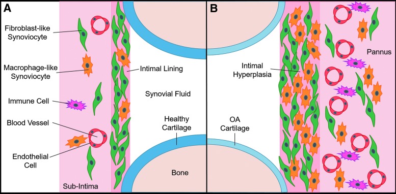
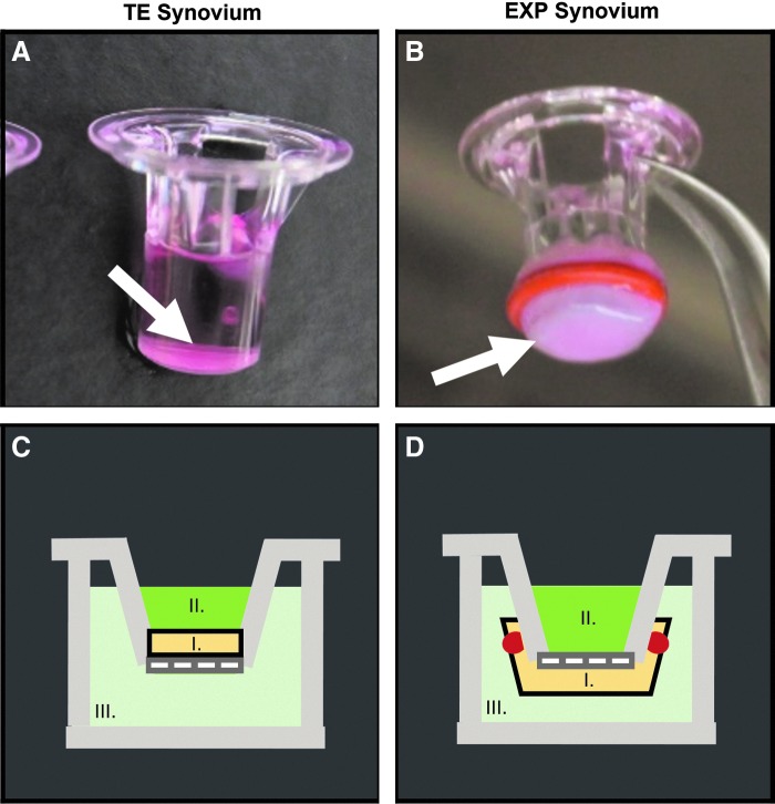
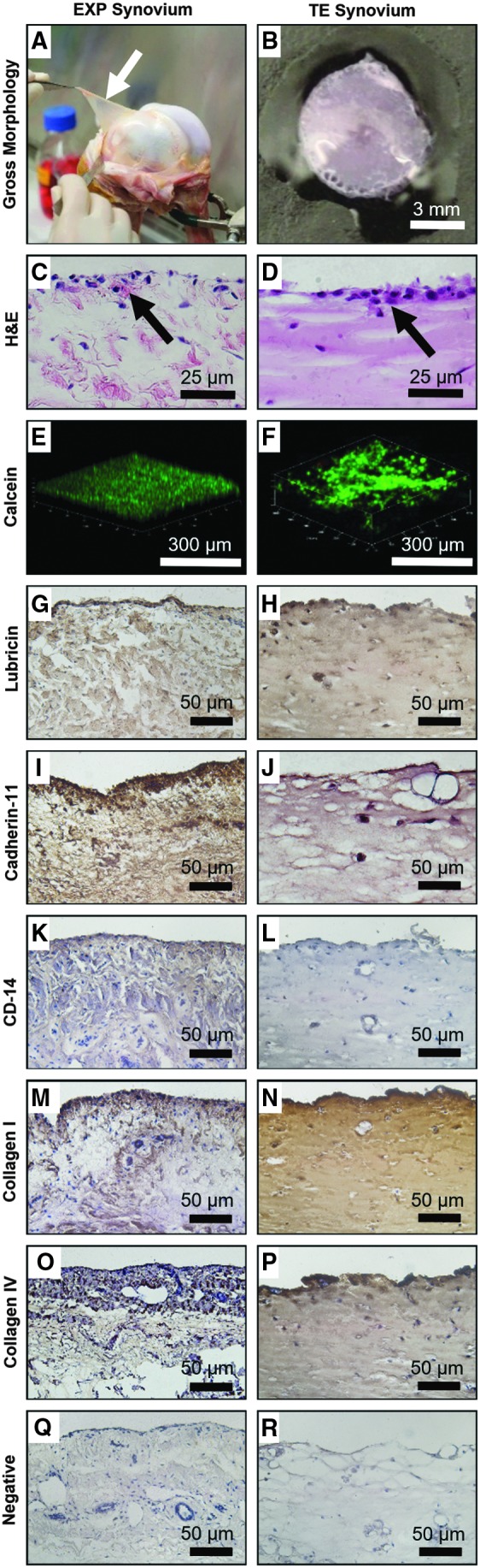
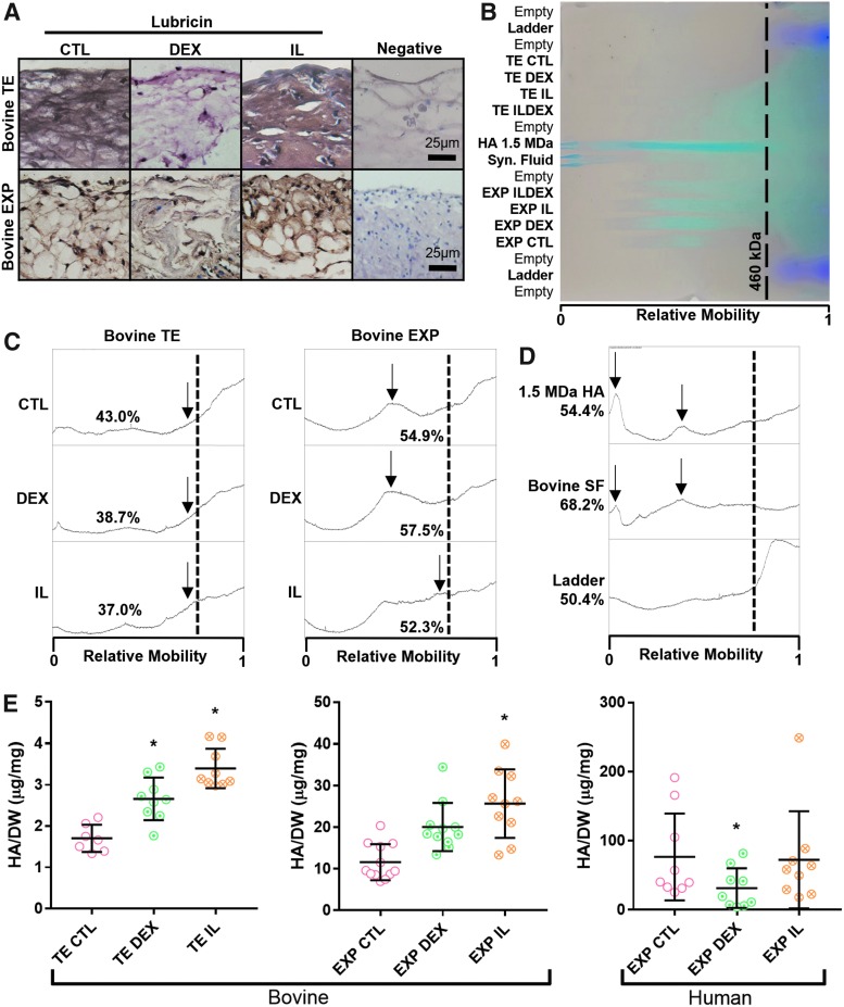
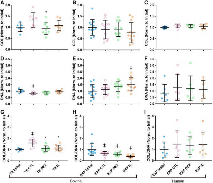
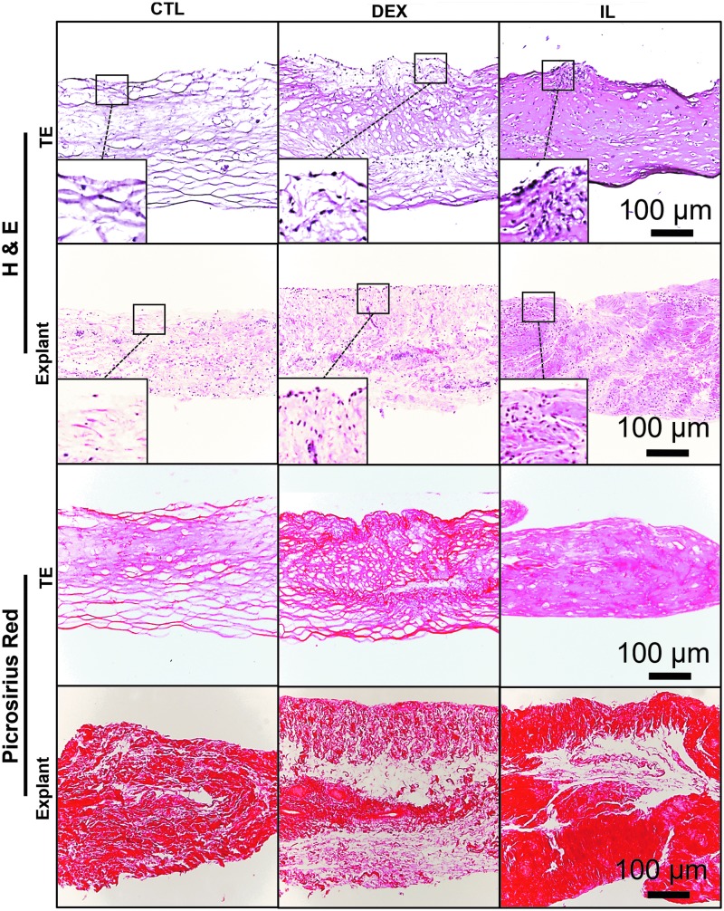
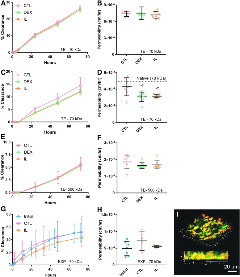
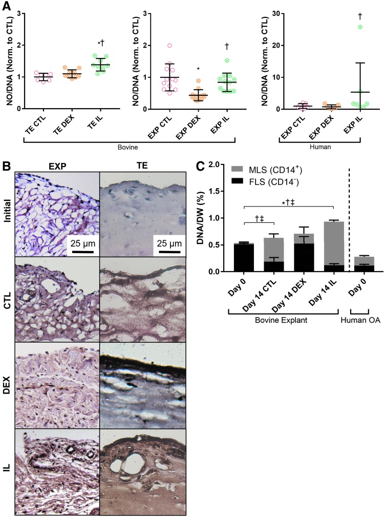
The synovium envelops the diarthrodial joint and plays a key regulatory role in defining the composition of the synovial fluid through filtration and biosynthesis of critical boundary lubricants. Synovium changes often precede cartilage damage in osteoarthritis. We describe a novel in vitro tissue engineered model, validated against native synovium explants, to investigate the structure-function of synovium through quantitative solute transport measures. Synovium was evaluated in the presence of a proinflammatory cytokine, interleukin-1, or the clinically relevant corticosteroid, dexamethasone. We anticipate that a better understanding of synovium transport would support efforts to develop more effective strategies aimed at restoring joint health.

Keywords: models; osteoarthritis; solute transport; synovium.


Conflict of interest statement

No competing financial interests exist.

Figures

<b>FIG. 1.</b>
FIG. 1.
Relevant synovium anatomy in an idealized knee joint. (A) Normal synovium comprises ∼90% FLS and ∼10% MLS. (B) OA synovium composition and structure change dramatically, coinciding with altered secretion of lubricants and inflammatory mediators. OA, osteoarthritis; FLS, fibroblast-like synoviocytes; MLS, macrophage-like synoviocytes.
<b>FIG. 2.</b>
FIG. 2.
Changes in permeability of FITC-labeled dextran in synovium were assessed using a Transwell® system. TE synovium (A, C) was cultured and tested in situ on the top of a 4.26 mm Transwell insert (arrow). EXP synovium (B, D) was secured to the underside of a 6.5 mm Transwell insert using a rubber gasket for transport measurements (arrow). For both tissues (I), the dextran suspension (II) was placed in the luminal compartment (i.e., the synovial cavity). Phosphate-buffered saline (III) was added to the basolateral compartment (i.e., the fibrous capsule and associated vasculature) and sampled at time points to quantify joint clearance. FITC, fluorescein isothiocyanate; TE, tissue engineered; EXP, explant.
<b>FIG. 3.</b>
FIG. 3.
Bovine EXP (white arrow) (day 0) and TE (mature, day 21) synovium exhibited similar structural characteristics (AF) and protein expression (GR). Both tissues (A, B) displayed an opaque, flat, and membranous morphology. A 1–4 cell thick intimal lining layer (black arrows) was observed using H&E (C, D); 3D z-stack reconstruction (E, F) with cells labeled with calcein-AM (green) showed similar cellular localization to the surface of the tissue with fewer subintimal cells. Positive immunohistochemical staining was visualized with 3,3′ diaminobenzidine Peroxidase and cells were counterstained with hematoxylin. Both tissues showed lubricin (G, H) and cadherin-11 expression (I, J). CD14 expression was extremely low in EXP synovium (K), as expected for “healthy” synovium containing primarily FLS, whereas TE synovium (L) had negative staining. Type I (M, N) and type IV (O, P) collagen, which are ECM components of the subintimal layer and intima, respectively, were also present in both tissues. Corresponding isotype controls (Q, R) showed the absence of nonspecific staining. 3D, three dimensional; H&E, hematoxylin and eosin; ECM, extracellular matrix.
<b>FIG. 4.</b>
FIG. 4.
(A) Robust lubricin expression (reddish brown) was observed in CTL and IL groups for both EXP and TE synovium, with comparatively weaker staining in DEX groups. (B) Agarose gel electrophoresis (HA, blue; ladder, purple) showed approximate HA size distribution of (C) pooled experimental groups and (D) standard molecules. (C) In both EXP and TE synovium, wide HA MW distributions were observed, with the highest proportion of high MW HA (lower relative mobility) being estimated in CTL- and DEX-treated groups. Peaks (arrows) represented mobility of the predominant high MW HA for a given group. (D) The upper size limit of the ladder was 460 kDa (dotted line), which was used as a cutoff point to approximate the relative amounts of small and large MW HA in the given sample. Lower MWs could not be accurately resolved in the given gel conditions (low agarose concentration and high voltage) that were otherwise necessary for HA mobility. (E) Overall, bovine TE and EXP synovium had elevated HA secretion in response to DEX and IL treatment. In human EXP synovium, HA secretion was depressed by DEX and unaltered by IL treatment, *p < 0.05 versus CTL. HA, hyaluronic acid; MW, molecular weight; IL, interleukin; DEX, dexamethasone.
<b>FIG. 5.</b>
FIG. 5.
(A) Change in total collagen was significantly lower in DEX-treated TE synovium relative to CTL (p < 0.05), and a similar trend was observed in IL-treated TE synovium (p = 0.2698). Total collagen was statistically unchanged in all (B) bovine and (C) human EXP groups. Change in DNA was elevated with IL treatment relative to CTL for (D) bovine TE synovium (p = 0.1631) and (E) EXP synovium (p = 0.1357). DNA significantly decreased compared to initial values in TE CTL and increased compared to initial values in EXP IL (p < 0.05). DNA was unchanged in (F) human EXP groups. Change in COL/DNA was significantly lower in DEX- and IL-treated groups compared to CTL for (G) bovine TE synovium (p < 0.05), and similar trends were seen for (H) EXP IL (p = 0.1035). COL/DNA was unchanged in (I) human EXP groups. *p < 0.05 versus CTL, p < 0.05 versus Initial. COL, collagen.
<b>FIG. 6.</b>
FIG. 6.
Similarities were observed in structural changes undergone by TE and EXP synovium, as visualized with H&E and Picrosirius red staining. CTL specimens were especially fibrous, with strong Picrosirius red staining and associated large gaps present in the ECM. DEX specimens had increased intimal cellularity and slightly denser matrix staining. IL specimens had further increased intimal cellularity and very dense collagen matrix present throughout the tissue.
<b>FIG. 7.</b>
FIG. 7.
Cumulative clearance of dextran over time and corresponding permeability values were presented for 10, 70, and 500 kDa sizes in TE specimens (AF) and for the 70 kDa size in bovine EXP specimens (G, H). (D) Both DEX- and IL-treated TE groups exhibited decreased permeability relative to CTL with 70 kDa dextran only. Permeability of 70 kDa dextran in CTL did not differ significantly from native tissue [dotted line, value from (H)], whereas DEX and IL specimens were significantly lower (*p < 0.05 vs. CTL, p < 0.05 vs. native). (G, H) In EXP synovium (N = 3–4), permeability in CTL increased over time and was higher than IL-treated tissue, but neither observation was statistically significant (p = 0.0941 and p = 0.3448, respectively). (I) Representative 3D confocal image of 70 kDa FITC-Dextran (green) and resident cells (red-orange) within a living synovium EXP showed diffusion occurring in intercellular spaces.
<b>FIG. 8.</b>
FIG. 8.
(A) Bovine TE and EXP synovium, as well as human OA EXPs, showed elevated nitric oxide production in response to IL treatment, *p < 0.05 versus CTL, p < 0.05 versus DEX. (B) Elevated CD14 expression (reddish brown) was observed in both TE and EXP tissue treated with IL. An increase was also seen in CTL groups, but CD14 staining was less intense for DEX specimens in both tissue types. (C) The phenotype ratio of MLS (CD14+) to FLS (CD14) in EXP tissue was maintained by DEX. In CTL- and IL-treated tissue, MLS content increased and FLS content decreased. Total cell content increased with IL treatment; *p < 0.05 (total DNA), p < 0.05 (FLS), p < 0.05 (MLS).
<b>FIG. 9.</b>
FIG. 9.
TE synovium with prelabeled MLS (DiI, red) and varying ratios of unlabeled FLS was cultured for 2 weeks in CTL- or IL-supplemented media and all cells subsequently counterstained with 4′,6-diamidino-2-phenylindole (blue). In both (A) CTL and (B) IL groups, DiI (red) was co-localized with intense CD-14 expression (green). Intimal lining formation was apparent in all groups (A, B), regardless of cell type, with intimal hyperplasia apparent in groups treated with IL (B).

Similar articles

Cited by

References

    1. Blewis M.E., Lao B.J., Jadin K.D., et al. . Semi-permeable membrane retention of synovial fluid lubricants hyaluronan and proteoglycan 4 for a biomimetic bioreactor. Biotechnol Bioeng 106, 149, 2010 - PMC - PubMed
    1. Blewis M.E., Lao B.J., Schumacher B.L., Bugbee W.D., Sah R.L., and Firestein G.S. Interactive cytokine regulation of synoviocyte lubricant secretion. Tissue Eng Part A 16, 1329, 2010 - PMC - PubMed
    1. Schmidt T.A., and Sah R.L. Effect of synovial fluid on boundary lubrication of articular cartilage. Osteoarthritis Cartilage 15, 35, 2007 - PubMed
    1. Kiener H.P., Watts G.F.M., Cui Y., et al. . Synovial fibroblasts self-direct multicellular lining architecture and synthetic function in three-dimensional organ culture. Arthritis Rheum 62, 742, 2010 - PubMed
    1. Smith M.D. The normal synovium. Open Rheumatol J 5, 100, 2011 - PMC - PubMed

Publication types

MeSH terms

LinkOut - more resources