Multicenter Study
. 2019 Apr;19(4):1086-1097.
doi: 10.1111/ajt.15116. Epub 2018 Oct 8.

Bidirectional transfer of Anelloviridae lineages between graft and host during lung transplantation

Affiliations
Multicenter Study

Bidirectional transfer of Anelloviridae lineages between graft and host during lung transplantation

Arwa A Abbas et al. Am J Transplant. 2019 Apr.

Abstract

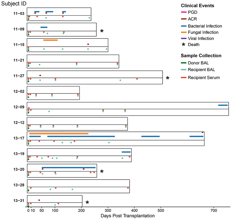
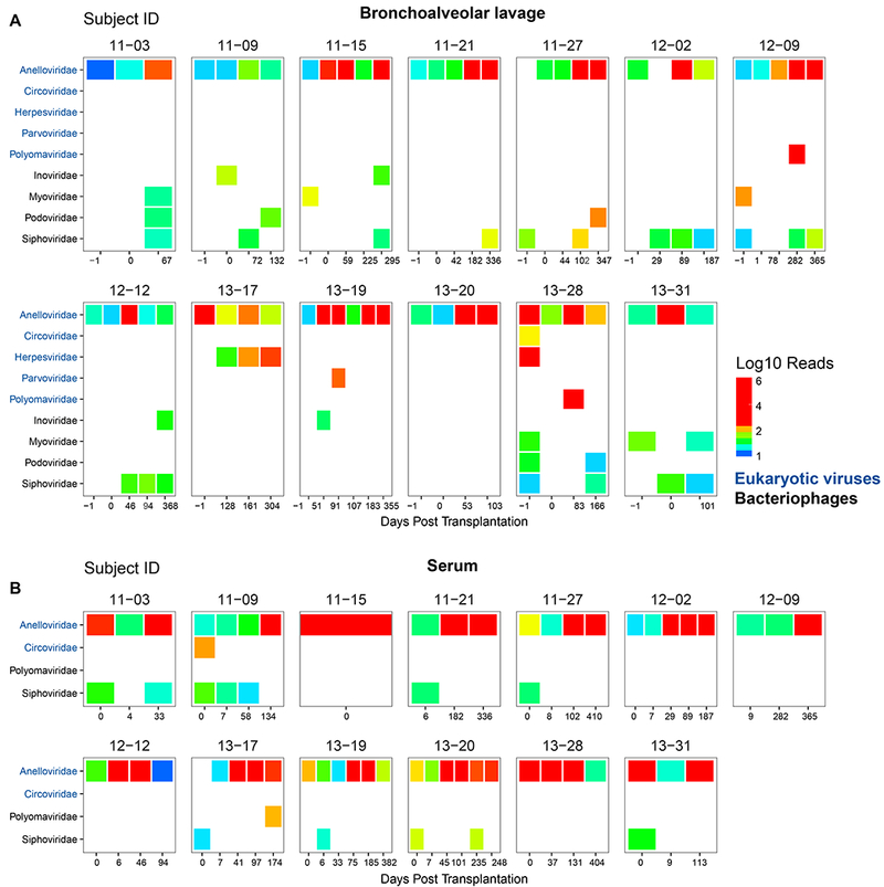
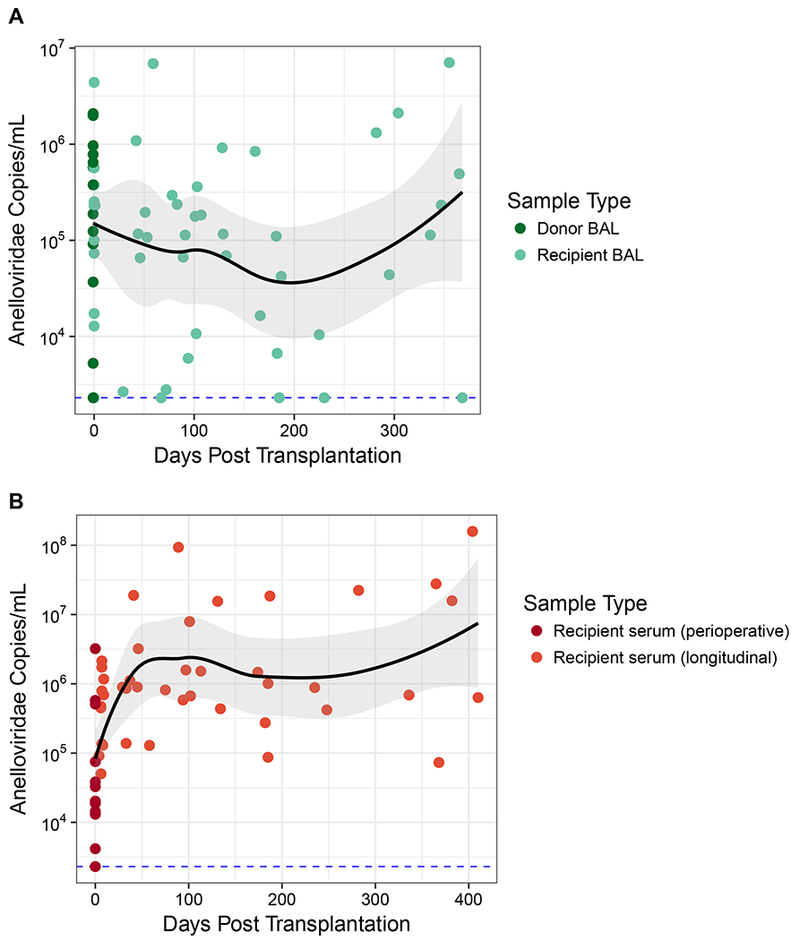
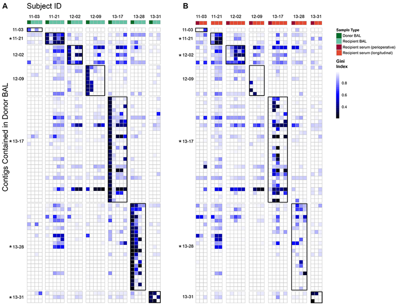
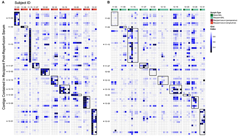
Solid organ transplantation disrupts virus-host relationships, potentially resulting in viral transfer from donor to recipient, reactivation of latent viruses, and new viral infections. Viral transfer, colonization, and reactivation are typically monitored using assays for specific viruses, leaving the behavior of full viral populations (the "virome") understudied. Here we sought to investigate the temporal behavior of viruses from donor lungs and transplant recipients comprehensively. We interrogated the bronchoalveolar lavage and blood viromes during the peritransplant period and 6-16 months posttransplant in 13 donor-recipient pairs using shotgun metagenomic sequencing. Anelloviridae, ubiquitous human commensal viruses, were the most abundant human viruses identified. Herpesviruses, parvoviruses, polyomaviruses, and bacteriophages were also detected. Anelloviridae populations were complex, with some donor organs and hosts harboring multiple contemporaneous lineages. We identified transfer of Anelloviridae lineages from donor organ to recipient serum in 4 of 7 cases that could be queried, and immigration of lineages from recipient serum into the allograft in 6 of 10 such cases. Thus, metagenomic analyses revealed that viral populations move between graft and host in both directions, showing that organ transplantation involves implantation of both the allograft and commensal viral communities.

Keywords: basic (laboratory) research/science; bronchoalveolar lavage (BAL); clinical research/practice; donors and donation: donor-derived infections; infection and infectious agents - viral; infectious disease; lung transplantation/pulmonology; microbiomics.


Figures

Figure 1:
Figure 1:. Sample Collection and Clinical Events
The duration of each subject’s enrollment in the study is shown. Sample collection (Bronchoalveolar lavage (BAL) and blood specimens) time points are displayed below the timeline and indicated by the color code. The timing and duration of adverse clinical events for each subject are displayed above the time line and similarly annotated. For subject 12–09, primary graft dysfunction (PGD) grade 3 occurred within the first 72 h following lung transplantation (55). Five subjects that experienced acute cellular rejection (ACR) had a maximum grade of A2 (56).
Figure 2:
Figure 2:. The Viral Microbiome before and after Transplantation
Displayed are the distribution and number of read pair assignments (on a log10 scale) from shotgun metagenomic sequencing of BAL (A) and serum (B). Shown are results from DNA sequences that match known viruses, filtered to remove spurious hits. Each box represents a different donor-recipient pair and each column a different BAL (A) or serum (B) sample. BAL of the donor lung prior to procurement and transplantation is the first column and indicated as “−1” days post-transplantation. Post transplantation BAL samples were taken during routine surveillance bronchoscopy or for other indications. Reads that could not be classified at the species level are not displayed. Only hits with a minimum of 10 reads assigned to a viral family per sample are included. Hits believed to be spurious or derived from environmental contamination are not displayed. Viral families are grouped according to target host. Sequencing, pre-processing of reads, classification and quality control was carried out as described in Supplemental Methods.
Figure 3:
Figure 3:. Anelloviridae Dynamics by Body Site
Anelloviridae species were quantified in BAL and serum by qPCR that targets torque teno viruses, torque teno midi viruses and torque teno mini viruses (Anelloviridae family members). The black line represents the local regression curve and standard error for all longitudinal samples. The dotted line represents the limit of detection for the qPCR assay (38 target copies/reaction). Samples at or below this limit were assigned this minimal value. Dots represent individual samples. (A) Longitudinal analysis of Anelloviridae genome copies in BAL. Donor BAL was taken prior to organ procurement. Recipient BAL was first obtained an hour after organ reperfusion and at various time points post transplantation. (B) Longitudinal analysis of Anelloviridae genome copies in serum from lung transplant recipients. Perioperative serum from the transplant recipients was obtained an hour after organ reperfusion.
Figure 4:
Figure 4:. Longitudinal Monitoring of Donor Lung Anelloviridae in Transplant Recipients’ Subsequent Lung and Serum Samples
Contigs >2000 bp that could be assembled from 7 organ donor BAL were aligned with reads found in post-transplantation BAL (A) and serum (B) samples. Each row is a contig representing an Anelloviridae partial genome in donor BAL. Rows are grouped according to the donor organ in which the contig was found, shown on the left. Columns represent individual samples of BAL (A) or serum (B) arranged chronologically, grouped by recipient ID and annotated based on the color key. Recipient perioperative serum in B was sampled one hour after organ reperfusion. The color in each block represents the Gini index of each comparison of initial sample contig to subsequent samples’ reads. A value of 1 is highly uneven coverage suggesting lack of detection, while 0 is even coverage across the genome suggesting highly confident detection. Alignments of samples to contigs in cognate donor-recipient pairs (donor lung and recipient of that specific donor organ) are outlined in black for ease of visualization. As a group, Anelloviridae lineages present in initial donor lungs were significantly more likely to be found in BAL and blood specimens from cognate recipients than in samples from unrelated subjects (p<0.001; Wilcoxon Rank Sum Test). Individual donor-recipient pairs where there was significant detection of donor lineages in either BAL (A) or serum (B) are indicated by the asterisk (p<0.05; Wilcoxon Rank Sum Test).
Figure 5:
Figure 5:. Longitudinal Monitoring of Initial Recipient Serum Anelloviridae in Transplant Recipients’ Subsequent Serum and Lung Samples
Contigs >2000 bp that could be assembled from 10 perioperative serum samples (obtained within an hour after transplantation and taken to represent recipient Anelloviridae at the time of transplantation) were aligned with reads found in all post-transplantation serum (A) and BAL (B) samples. Each row is a contig representing an Anelloviridae partial genome present in perioperative serum, grouped and annotated by subject. Columns represent individual serum (A) or BAL (B) samples arranged chronologically and grouped by subject (subject 11–15 only had serum from the perioperative time point available). In B, BAL of the donor organ is the first column. The color in each block represents the Gini index of each genome in each sample. A value of 1 is highly uneven coverage suggesting lack of detection while 0 is even coverage across the genome suggesting highly confident detection. Alignment results of only the 10 subjects with contigs from perioperative serum are shown here. Alignments of samples to contigs in cognate donor-recipient pairs are outlined in black for ease of visualization. As a group, Anelloviridae lineages present in initial recipient serum were significantly more likely to be found in BAL and blood specimens at later time points from that subject than in samples from unrelated subjects (p<0.001; Wilcoxon Rank Sum Test). Individual recipients where there was significant detection of these perioperative lineages in either BAL (A) or serum (B) are indicated by the asterisk (p<0.05; Wilcoxon Rank Sum Test).

Similar articles

Cited by

References

    1. Grossi PA, Fishman JA, of Practice AST. Donor-Derived Infections in Solid Organ Transplant Recipients. American Journal of Transplantation. 2009;9(s4). - PubMed
    1. Burguete SR, Elli DJ, Feandez JF, Levine SM. Lung transplant infection. Respirology. 2013;18(1):22–38. - PMC - PubMed
    1. Peghin M, Hirsch HH, Len Ó, Codina G, Berastegui C, Sáez B, et al. Epidemiology and Immediate Indirect Effects of Respiratory Viruses in Lung Transplant Recipients: A 5-Year Prospective Study. American journal of transplantation : official journal of the American Society of Transplantation and the American Society of Transplant Surgeons. 2017;17(5):1304–12. - PMC - PubMed
    1. Vu DLL, Bridevaux POO, Aubert JDD, Soccal PM, Kaiser L. Respiratory viruses in lung transplant recipients: a critical review and pooled analysis of clinical studies. American journal of transplantation : official journal of the American Society of Transplantation and the American Society of Transplant Surgeons. 2011;11(5):1071–8. - PMC - PubMed
    1. Lewandowska DW, Schreiber PW, Schuurmans MMM, Ruehe B, Zagordi O, Bayard C, et al. Metagenomic sequencing complements routine diagnostics in identifying viral pathogens in lung transplant recipients with unknown etiology of respiratory infection. PloS one. 2017;12(5). - PMC - PubMed

Publication types