

The association between lacunes and white matter hyperintensity features on MRI: The SMART-MR study
- PMID: 30204039
- PMCID: PMC6890997
- DOI: 10.1177/0271678X18800463
The association between lacunes and white matter hyperintensity features on MRI: The SMART-MR study
Abstract
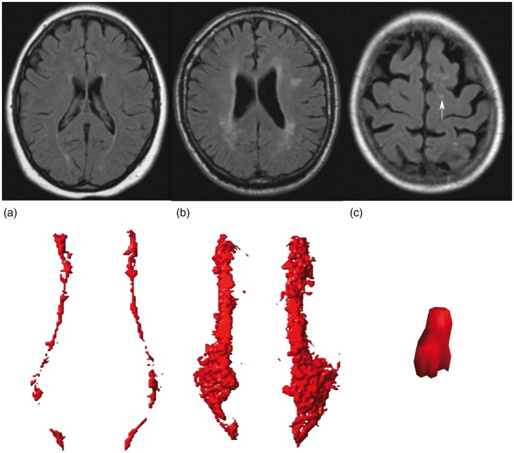
Lacunes and white matter hyperintensities (WMHs) are features of cerebral small vessel disease (CSVD) that are associated with poor functional outcomes. However, how the two are related remains unclear. In this study, we examined the association between lacunes and several WMH features in patients with a history of vascular disease. A total of 999 patients (mean age 59 ± 10 years) with a 1.5 T brain magnetic resonance imaging (MRI) scan were included from the SMART-MR study. Lacunes were scored visually and WMH features (volume, subtype and shape) were automatically determined. Analyses consisted of linear and Poisson regression adjusted for age, sex, and total intracranial volume (ICV). Patients with lacunes (n = 188; 19%) had greater total (B = 1.03, 95% CI: 0.86 to 1.21), periventricular/confluent (B = 1.08, 95% CI: 0.89 to 1.27), and deep (B = 0.71, 95% CI: 0.44 to 0.97) natural log-transformed WMH volumes than patients without lacunes. Patients with lacunes had an increased risk of confluent type WMHs (RR = 2.41, 95% CI: 1.98 to 2.92) and deep WMHs (RR = 1.41, 95% CI: 1.22 to 1.62) and had a more irregular shape of confluent WMHs than patients without lacunes, independent of total WMH volume. In conclusion, we found that lacunes on MRI were associated with WMH features that correspond to more severe small vessel changes, mortality, and poor functional outcomes.
Keywords: Small vessel disease; cerebrovascular disease; lacunes; magnetic resonance imaging; white matter hyperintensities.
Figures
Similar articles
-
White Matter Hyperintensities Are Associated with Slower Gait Speed in Older Adults without Dementia.Neurodegener Dis. 2024;24(3-4):97-105. doi: 10.1159/000538944. Epub 2024 Jul 18. Neurodegener Dis. 2024. PMID: 39025052 Free PMC article.
-
Spatial Relation Between White Matter Hyperintensities and Incident Lacunes of Presumed Vascular Origin: A 14-Year Follow-Up Study.Stroke. 2022 Dec;53(12):3688-3695. doi: 10.1161/STROKEAHA.122.039903. Epub 2022 Oct 3. Stroke. 2022. PMID: 36189679 Free PMC article.
-
Longitudinal relationship between cerebral small-vessel disease and cerebral blood flow: the second manifestations of arterial disease-magnetic resonance study.Stroke. 2015 May;46(5):1233-8. doi: 10.1161/STROKEAHA.114.008030. Epub 2015 Mar 24. Stroke. 2015. PMID: 25804924
-
Longitudinal Changes of White Matter Hyperintensities in Sporadic Small Vessel Disease: A Systematic Review and Meta-analysis.Neurology. 2022 Nov 29;99(22):e2454-e2463. doi: 10.1212/WNL.0000000000201205. Epub 2022 Sep 19. Neurology. 2022. PMID: 36123130 Free PMC article.
-
Cerebral small vessel disease and the risk of Alzheimer's disease: A systematic review.Ageing Res Rev. 2018 Nov;47:41-48. doi: 10.1016/j.arr.2018.06.002. Epub 2018 Jun 26. Ageing Res Rev. 2018. PMID: 29898422
Cited by
-
Identification of Distinct Brain MRI Phenotypes and Their Association With Long-Term Dementia Risk in Community-Dwelling Older Adults.Neurology. 2024 Apr 9;102(7):e209176. doi: 10.1212/WNL.0000000000209176. Epub 2024 Mar 12. Neurology. 2024. PMID: 38471053 Free PMC article.
-
Fibrinogen is an Independent Risk Factor for White Matter Hyperintensities in CADASIL but not in Sporadic Cerebral Small Vessel Disease Patients.Aging Dis. 2021 Jun 1;12(3):801-811. doi: 10.14336/AD.2020.1110. eCollection 2021 Jun. Aging Dis. 2021. PMID: 34094643 Free PMC article.
-
MRI phenotypes of the brain are related to future stroke and mortality in patients with manifest arterial disease: The SMART-MR study.J Cereb Blood Flow Metab. 2020 Feb;40(2):354-364. doi: 10.1177/0271678X18818918. Epub 2018 Dec 14. J Cereb Blood Flow Metab. 2020. PMID: 30547694 Free PMC article. Clinical Trial.
-
Impaired dynamic cerebral autoregulation is associated with the severity of neuroimaging features of cerebral small vessel disease.CNS Neurosci Ther. 2022 Feb;28(2):298-306. doi: 10.1111/cns.13778. Epub 2021 Dec 11. CNS Neurosci Ther. 2022. PMID: 34894087 Free PMC article.
-
Quantitative Analysis of White Matter Hyperintensities as a Predictor of 1-Year Risk for Ischemic Stroke Recurrence.Neurol Ther. 2024 Oct;13(5):1467-1482. doi: 10.1007/s40120-024-00652-3. Epub 2024 Aug 13. Neurol Ther. 2024. PMID: 39136813 Free PMC article.
References
-
- Pantoni L, Poggesi A, Inzitari D. Cognitive decline and dementia related to cerebrovascular diseases: some evidence and concepts. Cerebrovasc Dis 2009; 27(Suppl 1): 191–196. - PubMed
Publication types
MeSH terms
LinkOut - more resources
Full Text Sources
Other Literature Sources
Medical
