. 2019 Feb;33(2):1946-1962.
doi: 10.1096/fj.201800683RR. Epub 2018 Sep 11.

PERK regulates skeletal muscle mass and contractile function in adult mice

Affiliations

PERK regulates skeletal muscle mass and contractile function in adult mice

Yann S Gallot et al. FASEB J. 2019 Feb.

Abstract

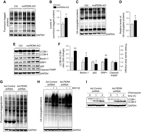
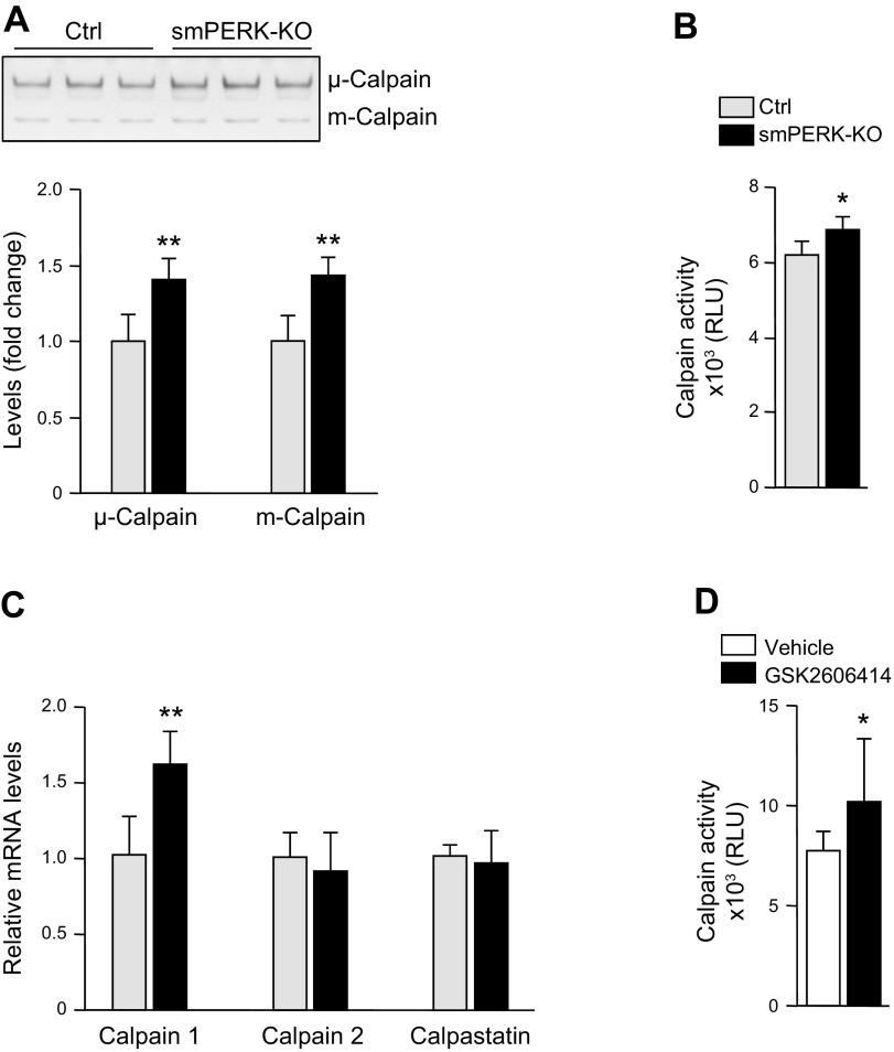
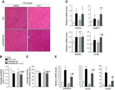
Skeletal muscle mass is regulated by the coordinated activation of several anabolic and catabolic pathways. The endoplasmic reticulum (ER) is a major site of protein folding and a reservoir for calcium ions. Accretion of misfolded proteins or depletion in calcium concentration causes stress in the ER, which leads to the activation of a signaling network known as the unfolded protein response (UPR). In the present study, we investigated the role of the protein kinase R-like endoplasmic reticulum kinase (PERK) arm of the UPR in the regulation of skeletal muscle mass and function in naive conditions and in a mouse model of cancer cachexia. Our results demonstrate that the targeted inducible deletion of PERK reduces skeletal muscle mass, strength, and force production during isometric contractions. Deletion of PERK also causes a slow-to-fast fiber type transition in skeletal muscle. Furthermore, short hairpin RNA-mediated knockdown or pharmacologic inhibition of PERK leads to atrophy in cultured myotubes. While increasing the rate of protein synthesis, the targeted deletion of PERK leads to the increased expression of components of the ubiquitin-proteasome system and autophagy in skeletal muscle. Ablation of PERK also increases the activation of calpains and deregulates the gene expression of the members of the FGF19 subfamily. Furthermore, the targeted deletion of PERK increases muscle wasting in Lewis lung carcinoma tumor-bearing mice. Our findings suggest that the PERK arm of the UPR is essential for the maintenance of skeletal muscle mass and function in adult mice.-Gallot, Y. S., Bohnert, K. R., Straughn, A. R., Xiong, G., Hindi, S. M., Kumar, A. PERK regulates skeletal muscle mass and contractile function in adult mice.

Keywords: ER stress; cancer cachexia; lewis lung carcinoma; proteostasis; unfolded protein response.


Conflict of interest statement

This work was supported by U.S. National Institutes of Health, National Institute of Arthritis and Musculoskeletal and Skin Disease Grants AG029623, AR059810 and AR068313 (to A.K.) Figure 1A was produced with the assistance of Servier Medical Art (https://smart.servier.com). The authors declare no conflicts of interest.

Figures

Figure 1
Figure 1
Ablation of PERK reduces body weight and diminishes muscle strength in adult mice. A) Schematic representation of the breeding strategy used for the generation of control (Ctrl) and smPERK-KO mice. B) Treatment protocol for tamoxifen-induced Cre-mediated recombination in smPERK-KO mice. C) Mean body weight of Ctrl and smPERK-KO mice 4 wk after the start of tamoxifen injections. D, E) Mean forelimb (D) and total 4-paw (E) grip strength per gram of body weight of Ctrl and smPERK-KO mice (n = 10–12/group). Contractile properties of the posterior compartment of the lower limb were measured in vivo. F) Representative traces of normalized specific twitch force (sPt). G) Quantification of mean specific twitch forces. H) Representative normalized traces for specific tetanic force (sPo) optimization. I) Normalized tetanic force to stimulation frequency relationship. J) Representative normalized traces for maximum tetanic force (sPo). K) Quantification of mean specific maximum tetanic forces. L) Fatigue traces over 180 s for each mouse. M) Percent fatigue index (forcet180/forcet0) for mice (n = 5/group). Error bars represent ± sd. *P < 0.05, **P < 0.01, ***P < 0.001 from corresponding littermate Ctrl mice (unpaired, 2-tailed Student’s t test).
Figure 2
Figure 2
Deletion of PERK reduces skeletal muscle mass in adult mice. A–C) Mean wet weight of isolated GA (A), TA (B), and soleus (C) muscle of control (Ctrl) and smPERK-KO, normalized with body weight, after 4 wk of the start of tamoxifen injection (n = 9–14/group). D) PCR analysis of Cre-mediated recombination performed on genomic DNA extracted from the GA muscle of WT, Ctrl (i.e., PERKf/f), and smPERK-KO mice. An agarose gel image of PCR products is shown. Arrows: to bands corresponding to PERK WT allele (400 bp), the PERK loxP-flanked allele (480 bp) and the PERK recombined loxP allele (700 bp). E) Relative mRNA levels of PERK in GA muscle from Ctrl and smPERK-KO mice (n = 3–4/group). F) Relative mRNA levels of ER-resident chaperones, GRP-78, GRP-94, calnexin, and calreticulin in GA muscle from Ctrl and smPERK-KO mice (n = 3–5/group). G) Relative mRNA levels of select ER stress/UPR markers: ATF4, CHOP, death receptor-5, GADD34, IRE1a, spliced XBP1, and ATF6 in GA muscle of Ctrl and smPERK-KO mice (n = 3–5/group). Error bars ± sd. *P < 0.05, **P < 0.01, ***P < 0.001 vs. corresponding littermate Ctrl mice (unpaired, 2-tailed Student’s t test).
Figure 3
Figure 3
Ablation of PERK causes skeletal muscle atrophy. A) Representative photomicrographs of TA and soleus muscle sections from control (Ctrl) and smPERK-KO mice after staining with anti-laminin. Nuclei were counterstained with DAPI. Scale bars, 50 μm. B, C) Quantification of mean myofiber CSA (B), and mean minimal Feret’s diameter (C) in TA muscle sections (n = 4–6/group). D, E) Quantification of mean myofiber CSA (D) and mean minimal Feret’s diameter (E) in soleus muscle sections (n = 5–6/group). Error bars ± sd. *P < 0.05 vs. corresponding Ctrl mice (unpaired, 2-tailed Student’s t test). F) C2C12 myotubes were transduced with adenoviral vectors expressing control shRNA or PERK shRNA for 24 h. The cells were washed and incubated in differentiation medium for an additional 48 h, with or without 1 µg/ml tunicamycin. Representative images of the cultures are presented. Scale bars, 50 µm. G) Quantification of mean diameter of myotubes in control and PERK shRNA-expressing cultures (n = 3/group). Error bars ± sd. *P < 0.05 vs. vehicle alone-treated cultures transduced with Ad.Control shRNA, #P < 0.05 vs. vehicle alone–treated cultures transduced with Ad.PERK shRNA (unpaired, 2-tailed Student’s t test). H) Immunoblots demonstrating levels of PERK and unrelated protein GAPDH in C2C12 myotube cultures transduced with Ad.Control or Ad.PERK shRNA vectors. Horizontal solid lines on the immunoblots indicate that intervening lanes have been spliced out.
Figure 4
Figure 4
Inactivation of PERK causes a slow-to-fast fiber-type transition in adult mice. A) Muscle sections prepared from soleus muscle of control (Ctrl) and smPERK-KO mice were subjected to triple immunostaining against MyHCI, IIa, and IIb proteins. Representative photomicrographs of triple-stained sections of soleus muscle. Scale bar, 50 μm. B, C) Quantification of percentage of each fiber type (B) and mean myofiber CSA of type I and IIa fibers (C) in soleus muscle of Ctrl and smPERK-KO mice (n = 6/group). D) Muscle sections prepared from TA muscle of Ctrl and smPERK-KO mice were subjected to triple immunostaining against MyHCI, IIa, and IIb proteins. Representative photomicrographs of triple-stained TA muscle sections are presented here. Scale bars, 50 μm. E, F) Quantification of percentage of each fiber type (E), and mean myofiber CSA of type IIa, and IIb (F) fibers in TA muscle of Ctrl and smPERK-KO mice (n = 3–5/group). Error bars ± sd. *P < 0.05 vs. corresponding littermate Ctrl (unpaired, 2-tailed Student’s t test).
Figure 5
Figure 5
Ablation of PERK disrupts protein turnover in skeletal muscle of adult mice. Control (Ctrl) and smPERK-KO mice were given an injection of puromycin (0.04 μM/g body weight, i.p.). The mice were euthanized 30 min later, and GA muscle was collected and processed for Western blot analysis. A) Immunoblots demonstrating levels of puromycin-tagged proteins and unrelated protein GAPDH in GA muscle of Ctrl and smPERK-KO mice. B) Densitometry quantification of relative levels of puromycin-tagged proteins in GA muscle of Ctrl and smPERK-KO mice (n = 3/group). C) Immunoblots showing relative amounts of ubiquitin-conjugated proteins and unrelated protein GAPDH in GA muscle of Ctrl and smPERK-KO mice. D) Densitometry quantification of relative levels of ubiquitin-conjugated proteins (n = 4–5/group). E) Representative immunoblots showing levels of autophagy markers: LC-3BI/II, Beclin-1, DRP1, p62, and an apoptosis marker, cleaved PARP, and unrelated protein GAPDH in GA muscle of Ctrl and smPERK-KO mice. F) Densitometry quantification of levels of LC-3BII/I, Beclin-1, DRP1, p62, and cleaved PARP (n = 3–5/group). Error bars ± sd. *P < 0.05, **P < 0.01 from corresponding Ctrl mice (unpaired, 2-tailed Student’s t test). G) Fully differentiated C2C12 myotubes were transduced with adenoviral vectors expressing control shRNA or PERK shRNA for 24 h. The cells were washed and incubated in differentiation medium for an additional 48 h. The myotubes were treated with 1 μM puromycin for exactly 30 min, followed by analysis for puromycin-tagged proteins. Immunoblot shows the levels of puromycin-tagged protein in Ad.Control shRNA and Ad.PERK shRNA cultures. H, I) In a separate experiment, control shRNA or PERK shRNA-expressing myotube cultures were treated with vehicle alone, 40 µM MG132 (for 1 h), or 100 µM chloroquine (for 1 or 2 h) followed by Western blot analysis to detect the levels of ubiquitinated protein or LC-3BI/II protein. Representative immunoblots from 2 independent experiments demonstrating levels of ubiquitin-conjugated proteins (H), and LC-3BI/II and unrelated protein GAPDH (I) in control shRNA- and PERK shRNA–expressing cultures. Black lines on the immunoblots indicate that intervening lanes have been spliced out.
Figure 6
Figure 6
Ablation of PERK activates calpains in skeletal muscle. A) Representative photograph and quantification of bands of μ- and m-calpain in a casein zymogram performed on GA muscle extracts from control (Ctrl) and smPERK-KO mice (n = 4/group). B) Enzymatic activity of calpains in GA muscle of Ctrl and smPERK-KO mice (n = 4 per group). C) Relative mRNA levels of calpain 1, calpain 2, and calpastatin in GA muscle of Ctrl and smPERK-KO mice (n = 4–5/group). D) Differentiated myotubes were incubated with vehicle alone (DMSO) or 2 µM GSK2606414 for 48 h, and protein extracts were analyzed for calpain activity (n = 8/group). Error bars ± sd. *P < 0.05, **P < 0.01 vs. corresponding littermate Ctrl mice (unpaired, 2-tailed Student’s t test).
Figure 7
Figure 7
Effect of ablation of PERK on skeletal muscle wasting during cancer-induced cachexia. Control (Ctrl) and smPERK-KO mice were inoculated with 2 × 10⁶ LLC cells in 100 μl of sterile PBS in the left flank. After 21 d, mice were euthanized for histologic and biochemical analysis. A) Representative photomicrographs of H&E-stained TA muscle sections from Ctrl and smPERK-KO mice. Scale bars, 50 μm. B, C) Quantification of mean myofiber CSA (B) and minimal Feret’s diameter (C) of myofibers in TA muscle sections of control (Ctrl) and smPERK-KO mice (n = 7–10/group). D) Relative mRNA levels of muscle atrophy F-box/Atrogin-1, MuRF1, MUSA1, and LC3B in GA muscle of Ctrl and smPERK-KO mice (n = 3–6/group). E) Relative mRNA levels of GADD34, ATF4, and CHOP in TA muscle of Ctrl and smPERK-KO mice (n = 3/group). Error bars ± sd. *P < 0.05 vs. Ctrl mice injected with saline alone, #P < 0.05 vs. smPERK-KO mice injected with saline alone, @P < 0.05 vs. Ctrl LLC-bearing mice (unpaired, 2-tailed Student’s t test).

Similar articles

Cited by

References

    1. Egerman, M. A., Glass, D. J. (2014) Signaling pathways controlling skeletal muscle mass. Crit. Rev. Biochem. Mol. Biol. 49, 59–68 - PMC - PubMed
    1. Bonaldo, P., Sandri, M. (2013) Cellular and molecular mechanisms of muscle atrophy. Dis. Model. Mech. 6, 25–39 - PMC - PubMed
    1. Kandarian, S. C., Jackman, R. W. (2006) Intracellular signaling during skeletal muscle atrophy. Muscle Nerve 33, 155–165 - PubMed
    1. Li, H., Malhotra, S., Kumar, A. (2008) Nuclear factor-kappa B signaling in skeletal muscle atrophy. J. Mol. Med. (Berl.) 86, 1113–1126 - PMC - PubMed
    1. Bohnert, K. R., McMillan, J. D., Kumar, A. (2018) Emerging roles of ER stress and unfolded protein response pathways in skeletal muscle health and disease. J. Cell. Physiol. 233, 67–78 - PMC - PubMed

Publication types

MeSH terms

LinkOut - more resources