. 2018 Sep 10;34(3):427-438.e6.
doi: 10.1016/j.ccell.2018.08.008.

The Genomic Landscape of Endocrine-Resistant Advanced Breast Cancers

Affiliations

The Genomic Landscape of Endocrine-Resistant Advanced Breast Cancers

Pedram Razavi et al. Cancer Cell. .

Abstract

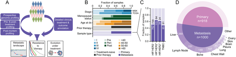
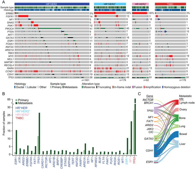
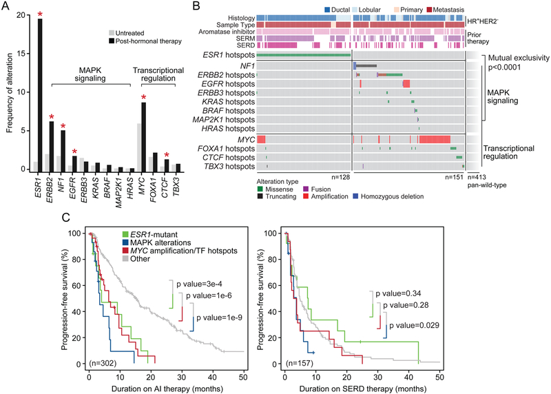
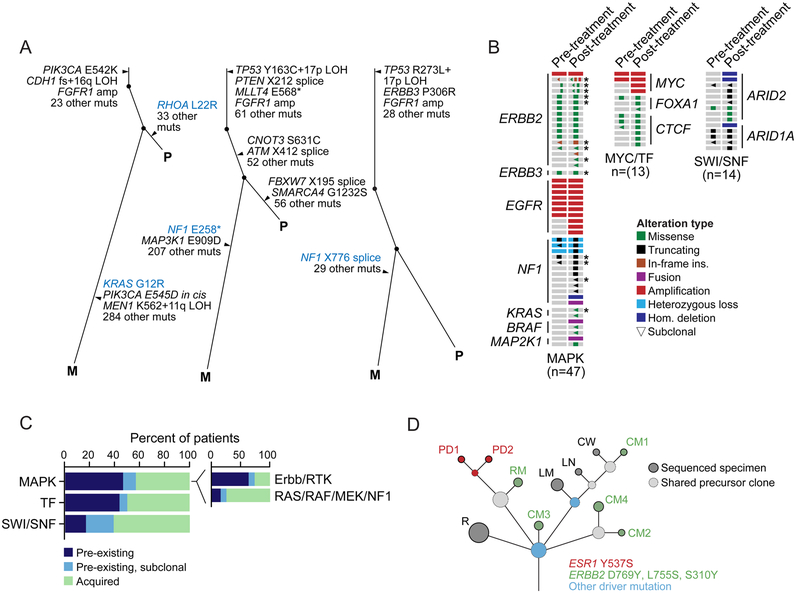
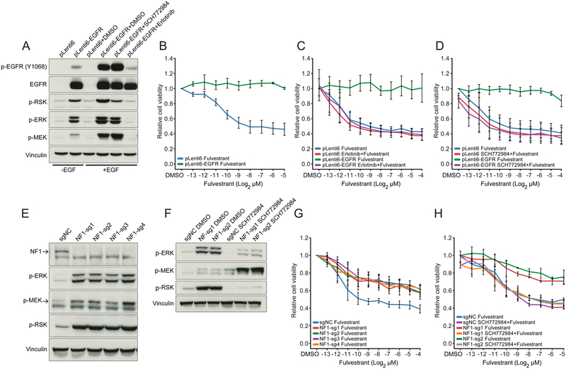
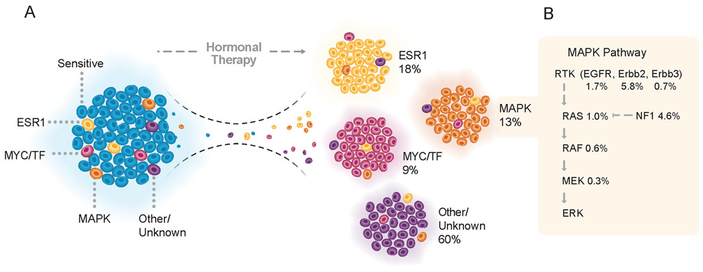
We integrated the genomic sequencing of 1,918 breast cancers, including 1,501 hormone receptor-positive tumors, with detailed clinical information and treatment outcomes. In 692 tumors previously exposed to hormonal therapy, we identified an increased number of alterations in genes involved in the mitogen-activated protein kinase (MAPK) pathway and in the estrogen receptor transcriptional machinery. Activating ERBB2 mutations and NF1 loss-of-function mutations were more than twice as common in endocrine resistant tumors. Alterations in other MAPK pathway genes (EGFR, KRAS, among others) and estrogen receptor transcriptional regulators (MYC, CTCF, FOXA1, and TBX3) were also enriched. Altogether, these alterations were present in 22% of tumors, mutually exclusive with ESR1 mutations, and associated with a shorter duration of response to subsequent hormonal therapies.

Keywords: breast cancer; cancer genomics; endocrine resistance; integrative genomics analysis; metastasis.


Figures

Figure 1:
Figure 1:. Clinical Characteristics of Prospectively Sequenced Advanced Breast Cancers
(A) Study schema. (B) Clinical features of this prospective cohort compared to a contemporary study of primary untreated breast cancers (TCGA). (C) Composition and cohort size of primary and metastatic breast cancers by receptor subtype. (D) Distribution of the biopsied metastatic disease sites and sample number in the study cohort. See also Figure S1 and Tables S1-S2.
Figure 2:
Figure 2:. Genomic Characteristics of Prospectively Sequenced Advanced Breast Cancers
(A) The pattern, frequency, and type of genomic alterations in key breast cancer genes by receptor type. (B) The frequency difference among genes with a statistically significant increase (q < 0.05) of alterations in metastatic specimens as compared with primary tumors. The color of the gene symbol indicates statistical significance by receptor status. (C) Recurrent genomic alterations (left) and their association with different organ sites of metastasis (right). Line thickness corresponds to the frequency of mutations arising in the indicated metastatic site. Shading identifies the relationships between genes and metastatic sites. Statistically significant associations are shown as asterisks (p < 0.05). See also Figures S2-S3 and Table S3.
Figure 3:
Figure 3:. Genomic Landscape of Endocrine Resistance after Treatment with Hormonal Therapy
(A) Frequency of recurrent functional genomic alterations as shown in (B) in tumors that were collected from patients that were either treatment-naïve or after treatment with hormonal therapy. Asterisks denote statistical significance between groups. (B) Pattern and frequency of known or putative functional mutations and focal amplifications or deletions targeting effectors of MAPK signaling or aberrant transcriptional regulation in ESR1-mutant and ESR1-wild-type tumors after endocrine therapy (mutual exclusivity p value < 0.0001). (C) Kaplan-Meier curves displaying progression-free survival of patients receiving aromatase inhibitor (AI) (left) or SERD (right) therapy based on genomic alterations. Tumors harboring hotspot mutations in ESR1 are displayed in green, those with known or putative functional alterations in effectors of MAPK signaling are shown in blue, and those with alterations in transcriptional regulators in red. Outcomes were compared with patients whose tumors were wild-type for such lesions (gray). All p values as indicated, log rank test. See also Figure S4.
Figure 4:
Figure 4:. Tumor Evolution Under Endocrine Therapy
(A) Evolutionary relationships among clones and somatic mutations inferred from whole-exome sequencing of three representative patients with paired treatment-naïve primary (P) and post-treatment metastatic (M) tumors indicating intra-tumoral heterogeneity and the acquisition of MAPK activating lesions after hormonal therapy (left, KRAS G12R; middle and right, two NF1 truncating mutations). (B) Comparison of treatment-naïve primary tumors and their patient-matched post-endocrine therapy specimens in 74 patients with ESR1-wild-type HR+HER2 disease with diverse lesions in MAPK effectors, MYC and transcription factors (MYC/TF), or components of the SWI/SNF complex (as labeled) indicated pre-existing or acquired lesions. (C) A summary of the rate of pre-existing and acquired lesions by pathway. Inset further subdivides MAPK alterations into receptor tyrosine kinase (Erbb/RTK) versus downstream RAS/RAF/MEK/NF1 lesions. Asterisks denote patients with multiple candidate resistance mutations. (D) The clonal evolution of a HR+ metastatic breast cancer inferred from the sequencing of an initial recurrence (R) and ten metastatic sites collected at the time of autopsy. Circles are colored based on the presence of one or more breast cancer driver mutations (blue) or candidate mechanisms of endocrine resistance (red and green, as indicated). The size of the circle is proportional to the number of mutations defining the clone. Dashed circles represent shared precursor clones and solid circles represent the sequenced specimens. PD, pleural disease; RM, retroperitoneal mass; CM, cutaneous metastasis; LM, liver metastasis; LN, lymph node; CW, chest wall. See also Figures S4-S5 and Tables S4-S5.
Figure 5:
Figure 5:. Functional Validation of MAPK Activation as Mechanism of Resistance to Endocrine Therapy
(A) Western blot showing expression and phosphorylation of EGFR and activation of MAPK pathway effectors in MCF7 cells transduced with pLenti6 mock control and pLenti6-EGFR vector. (B and D) Cell viability of MCF7 cells stably transfected with pLenti6 mock control or pLenti6-EGFR treated with fulvestrant (B), fulvestrant + 5 mM erlotinib (C), or fulvestrant + 500 nM SCH772984 (D). (E) Western blot showing activation of MAPK pathway effectors in four independent pools of NF1-knockout and control (scrambled-sgNC) MCF7 cells. (F) Change in expression of phosphorylated ERK, MEK and RSK in cells treated with 500 nM SCH772984 versus control (DMSO). Results are shown for two independent MCF7 pools expressing NF1-targeted guide RNAs and a control (scrambled-sgNC) guide RNA. (G and H) Cell viability following treatment with fulvestrant (G), or fulvestrant + 500 nM SCH772984 (H). Results are shown for cells in which NF1 was knocked out using four different NF1-targeted guide RNAs. Error bars represent standard deviations. See also Figure S6.
Figure 6:
Figure 6:. A Taxonomy of Mechanisms of Resistance to Endocrine Therapy
(A) Tumors after hormonal therapy failure may be divided into four categories, depending on the genomic aberrations present in the refractory lesions. Tumors harboring ESR1 mutations (yellow) were responsible for 18% of the tumors relapsing after endocrine therapy. Functional lesions in the MAPK pathway (orange) and mutations in the machinery of transcriptional regulation (MYC/TF, red) were responsible for 13% and 9% of the resistant cases, respectively. Pan-wild-type tumors with a still occult mechanism of resistance to hormonal therapy (purple) accounted for the remaining 60% of cases. These alterations may preexist in the pre-treatment tumors and expand or be acquired under the selective pressure of endocrine therapy. (B) Frequency of alterations in the MAPK signaling pathway in tumors following hormonal therapy.

References

    1. Al-Ahmadie HA, Iyer G, Lee BH, Scott SN, Mehra R, Bagrodia A, Jordan EJ, Gao SP, Ramirez R, Cha EK, et al. (2016). Frequent somatic CDH1 loss-of-function mutations in plasmacytoid variant bladder cancer. Nat Genet 48, 356–358. - PMC - PubMed
    1. Andre F, Campone M, Ciruelos EM, Iwata H, Liobl S, Rugo HS, Wilke C, Mills D, Chol M, Longin A, et al. (2016). SOLAR-1: A phase III study of alpelisib + fulvestrant in men and postmenopausal women with HR+/HER2− advanced breast cancer (BC) progressing on or after prior aromatase inhibitor therapy. J Clin Oncol 34, TPS618–TPS618.
    1. Augello MA, Hickey TE, and Knudsen KE (2011). FOXA1: master of steroid receptor function in cancer. EMBO J 30, 3885–3894. - PMC - PubMed
    1. Banerji S, Cibulskis K, Rangel-Escareno C, Brown KK, Carter SL, Frederick AM, Lawrence MS, Sivachenko AY, Sougnez C, Zou L, et al. (2012). Sequence analysis of mutations and translocations across breast cancer subtypes. Nature 486, 405–409. - PMC - PubMed
    1. Barbieri CE, Baca SC, Lawrence MS, Demichelis F, Blattner M, Theurillat JP, White TA, Stojanov P, Van Allen E, Stransky N, et al. (2012). Exome sequencing identifies recurrent SPOP, FOXA1 and MED12 mutations in prostate cancer. Nat Genet 44, 685–689. - PMC - PubMed

Publication types

MeSH terms