Review
. 2018 Sep 10;28(17):R1059-R1073.
doi: 10.1016/j.cub.2018.04.057.

The Neurocognitive Basis of Spatial Reorientation

Affiliations
Review

The Neurocognitive Basis of Spatial Reorientation

Joshua B Julian et al. Curr Biol. .

Abstract

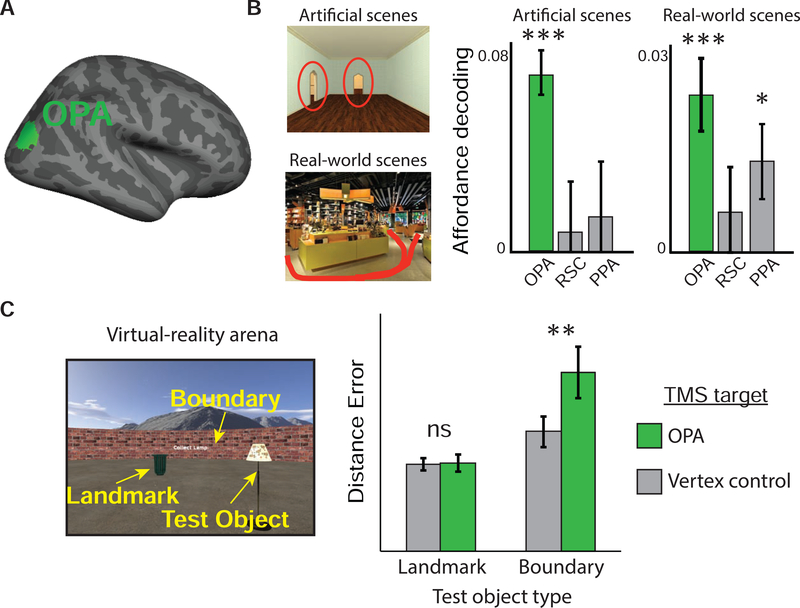
The ability to recover one's bearings when lost is a skill that is fundamental for spatial navigation. We review the cognitive and neural mechanisms that underlie this ability, with the aim of linking together previously disparate findings from animal behavior, human psychology, electrophysiology, and cognitive neuroscience. Behavioral work suggests that reorientation involves two key abilities: first, the recovery of a spatial reference frame (a cognitive map) that is appropriate to the current environment; and second, the determination of one's heading and location relative to that reference frame. Electrophysiological recording studies, primarily in rodents, have revealed potential correlates of these operations in place, grid, border/boundary, and head-direction cells in the hippocampal formation. Cognitive neuroscience studies, primarily in humans, suggest that the perceptual inputs necessary for these operations are processed by neocortical regions such as the retrosplenial complex, occipital place area and parahippocampal place area, with the retrosplenial complex mediating spatial transformations between the local environment and the recovered spatial reference frame, the occipital place area supporting perception of local boundaries, and the parahippocampal place area processing visual information that is essential for identification of the local spatial context. By combining results across these various literatures, we converge on a unified account of reorientation that bridges the cognitive and neural domains.


Figures

Figure 1.
Figure 1.. Components of reorientation.
A lost navigator must solve three tasks in order to regain her bearings: recognize her current context (context recognition), determine where she is located in that context (self-location) and which way she is facing (heading retrieval).
Figure 2.
Figure 2.. Cognitive mechanisms underlying reorientation.
(A) In the standard reorientation task, a navigator is allowed to locate a reward in one corner (R, correct corner) of a small rectangular chamber with polarizing features along the walls (for example, black stripes). They are then removed from the chamber and disoriented. When placed back in the chamber at test, navigators typically use boundary geometry to reorient, searching equally often at the rewarded and geometrically equivalent opposite (G) corners, while ignoring non-geometric features. (B) Judgment of relative direction (JRD) tasks reveal the reference frames underlying spatial representations. Memory for the relative locations of buildings on a college campus (map shown on left) was reported more accurately when imagining headings that were aligned to cardinal directions (N, W, S, E) compared to diagonal directions (NW, SW, SE, NE), with the best performance for imagined headings facing North (adapted with permission from [29]). (C) To test whether context recognition is dissociable from heading retrieval, mice were trained to locate a hidden reward in two rectangular chambers that had identical geometry but were distinguishable by vertical versus horizontal stripes along one wall. Mice dug more often in the corners that were geometrically appropriate for each chamber, indicating that they used the features to distinguish between the chambers (adapted with permission from [35]). However, they failed to use the features to distinguish between geometrically appropriate corners within each chamber, instead relying exclusively on geometry (data not shown).
Figure 3.
Figure 3.. Neural mechanisms for reorientation.
(A) Following disorientation, the hippocampal map reorients based on chamber geometry rather than featural cues (adapted with permission from [55]). Mice in this experiment were disoriented between each experimental trial. For each chamber shape (rectangle, square, and isosceles triangle), trial-by-trial place fields reflected the geometric symmetries of the chamber (2×, 4×, 1×), despite the presence of a disambiguating feature cue along one wall. Simultaneously recorded place cells had fields that aligned coherently. (B) When trained to perform a classic reorientation task, there was a strong correspondence between the hippocampal map alignment on each trial and the location that the animal searched (adapted with permission from [55]; note that counter to this example, geometrically aligning place fields were found throughout the chamber, not just at the goal location). (C) Like place cells, head-direction cells tend to exhibit two preferred firing directions in a rectangular chamber from block-to-block following disorientation, oriented 180 degrees from each other (adapted with permission from [63]).
Figure 4.
Figure 4.. The retrosplenial complex supports spatial transformations necessary for reorientation.
(A) Left: retrosplenial complex/medial place area (RSC/MPA) and retrosplenial cortex (BA 29/30) shown on the human cortical surface. Right: retrosplenial cortex shown on the rodent brain. (B) fMRI evidence that the retrosplenial complex represents heading in a local reference frame. During training before scanning, participants learned the locations of objects (denoted by circles) inside virtual reality ‘museums’ that were oriented at 90 degrees from each other within a larger navigable space. During scanning, participants performed a judgement-of-relativedirection task that required them to imagine facing each object encountered during training. Multivoxel activity patterns in RSC were similar for imagined views across the two museums that had the same heading as defined by the local (museum-centered) reference frame indicated by the red arrows (adapted with permission from [116]). (C) In rodents, retrosplenial cortex contains ‘bidirectional’ (BD) cells that have preferred firing directions that represent heading in a local reference frame tied to each subchamber (adapted with permission from [121]). These bidirectional cells were interspersed with classical head direction cells that represented heading in a global reference frame that was consistent across both subchambers.
Figure 5.
Figure 5.. occipital place area is involved in perception of boundaries and local navigational affordances.
(A) Occipital place area (OPA) shown on the human cortical surface. (B) occipital place area multivoxel activity patterns reflect the locations of fine-grained navigational affordances in both artificial scenes (the position of doorways) and real-world scenes (locations of pathways), irrespective of other perceptual details present in the scenes [142]. (C) To test the causal role of occipital place area in boundary perception, participants learned the locations of test objects inside a virtual-reality arena containing a ‘landmark’ object and surrounded by a boundary wall. Half of the test objects had locations defined relative to the location of the ‘landmark’ object, and the other half had locations defined relative to a boundary. Compared to a vertex control stimulation site, TMS to the occipital place area impaired memory for the boundary-tethered object locations, but not the landmark-tethered objects (adapted with permission from [146]).
Figure 6.
Figure 6.. Parahippocampal place area is involved in identification of place and contexts.
(A) Parahippocampal place area (PPA) shown on the human cortical surface. (B) Postrhinal cortex (POR), a putative homologue of parahippocampal cortex shown on the rodent brain. (C) Lesions to POR result in context recognition impairments. Sham-operated control rats explore familiar objects appearing in incongruent but familiar contexts more than those appearing in congruent contexts. Postrhinal cortex lesions eliminate this preference. In a comparable noncontextual object recognition task, postrhinal cortex lesions had no effect (adapted with permission from [158]). (D) parahippocampal place area shows similar multivoxel activity patterns for images of the interior and exterior of the same buildings, thus reflecting the same navigational context, but only in participants who have navigational experience with the buildings (Penn students), not in participants who do not (Temple students) (adapted from ref. [163]).

Similar articles

Cited by

References

    1. O’Keefe J, and Nadel L (1978). The hippocampus as a cognitive map (Clarendon Press Oxford; ).
    1. Gallistel CR (1990). The organization of learning (The MIT Press; ).
    1. Trullier O, Wiener SI, Berthoz A, and Meyer JA (1997). Biologically based artificial navigation systems: Review and prospects. Prog. Neurobiol 51, 483–544. - PubMed
    1. Kuipers B (2000). The spatial semantic hierarchy. Artif. Intell 119, 191–233.
    1. Chrastil ER, and Warren WH (2014). From cognitive maps to cognitive graphs. PloS One 9, e112544. - PMC - PubMed

Publication types

LinkOut - more resources