. 2018 Dec:525:1-9.
doi: 10.1016/j.virol.2018.08.022. Epub 2018 Sep 8.

Characterizing replication kinetics and plaque production of type I feline infectious peritonitis virus in three feline cell lines

Affiliations

Characterizing replication kinetics and plaque production of type I feline infectious peritonitis virus in three feline cell lines

Amornrat O'Brien et al. Virology. 2018 Dec.

Abstract

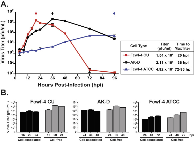
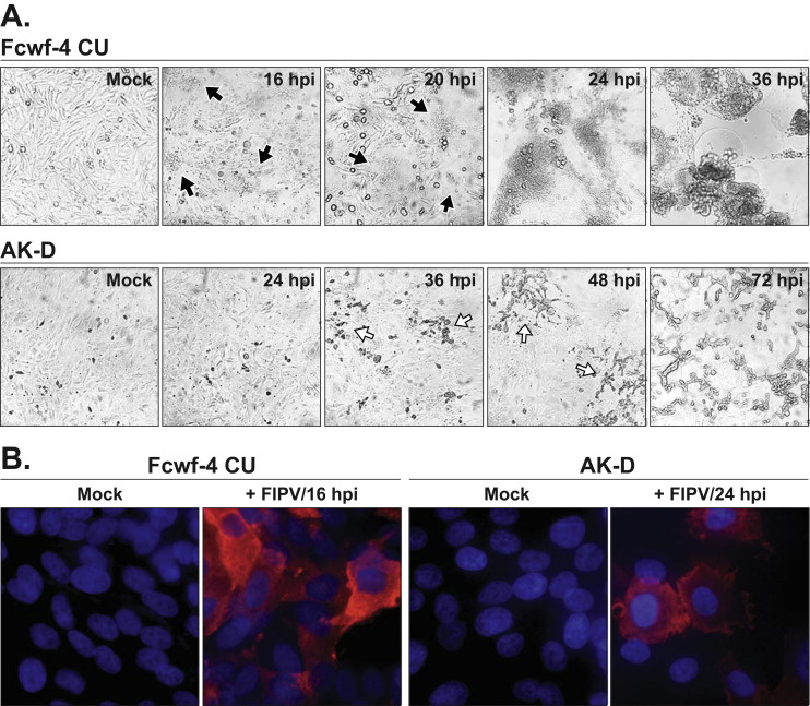
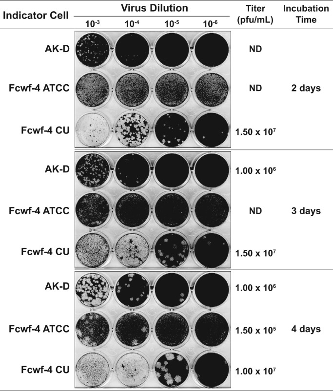
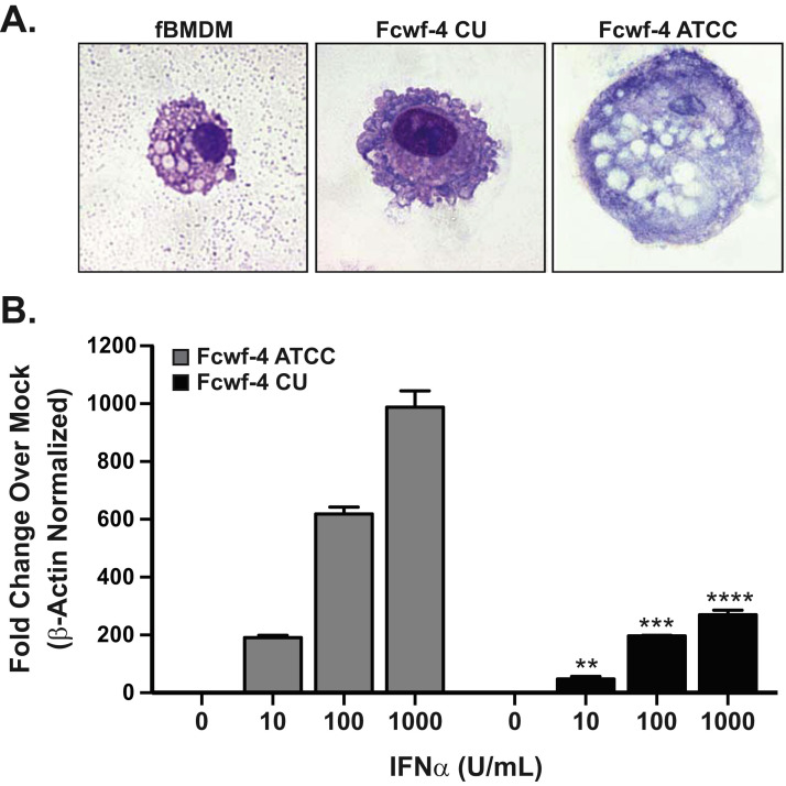
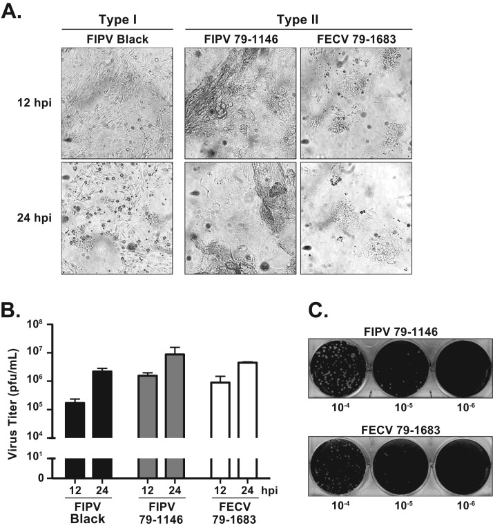
Investigating type I feline coronaviruses (FCoVs) in tissue culture is critical for understanding the basic virology, pathogenesis, and virus-host interactome of these important veterinary pathogens. This has been a perennial challenge as type I FCoV strains do not easily adapt to cell culture. Here we characterize replication kinetics and plaque formation of a model type I strain FIPV Black in Fcwf-4 cells established at Cornell University (Fcwf-4 CU). We determined that maximum virus titers (>107 pfu/mL) were recoverable from infected Fcwf-4 CU cell-free supernatant at 20 h post-infection. Type I FIPV Black and both biotypes of type II FCoV formed uniform and enumerable plaques on Fcwf-4 CU cells. Therefore, these cells were employable in a standardized plaque assay. Finally, we determined that the Fcwf-4 CU cells were morphologically distinct from feline bone marrow-derived macrophages and were less sensitive to exogenous type I interferon than were Fcwf-4 cells purchased from ATCC.

Keywords: AK-D cells; FIPV; Fcwf-4 cells; Feline coronavirus; Feline macrophage-like cell line; Plaque assay.


Figures

Fig. 1
Fig. 1
Fcwf-4 Cornell University (CU) cells produce high titers of cell-free type I FIPV with rapid growth kinetics. A) Virus growth kinetics measured from cell-free supernatants of AK-D (black), Fcwf-4 ATCC (blue), and Fcwf-4 CU (red) cells infected with FIPV Black (MOI=0.1) over 96 h. Arrows indicate time of peak titer presented as plaque-forming units (pfu)/mL. Titer determined by plaque assay on AK-D cells in triplicate; error bars ± SD. B) Cell-associated and cell-free virus titers were determined following infection of Fcwf-4 CU, AK-D, and Fcwf-4 ATCC cells with FIPV Black (MOI=0.1). Cell-free titer was determined from cell-clarified supernatants; cell-associated titer was determined from suspended cell monolayers following three freeze-thaw cycles alternating between −80 °C and 37 oC. Samples were taken at hours post-infection (hpi) just prior to, at, and following the maximum (max) virus titer for each cell type. Titers determined by plaque assay on AK-D cells in triplicate; error bars ± SD.
Fig. 2
Fig. 2
FIPV Black-induced cell cytopathic effect varies between infected cell types. A) Progression of cell cytopathic effects (CPE) induced by FIPV Black over time in Fcwf-4 and AK-D cells (MOI=0.1). Localized cell syncytia formation (black, closed arrows) and discrete cell death (white, open arrows) indicated on brightfield images taken at 20x magnification. B) Immunofluorescence staining of FIPV Black-infected Fcwf-4 CU and AK-D cells (MOI=0.1) at indicated hours post-infection (hpi). Nucleocapsid protein staining (Red; CCV2-2) indicates infected cells; nuclei stained in blue (Hoesch3342). Images taken at 60x magnification.
Fig. 3
Fig. 3
Evaluating FIPV Black plaque development and titer over time in different feline cell lines. Tenfold serial dilutions (10-3-10-6) of virus inoculum, derived from 24 h cell-free supernatant of Fcwf-4 CU cells infected with FIPV Black (MOI=0.1), were applied to AK-D, Fcwf-4 ATCC, and Fcwf-4 CU indicator cells. Following Oxoid agar/media overlay, plates were incubated for 2 (top), 3 (middle), or 4 (bottom) days before cells were fixed with 3.7% formaldehyde and stained with 0.1% crystal violet. Titers were calculated by plaque counts and are presented as plaque forming units (pfu)/mL. ND indicates that titers were not determined.
Fig. 4
Fig. 4
Fcwf-4 CU cells differ in morphology and IFN-responsiveness from Fcwf-4 ATCC. A) Single-cell morphology of feline bone marrow-derived macrophages (fBMDMs), Fcwf-4 CU, and Fcwf-4 ATCC cells determined by Wright-Giemsa stain following cytospin preparation. Images were taken at 100 × . B) Interferon stimulated gene (ISG) 54 transcript levels from Fcwf-4 ATCC (grey) and Fcwf-4 CU (black) cell lines stimulated with increasing concentrations of feline type I interferon (IFNα). After 6 h, total RNA was extracted and analyzed by qPCR for ISG54 and feline beta-actin. ISG54 mRNA expression was normalized to β-actin via the delta Ct method (2-ΔCt[ΔCt=Ct(gene of interest)-Ct(β-actin)]), then presented as relative expression over corresponding mock (basal) expression. Data are representative of three independent experiments performed in triplicate and presented as means ± SD. Values were analyzed by unpaired t-test. * * P < 0.01; * ** P < 0.001; * ** * P < 0.0001.
Fig. 5
Fig. 5
Fcwf-4 CU cells and are permissive to both biotypes of type II FCoV. A) Cell cytopathic effect at 12 and 24 h post-infection (hpi) of Fcwf-4 CU cells infected with indicated FCoV strains (MOI=0.1). B) FCoV growth kinetics in Fcwf-4 CU cells (MOI=0.1) determined by C) plaque assay on Fcwf-4 CU indicator cells. Tenfold serial dilutions of cell-free virus inoculum were applied to cells. Following Oxoid agar/media overlay, cells were fixed in 3.7% formaldehyde and stained using 0.1% crystal violet after 24 h. FCoV used were the type I strain FIPV Black and the type II strains FIPV WSU 79–1146 and FECV WSU 79–1683. Plaque assay performed in triplicate; error bars ± SD.

References

    1. Addie D.D. Feline coronaviral infections. In: Greene C., editor. Infectious Diseases of the Dog and Cat. Saunders; 2011. pp. 92–108.
    1. Addie D.D., Jarrett O. A study of naturally occurring feline coronavirus infections in kittens. Vet. Rec. 1992;130:133–137. doi: 10.1136/vr.130.7.133. - DOI - PubMed
    1. Addie D.D., Schaap I.A.T., Nicolson L., Jarrett O. Persistence and transmission of natural type I feline coronavirus infection. J. Gen. Virol. 2003;84:2735–2744. doi: 10.1099/vir.0.19129-0. - DOI - PubMed
    1. American Type Culture Collection, 2013. Fcwf­4 [Fcwf] (ATCC ® CRL­ 2787 ™) Product Sheet.
    1. Benetka V., Kübber-Heiss A., Kolodziejek J., Nowotny N., Hofmann-Parisot M., Möstl K. Prevalence of feline coronavirus types I and II in cats with histopathologically verified feline infectious peritonitis. Vet. Microbiol. 2004;99:31–42. doi: 10.1016/j.vetmic.2003.07.010. - DOI - PMC - PubMed

Publication types

LinkOut - more resources