Review
. 2018 Sep 8;19(9):2668.
doi: 10.3390/ijms19092668.

Clinical and Biological Characterization of Skin Pigmentation Diversity and Its Consequences on UV Impact

Affiliations
Review

Clinical and Biological Characterization of Skin Pigmentation Diversity and Its Consequences on UV Impact

Sandra Del Bino et al. Int J Mol Sci. .

Abstract

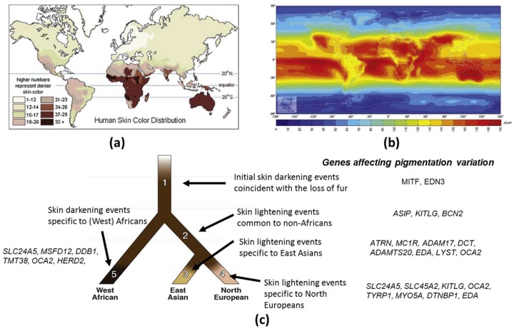
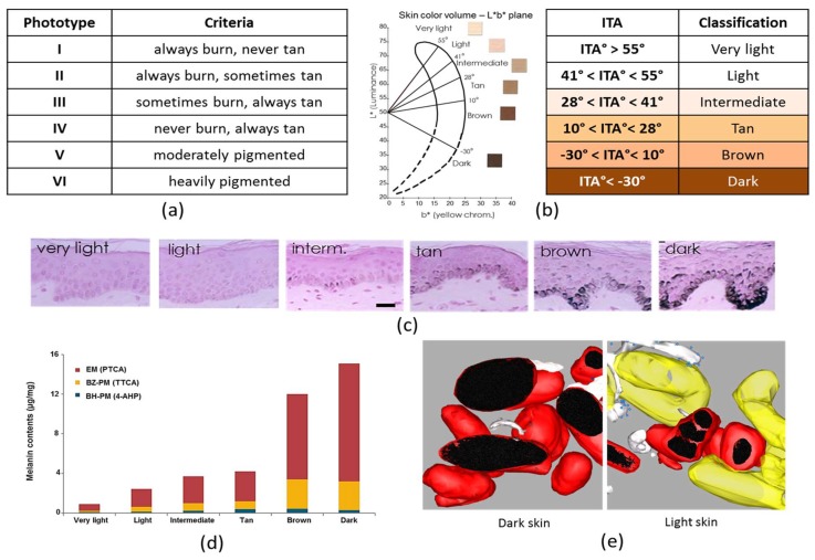
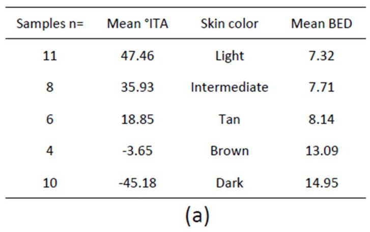
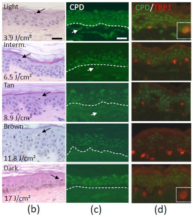
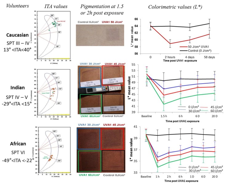
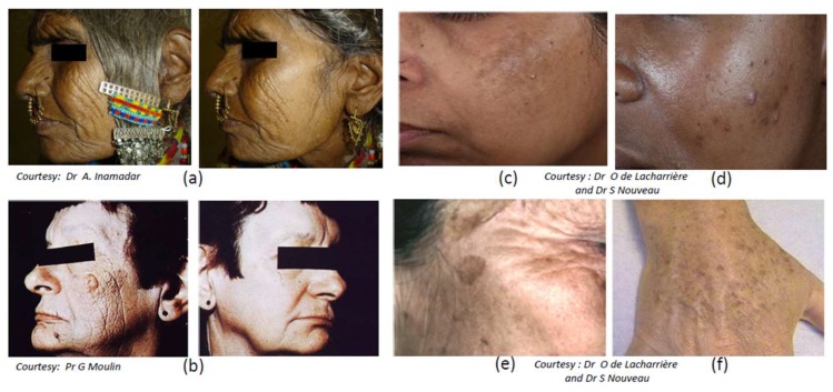
Skin color diversity is the most variable and noticeable phenotypic trait in humans resulting from constitutive pigmentation variability. This paper will review the characterization of skin pigmentation diversity with a focus on the most recent data on the genetic basis of skin pigmentation, and the various methodologies for skin color assessment. Then, melanocyte activity and amount, type and distribution of melanins, which are the main drivers for skin pigmentation, are described. Paracrine regulators of melanocyte microenvironment are also discussed. Skin response to sun exposure is also highly dependent on color diversity. Thus, sensitivity to solar wavelengths is examined in terms of acute effects such as sunburn/erythema or induced-pigmentation but also long-term consequences such as skin cancers, photoageing and pigmentary disorders. More pronounced sun-sensitivity in lighter or darker skin types depending on the detrimental effects and involved wavelengths is reviewed.

Keywords: UV sensitivity; constitutive skin pigmentation; melanocyte; phototype; pigmentary disorders.


Conflict of interest statement

The authors declare a conflict of interest: all authors are full employees of a cosmetic industry.

Figures

Figure 1
Figure 1
Skin color and UV distribution: (a) World map of skin color. Data for native populations collected by R. Biasutti prior to 1940 (http://anthro.palomar.edu/vary/vary_1.htm) show that darker skin types can be found mostly between 20° north and south of the equator; (b) World map of yearly mean of daily irradiation in UV (280–400 nm) on horizontal plane in J/cm2 averaged over the period (1990–2004) (http://www.soda-is.com/eng/map/maps_for_free.html) computed from satellite imagery, Mines ParisTech/Armines 2006; (c) Speculative evolutionary tree model for human skin pigmentation in three populations (adapted from [3]). Shading on the branches shows deduced pigmentation levels of populations. Genes hypothesized to have been subject to positive selection are listed.
Figure 2
Figure 2
Classification of skin pigmentation: (a) Fitzpatrick classification; and (b) Individual typology angle (ITA)-based classification; (c) Melanin content in Fontana–Masson stained skin sections classified according to their ITA. scale bar = 50 µm; (d) Eumelanin (PTCA) and pheomelanin (TTCA and 4-AHP) content in each skin color group defined by ITA (adapted from [78]); (e) 3D models of isolated melanosomes in dark skin and clusters in light skin (adapted from [79]).
Figure 3
Figure 3
(a) Number, mean individual typology angle (ITA) value, skin color and mean biologically efficient dose (BED) of 39 skin samples; (b) Hematoxylin, eosin, saffron staining at the BED 24 h after UVR exposure. Typical sunburn cells (arrows) are shown; (c) Cyclobutane pyrimidine dimer (CPD) immunostaining at the BED immediately after UVR exposure. Nuclear accumulation of DNA damage in all the epidermal layers and the dermis is shown (arrows) for Light, Intermediate (Interm.) and Tan skin. Lesions are found in the suprabasal epidermal layers for Brown and Dark skin. Dotted lines delimitate the dermal-epidermal junction; (d) CPD (green) and tyrosinase-related protein (TRP1) (red) double detection at the BED show CPD-positive melanocytes in Light, Intermediate and Tan skin and a majority of CPD negative melanocytes in Brown and Dark skin. Dotted frames are magnifications of melanocytes. Bar = 25 µm.
Figure 3
Figure 3
(a) Number, mean individual typology angle (ITA) value, skin color and mean biologically efficient dose (BED) of 39 skin samples; (b) Hematoxylin, eosin, saffron staining at the BED 24 h after UVR exposure. Typical sunburn cells (arrows) are shown; (c) Cyclobutane pyrimidine dimer (CPD) immunostaining at the BED immediately after UVR exposure. Nuclear accumulation of DNA damage in all the epidermal layers and the dermis is shown (arrows) for Light, Intermediate (Interm.) and Tan skin. Lesions are found in the suprabasal epidermal layers for Brown and Dark skin. Dotted lines delimitate the dermal-epidermal junction; (d) CPD (green) and tyrosinase-related protein (TRP1) (red) double detection at the BED show CPD-positive melanocytes in Light, Intermediate and Tan skin and a majority of CPD negative melanocytes in Brown and Dark skin. Dotted frames are magnifications of melanocytes. Bar = 25 µm.
Figure 4
Figure 4
UVA1-induced pigmentation in volunteers with different skin color type. Three groups of volunteers with European (n = 24), Indian (n = 16) or African (n = 22) origin were included. Each volunteer was characterized by its Fitzpatrick phototype (SPT) and ITA value calculated using the colorimetric parameters L* and b* according to the formula ITA° = [ArcTangent((L*− 50)/b*)]180/π. After a UVA1 exposure, pigmentation was visually followed and scored, and measured using colorimetric parameters comparing UVA1-exposed site to non-exposed site. The UVA1-induced pigmentation with a long lasting effect could be observed in the three groups whatever the constitutive pigmentation (adapted from [208]).
Figure 5
Figure 5
Photoaging and pigmentary disorders in skin of various phenotypes. Photoaging: (a) in Indian and (b) Caucasian (North European) women. Four types of major hyperpigmented disorders linked to sun exposure: (c) melasma; (d) post-inflammatory hyperpigmentation (PIH) (acne marks); (e) seborrheic keratosis; and (f) solar lentigo.

References

    1. Jablonski N.G., Chaplin G. Colloquium paper: Human skin pigmentation as an adaptation to UV radiation. Proc. Natl. Acad. Sci. USA. 2010;107(Suppl. 2):8962–8968. doi: 10.1073/pnas.0914628107. - DOI - PMC - PubMed
    1. Holick M.F. Vitamin D: A D-Lightful health perspective. Nutr. Rev. 2008;66:S182–S194. doi: 10.1111/j.1753-4887.2008.00104.x. - DOI - PubMed
    1. McEvoy B., Beleza S., Shriver M.D. The genetic architecture of normal variation in human pigmentation: An evolutionary perspective and model. Hum. Mol. Genet. 2006;15:R176–R181. doi: 10.1093/hmg/ddl217. (Spec No 2) - DOI - PubMed
    1. Alaluf S., Heath A., Carter N., Atkins D., Mahalingam H., Barrett K., Kolb R., Smit N. Variation in melanin content and composition in type V and VI photoexposed and photoprotected human skin: The dominant role of DHI. Pigment Cell Res. 2001;14:337–347. doi: 10.1034/j.1600-0749.2001.140505.x. - DOI - PubMed
    1. Slominski A., Tobin D.J., Shibahara S., Wortsman J. Melanin pigmentation in mammalian skin and its hormonal regulation. Physiol. Rev. 2004;84:1155–1228. doi: 10.1152/physrev.00044.2003. - DOI - PubMed

MeSH terms