Review
. 2018 Sep 9;19(9):2677.
doi: 10.3390/ijms19092677.

Targeting Amyloid Aggregation: An Overview of Strategies and Mechanisms

Affiliations
Review

Targeting Amyloid Aggregation: An Overview of Strategies and Mechanisms

Sofia Giorgetti et al. Int J Mol Sci. .

Abstract

Amyloids result from the aggregation of a set of diverse proteins, due to either specific mutations or promoting intra- or extra-cellular conditions. Structurally, they are rich in intermolecular β-sheets and are the causative agents of several diseases, both neurodegenerative and systemic. It is believed that the most toxic species are small aggregates, referred to as oligomers, rather than the final fibrillar assemblies. Their mechanisms of toxicity are mostly mediated by aberrant interactions with the cell membranes, with resulting derangement of membrane-related functions. Much effort is being exerted in the search for natural antiamyloid agents, and/or in the development of synthetic molecules. Actually, it is well documented that the prevention of amyloid aggregation results in several cytoprotective effects. Here, we portray the state of the art in the field. Several natural compounds are effective antiamyloid agents, notably tetracyclines and polyphenols. They are generally non-specific, as documented by their partially overlapping mechanisms and the capability to interfere with the aggregation of several unrelated proteins. Among rationally designed molecules, we mention the prominent examples of β-breakers peptides, whole antibodies and fragments thereof, and the special case of drugs with contrasting transthyretin aggregation. In this framework, we stress the pivotal role of the computational approaches. When combined with biophysical methods, in several cases they have helped clarify in detail the protein/drug modes of interaction, which makes it plausible that more effective drugs will be developed in the future.

Keywords: amyloid diseases; biocomputing; drug design; natural antiamyloids.


Conflict of interest statement

The authors declare no conflict of interest.

Figures

Figure 1
Figure 1
Schematic representation showing the intermediates of a generic amyloid aggregation pathway (monomers, oligomers, protofibrils and fibrils). The scheme includes a membrane as well, which in some cases can play a role in the process, such as for α-syn. In the figure, the main classes of anti-aggregation molecules discussed in this review are connected to the aggregated species to which they have been reported to preferentially bind.
Figure 2
Figure 2
Chemical structures of the antiamyloid compounds discussed in the present review.
Figure 3
Figure 3
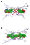
Three representative complexes of anti-amyloyd molecules with their respective targets: (A) TTR binding sites in the presence of tolcapone, with ligand shown as solvent accessible surface. For clarity, H2O oxygen atoms are shown as spheres with 50% of the van der Waals radius (PDB code 4D7B); (B) TTR binding sites in the presence of mds84, with ligand shown as solvent accessible surface as above (PDB code 3IPE); (C) crenezumab Fab in complex with Aβ, with backbones of the interactors coloured in red and green, respectively. Carbon atoms of selected side chains in the Fab are coloured in grey, whereas those belonging to Aβ are green (O, red; N, blue; S, yellow; PDB code: 5VZY). The pictorial representations in panels (A), (B) were taken from Ref. 134 and licensed under a Creative Commons Attribution 4.0. The image in panel (C) was created by means of PyMOL (v. 0.98), using the set of atomic coordinates available in the Protein Data Bank.
Figure 3
Figure 3
Three representative complexes of anti-amyloyd molecules with their respective targets: (A) TTR binding sites in the presence of tolcapone, with ligand shown as solvent accessible surface. For clarity, H2O oxygen atoms are shown as spheres with 50% of the van der Waals radius (PDB code 4D7B); (B) TTR binding sites in the presence of mds84, with ligand shown as solvent accessible surface as above (PDB code 3IPE); (C) crenezumab Fab in complex with Aβ, with backbones of the interactors coloured in red and green, respectively. Carbon atoms of selected side chains in the Fab are coloured in grey, whereas those belonging to Aβ are green (O, red; N, blue; S, yellow; PDB code: 5VZY). The pictorial representations in panels (A), (B) were taken from Ref. 134 and licensed under a Creative Commons Attribution 4.0. The image in panel (C) was created by means of PyMOL (v. 0.98), using the set of atomic coordinates available in the Protein Data Bank.

References

    1. Dobson C.M. Protein folding and misfolding. Nature. 2003;426:884–890. doi: 10.1038/nature02261. - DOI - PubMed
    1. Hartl F.U. Protein misfolding diseases. Annu. Rev. Biochem. 2017;86:21–26. doi: 10.1146/annurev-biochem-061516-044518. - DOI - PubMed
    1. Chiti F., Dobson C.M. Protein misfolding, functional amyloid, and human disease. Annu. Rev. Biochem. 2006;75:333–366. doi: 10.1146/annurev.biochem.75.101304.123901. - DOI - PubMed
    1. Smith J.F., Knowles T.P., Dobson C.M., MacPhee C.E., Welland M.E. Characterization of the nanoscale properties of individual amyloid fibrils. Proc. Natl. Acad. Sci. USA. 2006;103:15806–15811. doi: 10.1073/pnas.0604035103. - DOI - PMC - PubMed
    1. Morel B., Varela L., Conejero-Lara F. The thermodynamic stability of amyloid fibrils studied by differential scanning calorimetry. J. Phys. Chem. B. 2010;114:4010–4019. doi: 10.1021/jp9102993. - DOI - PubMed