. 2018 Nov;178(3):1154-1169.
doi: 10.1104/pp.18.00329. Epub 2018 Sep 11.

Arabidopsis PIRL6 Is Essential for Male and Female Gametogenesis and Is Regulated by Alternative Splicing

Affiliations

Arabidopsis PIRL6 Is Essential for Male and Female Gametogenesis and Is Regulated by Alternative Splicing

Nancy R Forsthoefel et al. Plant Physiol. 2018 Nov.

Abstract

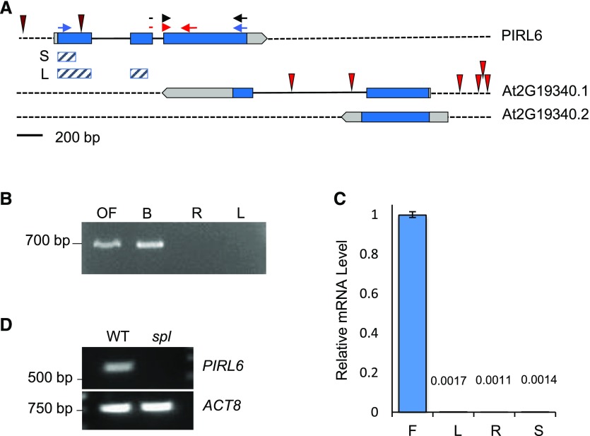
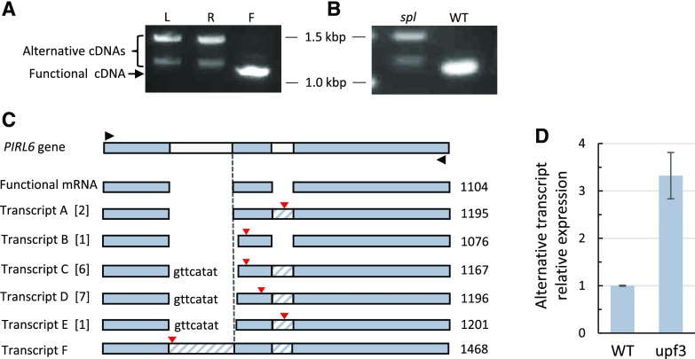
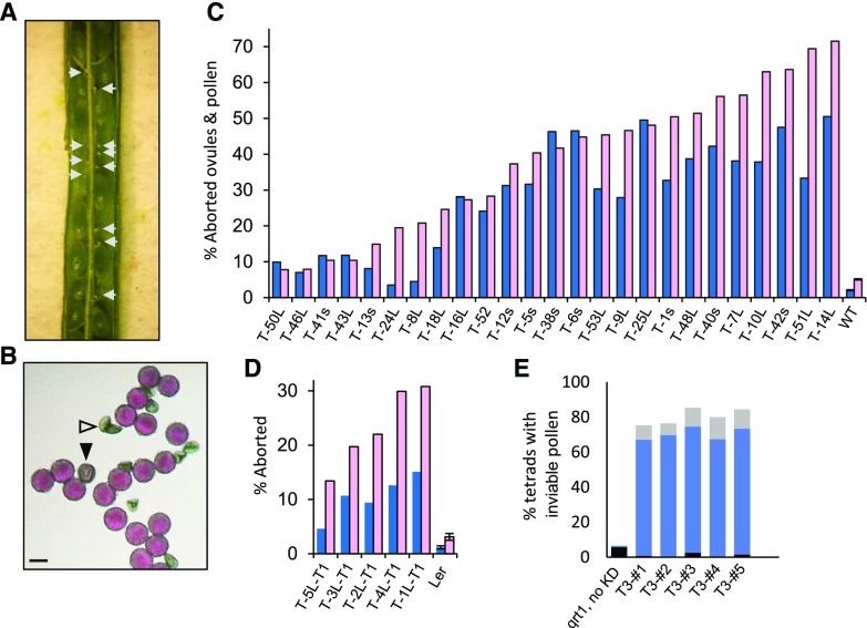
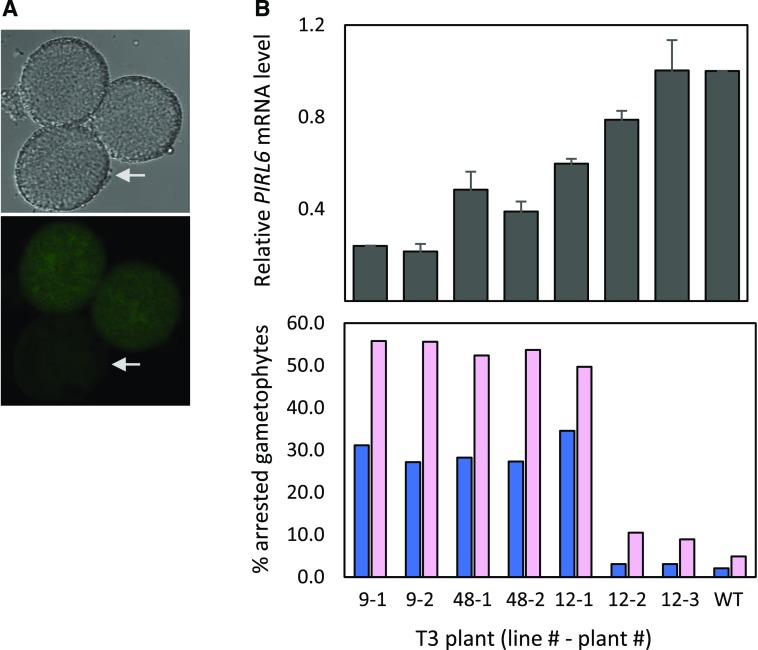
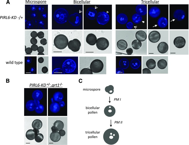
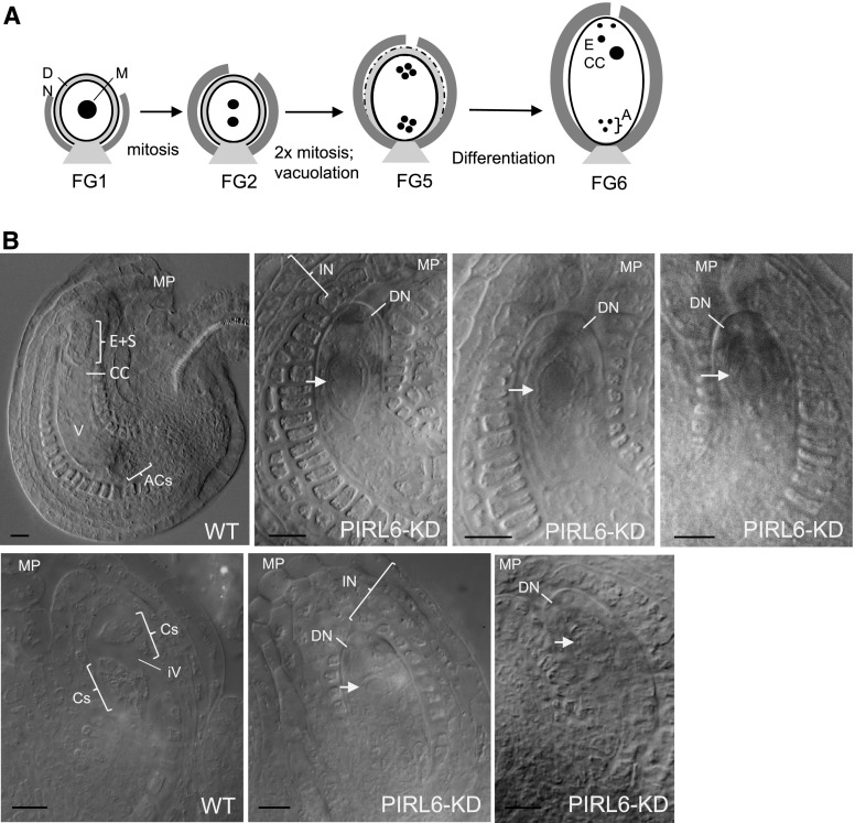
Plant intracellular Ras-group leucine-rich repeat (LRR) proteins (PIRLs) are related to Ras-interacting animal LRR proteins that participate in developmental cell signaling. Systematic knockout analysis has implicated some members of the Arabidopsis (Arabidopsis thaliana) PIRL family in pollen development. However, for PIRL6, no bona fide knockout alleles have been recovered, suggesting that it may have an essential function in both male and female gametophytes. To test this hypothesis, we investigated PIRL6 expression and induced knockdown by RNA interference. Knockdown triggered defects in gametogenesis, resulting in abnormal pollen and early developmental arrest in the embryo sac. Consistent with this, PIRL6 was expressed in gametophytes: functional transcripts were detected in wild-type flowers but not in sporocyteless (spl) mutant flowers, which do not produce gametophytes. A genomic PIRL6-GFP fusion construct confirmed expression in both pollen and the embryo sac. Interestingly, PIRL6 is part of a convergent overlapping gene pair, a scenario associated with an increased likelihood of alternative splicing. We detected multiple alternative PIRL6 mRNAs in vegetative organs and spl mutant flowers, tissues that lacked the functionally spliced transcript. cDNA sequencing revealed that all contained intron sequences and premature termination codons. These alternative mRNAs accumulated in the nonsense-mediated decay mutant upf3, indicating that they are normally subjected to degradation. Together, these results demonstrate that PIRL6 is required in both male and female gametogenesis and suggest that sporophytic expression is negatively regulated by unproductive alternative splicing. This posttranscriptional mechanism may function to minimize PIRL6 protein expression in sporophyte tissues while allowing the overlapping adjacent gene to remain widely transcribed.


Figures

Figure 1.
Figure 1.
PIRL6 gene structure and mRNA expression. A, PIRL6 (At2g19330) overlaps with the neighboring gene At2g19340, which generates two transcripts (0.1 and 0.2) with different polyadenylation sites (www.arabidopsis.org). Blue, Translated regions; light gray, untranslated regions; solid black lines, introns. T-DNA insertion sites in both loci are indicated by red spikes; neither PIRL6 insertion is a bona fide knockout (Forsthoefel et al., 2013). Small arrows indicate primer positions for PCR experiments shown in subsequent figures. Red arrows represent the primer pair used for qPCR; the forward primer straddles the exon II-III splice junction. Blue arrows represent primers used in full-length RT-PCR to detect the alternatively spliced cDNA species shown in Figure 4. Black arrows represent the primer pair used for RT-PCR experiments shown in Figure 2, A and C. Cross-hatched bars indicate exon regions included in short (S) and long (L) inverted repeat constructs for RNAi knockdown. B, PIRL6 RT-PCR carried out using a forward primer specific to the exon II-III splice junction, with RNA from open flowers (OF), developing inflorescences (buds; B), roots (R), and rosette leaves (L). C, PIRL6 RT-qPCR carried out on RNA from inflorescence (F), leaf (L), root (R), and germinated seedling (S) using the splice junction-specific primer used in B. Values are means from three replicate reactions; means for leaf, root, and seedling samples are provided above the bars. se is shown. D, RT-PCR of PIRL6 in flowers from wild-type (WT) and sterile spl homozygotes, which do not produce gametophytes. ACTIN8 (ACT8) was included as a positive control.
Figure 2.
Figure 2.
Expression of PIRL6-GFP in both male and female gametophytes. Gametophytes segregating for a full-length PIRL6-GFP fusion construct were viewed at the indicated developmental stages by confocal and differential interference contrast (DIC) microscopy. Plants were hemizygous for the reporter construct, producing pollen and embryo sacs that segregated 1:1 for the PIRL6-GFP construct; for each developmental stage shown above, pollen and ovules were from the same anthers and ovaries, respectively. A, Expression in bicellular and tricellular stage pollen; the right-most image is a single image illustrating the meiotic segregation of the reporter construct in pollen from a hemizygous anther. B, Expression during female gametophyte development. Triangles mark locations of PIRL-GFP within the embryo sac. Female gametophyte (FG) developmental stages were defined by Christensen et al. (1998). To provide a reference point, the micropylar end of each ovule is labeled on the DIC panels (MP). The FG6 stage image is a single image illustrating the segregation of the reporter construct in adjacent ovules. Bars = 10 μm.
Figure 3.
Figure 3.
PIRL6 undergoes unproductive alternative splicing outside of gametophytes. PIRL6 transcripts were amplified from polyadenylated RNA from wild-type and spl mutant homozygotes using primers designed to amplify the full-length coding region. A, RT-PCR products from wild-type leaves (L), roots (R), and flowers (F). Sequencing confirmed that the predominant flower cDNA was derived from the translatable, spliced PIRL6 mRNA. Larger alternative products are evident in leaves and roots; the largest band corresponds in size to the unspliced transcript (1,468 bp). B, RT-PCR of PIRL6 transcripts from wild-type (WT) and sterile spl mutant flowers, showing the predominance of alternative transcripts in flowers that lack gametophytes. To detect transcripts present in spl, the spl lane was loaded with 10× the volume of the reaction product than the wild-type lane. C, Structures of alternative polyadenylated PIRL6 transcripts sequenced from roots and leaves. Gel-resolved leaf and root RT-PCR products between approximately 1.1 and 1.4 kb were cloned in Escherichia coli, and 17 independent cDNAs were sequenced. Black triangles indicate primer positions used for PCR following cDNA synthesis primed with oligo(dT). Functional mRNA represents the flower RT-PCR product, corresponding to the annotated spliced PIRL6 mRNA. Transcripts A to F are alternative mRNA species. Numbers in brackets indicate the number of independent cDNA clones obtained for each transcript species. Lowercase letters indicate residual unspliced nucleotides from intron I; cross-hatched regions indicate unspliced introns. Red triangles mark the positions of premature termination codons. The dashed line marks the position of the normal 5′ end of exon II. The sizes of RT-PCR products corresponding to each mRNA species are indicated in bp at far right. D, qPCR showing increased accumulation of PIRL6 alternative transcripts in the NMD-deficient mutant upf3. qPCR was carried out on leaf RNA using an intron II-specific forward primer, allowing the detection of alternative transcripts A, C, D, E, and F. Values are means of two biological replicates, with three reaction replicates for each; se is shown.
Figure 4.
Figure 4.
Aborted ovules and pollen resulting from PIRL6 knockdown. PIRL6 inverted repeat constructs were introduced into wild-type WS Arabidopsis by A. tumefaciens-mediated transformation; transgenic T1 plants harboring the RNAi construct were identified by glufosinate resistance and confirmed by genomic PCR. T1 plants were hemizygous and were scored directly for segregating ovule and pollen defects. A, A silique from a T1 plant (T-48L-T1) containing seeds from successfully fertilized ovules segregating with aborted ovules (white arrows). B, Alexander-stained pollen produced by T1 plant T-48L-T1, showing the segregation of aborted (white triangle) and stunted (black triangle) grains. Bar = 20 µm. C, Percentages of abnormal or aborted pollen (blue) and aborted ovules (pink) in 25 T1 plants independently transformed with PIRL6 RNAi constructs and in wild-type controls (WT). The ranges of n values are as follows: pollen, 61 to 1,468; ovules, 105 to 191. L or s in plant line labels indicates the PIRL6-RNAi construct introduced in that line (see text). D, Replication of the RNAi-induced phenotype in the Ler ecotype. Percentages are shown for abnormal or aborted pollen (blue) and aborted ovules (pink) in five Ler T1 plants independently transformed with the PIRL6-RNAi(L) construct. The n value ranges are as follows: pollen, 352 to 1,124; ovules, 142 to 178. In C and D, means and se are shown only for untransformed controls; se was not determined for T1 samples because values were obtained, by definition, from individual transformed plants. E, Gametophytic basis of PIRL6-KD pollen defects, shown by meiotic segregation in pollen tetrads produced by five individual PIRL6-KD hemizygotes in a qrt1 background. Black bars, percentage of tetrads with one dead pollen grain; solid blue bars, percentage of tetrads with two dead pollen grains; light gray bars, percentage of tetrads with more than two dead pollen grains (the range of n values is as follows: 89–198 tetrads per plant).
Figure 5.
Figure 5.
Reduction of PIRL6 mRNA levels by PIRL6-KD. A, Reduced PIRL6-GFP expression in pollen produced by a plant homozygous for the genomic PIRL6-GFP reporter construct and hemizygous for the PIRL6-KD(L) construct, such that pollen all contained the GFP reporter but were segregating for the knockdown construct. Mature pollen were viewed by DIC (top) and confocal fluorescence (bottom) microscopy. Arrows indicate pollen with visibly reduced PIRL6-GFP signal. B, Relationship between PIRL6 mRNA expression and phenotype severity in wild-type controls (WT) and individual PIRL6-KD plants from three independent transgenic lines. Top graph, PIRL6 RT-qPCR carried out on developing flowers from individual T3 plants from three independent transgenic lines. Plants were homozygous for the PIRL6-KD construct; means and se from three replicate RT-qPCRs are shown. Bottom graph, Phenotype severity in the same individual T3 plants, gauged by the percentage of inviable pollen (dark blue) or aborted ovules (pink). The range of n values is as follows: 124 to 1,773 (pollen); 87 to 192 (ovules)
Figure 6.
Figure 6.
PIRL6 knockdown disrupts male gametogenesis. Microspores and pollen produced by plants hemizygous for the PIRL6-KD construct, which segregate at 50% for PIRL6 knockdown, were stained with DAPI and viewed by confocal fluorescence or DIC microscopy. A, Microspores and pollen from anthers at the indicated developmental stages, viewed by confocal fluorescence (top row) or DIC (middle row). Arrows indicate pollen segregating for developmental defects of varying severity, including arrested microspores (broad arrows), enlargement with absence of germinal nucleus (hollow triangle), arrest after PM1 (short arrow), or abnormal male germ unit configuration (long arrows). The bottom row shows wild-type control samples at corresponding developmental stages. Bars = 10 µm. B, Tetrads from tricellular stage anthers, showing meiotic segregation of developmentally arrested PIRL6-KD pollen in the qrt1 background. Bars = 10 µm. C, Simplified diagram of the major stages of wild-type Arabidopsis pollen development, provided for reference. PM, Pollen mitoses. White ovals represent idealized nuclear configurations observable with DAPI staining: large ovals, vegetative cell nuclei; small ovals, generative nucleus (bicellular stage) and sperm nuclei (tricellular stage). The tricellular stage nuclei illustrate the triangular configuration of the male germ unit characteristic of mature Arabidopsis pollen.
Figure 7.
Figure 7.
PIRL6 knockdown disrupts female gametogenesis. A, Simplified diagram summarizing selected aspects of Arabidopsis female gametophyte development at the indicated developmental stages (Christensen et al., 1998), pictured with the micropyle at top and the chalazal end at bottom. The embryo sac is represented as a white oval and constituent nuclei as black circles. Adjacent sporophytic tissues are included: light gray, nucellus; dark gray, inner integument. A, Antipodal cell nuclei in their characteristic triangular configuration; CC, central cell nucleus; DN, distal nucellus region; M, megaspore nucleus. B, Ovules from ovaries at postmitotic developmental stages, cleared as whole mounts in Hoyer’s solution and viewed by DIC microscopy. The micropylar region (MP) of each ovule is labeled to indicate the orientation of the image. Parent plants were hemizygous for the PIRL6-KD construct, and embryo sacs segregated approximately 50% for PIRL6 knockdown. The top row shows a wild-type ovule (WT) and three examples of segregating PIRL6-KD ovules from FG6 stage ovaries. Antipodal cells (ACs), a large vacuole (V), the central cell (CC), and the cluster of egg cell and synergids (E+S) were discernible in segregating wild-type ovules (nuclei are not visible in the selected focal plane). PIRL6-KD embryo sacs each feature single, apparently mitotically arrested cells (white arrows) with large nuclei, surrounded by an abnormally persistent, sharply defined distal nucellus layer (DN). Integument (IN) layers fully envelope the PIRL6-KD gametophytes, despite their developmental arrest as single cells (in contrast to the integuments flanking the wild-type single-cell FG1 ovule shown in Fig. 2B). The bottom row shows wild-type and PIRL6-KD ovules from FG5 stage ovaries. Yet-undifferentiated cells produced by mitotic divisions (Cs) and an initiating central vacuole (iV) are discernible in the FG5 wild-type ovule. PIRL6-KD embryo sacs resembled those observed in PIRL6-KD FG6 ovaries, with single, arrested cells (white arrows) enveloped by a persistent distal nucleus (DN) and expanded integuments (IN) that resembled those of wild-type ovules. Bars = 10 µm.

Similar articles

Cited by

References

    1. Arciga-Reyes L, Wootton L, Kieffer M, Davies B (2006) UPF1 is required for nonsense-mediated mRNA decay (NMD) and RNAi in Arabidopsis. Plant J 47: 480–489 - PubMed
    1. Bencivenga S, Colombo L, Masiero S (2011) Cross talk between the sporophyte and the megagametophyte during ovule development. Sex Plant Reprod 24: 113–121 - PubMed
    1. Berg M, Rogers R, Muralla R, Meinke D (2005) Requirement of aminoacyl-tRNA synthetases for gametogenesis and embryo development in Arabidopsis. Plant J 44: 866–878 - PubMed
    1. Berger F, Twell D (2011) Germline specification and function in plants. Annu Rev Plant Biol 62: 461–484 - PubMed
    1. Boavida LC, Shuai B, Yu HJ, Pagnussat GC, Sundaresan V, McCormick S (2009) A collection of Ds insertional mutants associated with defects in male gametophyte development and function in Arabidopsis thaliana. Genetics 181: 1369–1385 - PMC - PubMed

Publication types

MeSH terms

LinkOut - more resources