

Sprague Dawley Rag2-Null Rats Created from Engineered Spermatogonial Stem Cells Are Immunodeficient and Permissive to Human Xenografts
- PMID: 30206106
- PMCID: PMC6215516
- DOI: 10.1158/1535-7163.MCT-18-0156
Sprague Dawley Rag2-Null Rats Created from Engineered Spermatogonial Stem Cells Are Immunodeficient and Permissive to Human Xenografts
Abstract
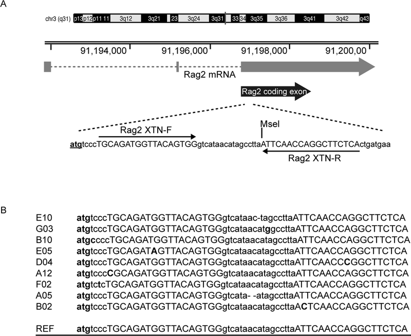
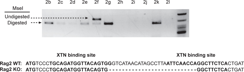
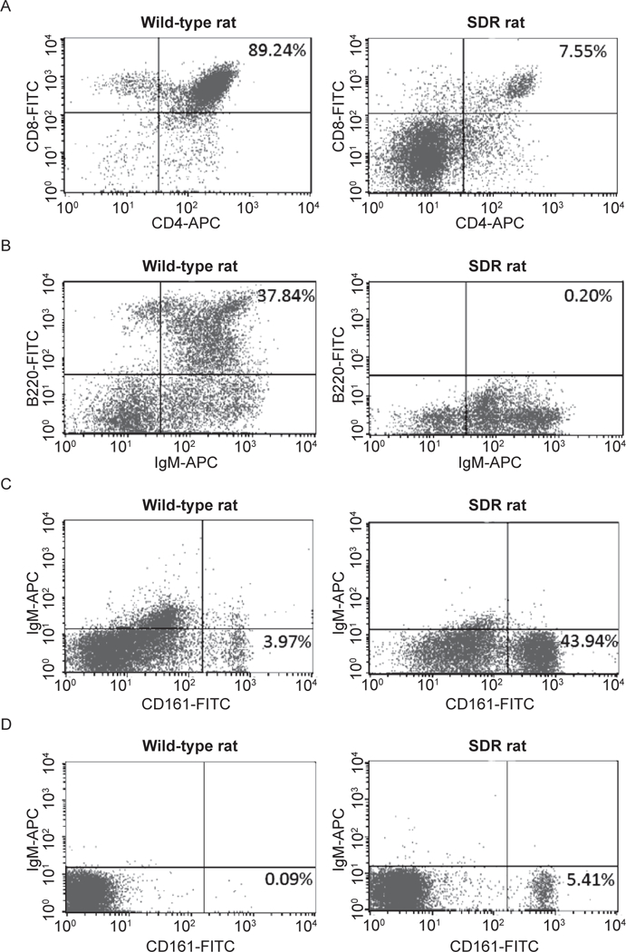
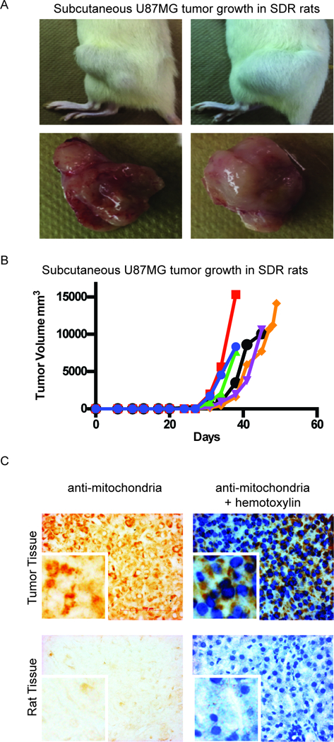
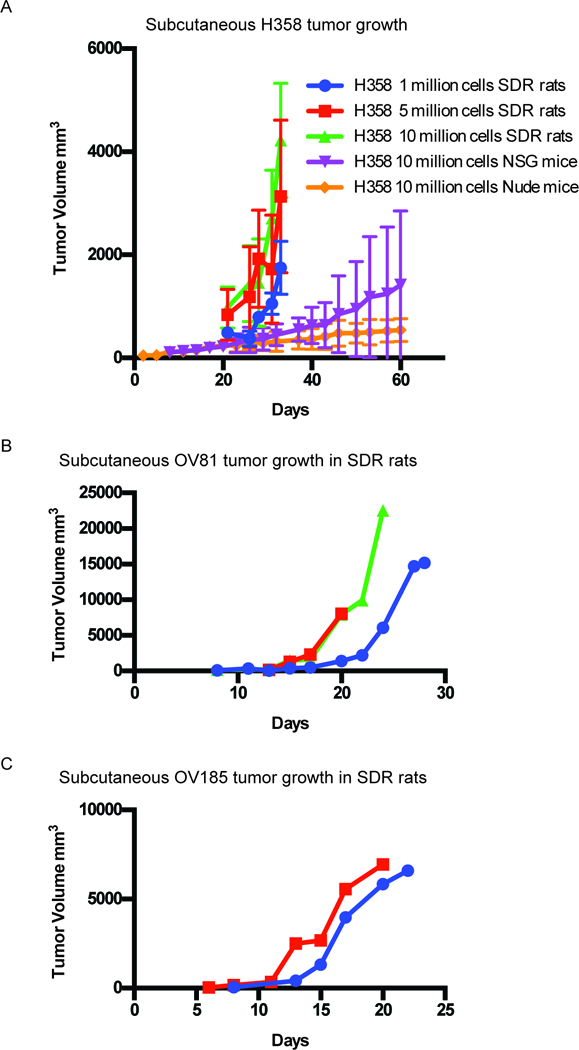
The rat is the preferred model for toxicology studies, and it offers distinctive advantages over the mouse as a preclinical research model including larger sample size collection, lower rates of drug clearance, and relative ease of surgical manipulation. An immunodeficient rat would allow for larger tumor size development, prolonged dosing and drug efficacy studies, and preliminary toxicologic testing and pharmacokinetic/pharmacodynamic studies in the same model animal. Here, we created an immunodeficient rat with a functional deletion of the Recombination Activating Gene 2 (Rag2) gene, using genetically modified spermatogonial stem cells (SSC). We targeted the Rag2 gene in rat SSCs with TALENs and transplanted these Rag2-deficient SSCs into sterile recipients. Offspring were genotyped, and a founder with a 27 bp deletion mutation was identified and bred to homozygosity to produce the Sprague-Dawley Rag2 - Rag2 tm1Hera (SDR) knockout rat. We demonstrated that SDR rat lacks mature B and T cells. Furthermore, the SDR rat model was permissive to growth of human glioblastoma cell line subcutaneously resulting in successful growth of tumors. In addition, a human KRAS-mutant non-small cell lung cancer cell line (H358), a patient-derived high-grade serous ovarian cancer cell line (OV81), and a patient-derived recurrent endometrial cancer cell line (OV185) were transplanted subcutaneously to test the ability of the SDR rat to accommodate human xenografts from multiple tissue types. All human cancer cell lines showed efficient tumor uptake and growth kinetics indicating that the SDR rat is a viable host for a range of xenograft studies. Mol Cancer Ther; 17(11); 2481-9. ©2018 AACR.
©2018 American Association for Cancer Research.
Conflict of interest statement
Conflicts of interest
None.
Figures
Similar articles
-
The SRG rat, a Sprague-Dawley Rag2/Il2rg double-knockout validated for human tumor oncology studies.PLoS One. 2020 Oct 7;15(10):e0240169. doi: 10.1371/journal.pone.0240169. eCollection 2020. PLoS One. 2020. PMID: 33027304 Free PMC article.
-
A novel immunodeficient rat model supports human lung cancer xenografts.FASEB J. 2019 Jan;33(1):140-150. doi: 10.1096/fj.201800102RR. Epub 2018 Jun 26. FASEB J. 2019. PMID: 29944447
-
Bioluminescent imaging of vaccinia virus infection in immunocompetent and immunodeficient rats as a model for human smallpox.Sci Rep. 2015 Aug 3;5:11397. doi: 10.1038/srep11397. Sci Rep. 2015. PMID: 26235050 Free PMC article.
-
Advances in Isolation Methods for Spermatogonial Stem Cells.Stem Cell Rev Rep. 2016 Feb;12(1):15-25. doi: 10.1007/s12015-015-9632-6. Stem Cell Rev Rep. 2016. PMID: 26498644 Review.
-
The nature and dynamics of spermatogonial stem cells.Development. 2017 Sep 1;144(17):3022-3030. doi: 10.1242/dev.146571. Development. 2017. PMID: 28851723 Review.
Cited by
-
Generation of RAG2 Knockout Immune-Deficient Miniature Pigs.Animals (Basel). 2024 Sep 6;14(17):2597. doi: 10.3390/ani14172597. Animals (Basel). 2024. PMID: 39272382 Free PMC article.
-
The SRG rat, a Sprague-Dawley Rag2/Il2rg double-knockout validated for human tumor oncology studies.PLoS One. 2020 Oct 7;15(10):e0240169. doi: 10.1371/journal.pone.0240169. eCollection 2020. PLoS One. 2020. PMID: 33027304 Free PMC article.
-
Advances in Genome Editing and Application to the Generation of Genetically Modified Rat Models.Front Genet. 2021 Apr 20;12:615491. doi: 10.3389/fgene.2021.615491. eCollection 2021. Front Genet. 2021. PMID: 33959146 Free PMC article. Review.
-
In Vivo Analysis of Human Immune Responses in Immunodeficient Rats.Transplantation. 2020 Apr;104(4):715-723. doi: 10.1097/TP.0000000000003047. Transplantation. 2020. PMID: 31764762 Free PMC article.
-
Targeting Ribonucleotide Reductase Induces Synthetic Lethality in PP2A-Deficient Uterine Serous Carcinoma.Cancer Res. 2022 Feb 15;82(4):721-733. doi: 10.1158/0008-5472.CAN-21-1987. Cancer Res. 2022. PMID: 34921012 Free PMC article.
References
-
- Anderson S, Luffer-Atlas D, Knadler MP. Predicting circulating human metabolites: how good are we? Chem Res Toxicol. 2009;22(2):243–56. - PubMed
-
- Leclercq L, Cuyckens F, Mannens GS, de Vries R, Timmerman P, Evans DC. Which human metabolites have we MIST? Retrospective analysis, practical aspects, and perspectives for metabolite identification and quantification in pharmaceutical development. Chem Res Toxicol. 2009;22(2):280–93. - PubMed
-
- Walker D, Brady J, Dalvie D, Davis J, Dowty M, Duncan JN, et al. A holistic strategy for characterizing the safety of metabolites through drug discovery and development. Chem Res Toxicol. 2009;22(10):1653–62. - PubMed
Publication types
MeSH terms
Substances
Grants and funding
LinkOut - more resources
Full Text Sources
Other Literature Sources
Medical
Miscellaneous
