. 2018 Oct 26;293(43):16709-16723.
doi: 10.1074/jbc.RA117.000733. Epub 2018 Sep 11.

Structural analyses of 4-phosphate adaptor protein 2 yield mechanistic insights into sphingolipid recognition by the glycolipid transfer protein family

Affiliations

Structural analyses of 4-phosphate adaptor protein 2 yield mechanistic insights into sphingolipid recognition by the glycolipid transfer protein family

Borja Ochoa-Lizarralde et al. J Biol Chem. .

Abstract

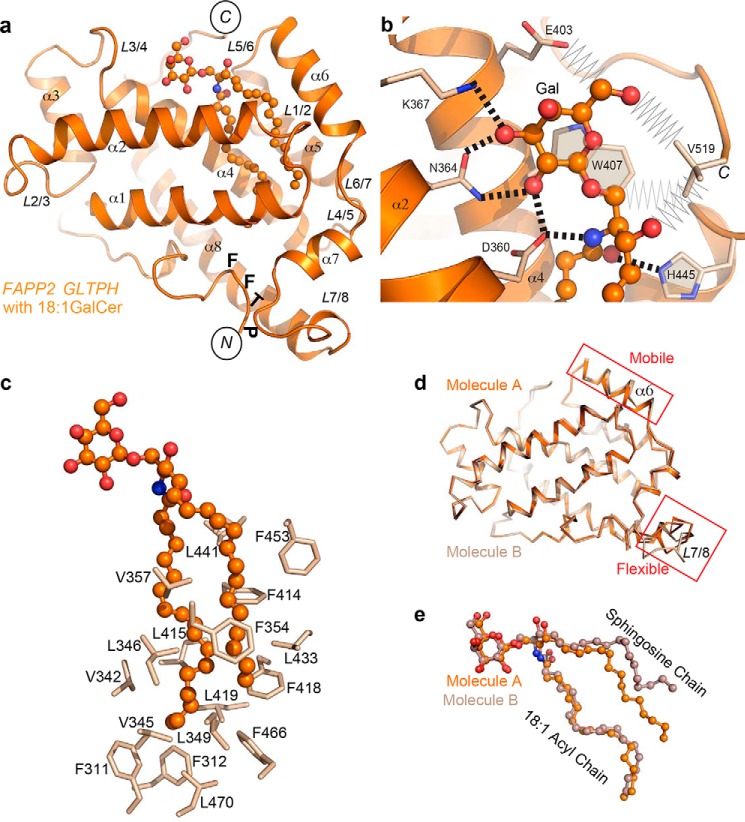
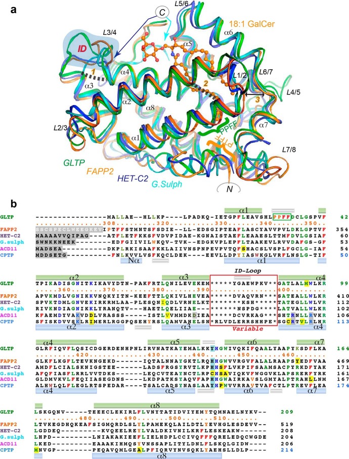
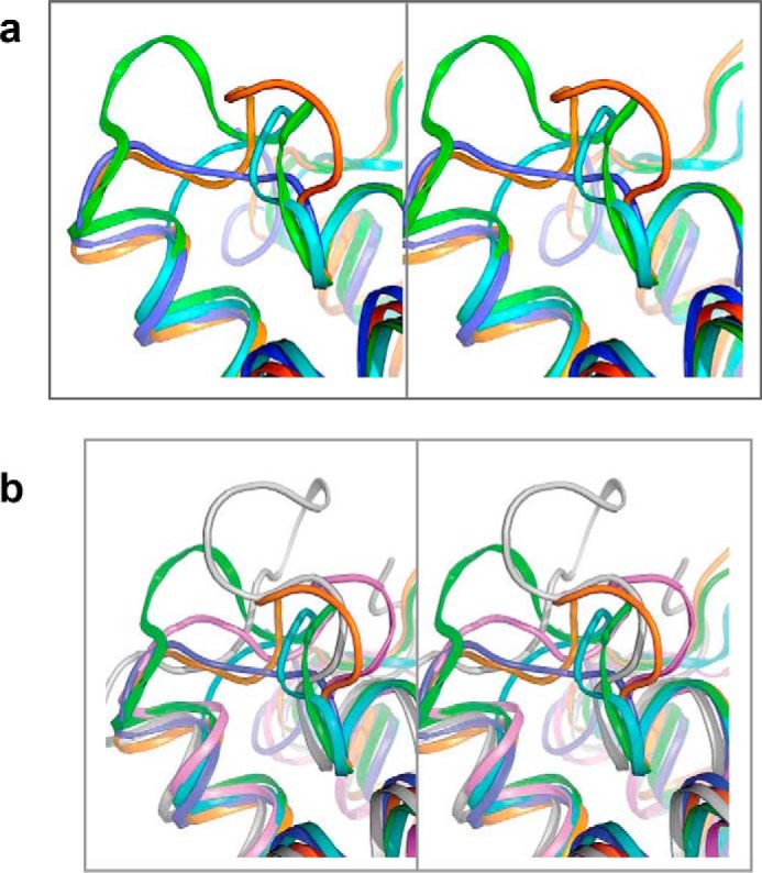
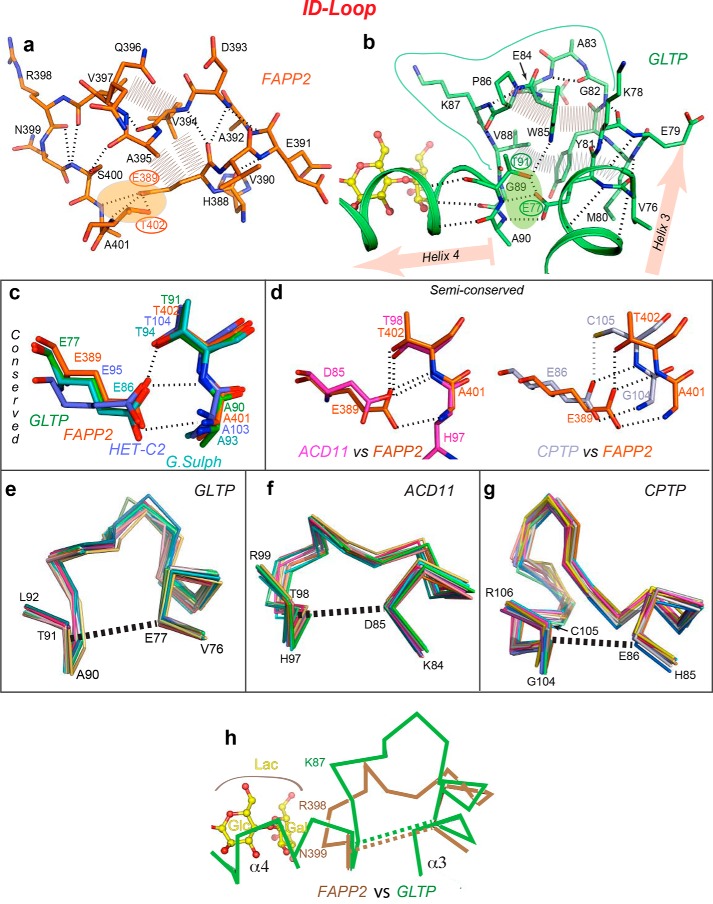
The glycolipid transfer protein (GLTP) fold defines a superfamily of eukaryotic proteins that selectively transport sphingolipids (SLs) between membranes. However, the mechanisms determining the protein selectivity for specific glycosphingolipids (GSLs) are unclear. Here, we report the crystal structure of the GLTP homology (GLTPH) domain of human 4-phosphate adaptor protein 2 (FAPP2) bound with N-oleoyl-galactosylceramide. Using this domain, FAPP2 transports glucosylceramide from its cis-Golgi synthesis site to the trans-Golgi for conversion into complex GSLs. The FAPP2-GLTPH structure revealed an element, termed the ID loop, that controls specificity in the GLTP family. We found that, in accordance with FAPP2 preference for simple GSLs, the ID loop protrudes from behind the SL headgroup-recognition center to mitigate binding by complex GSLs. Mutational analyses including GLTP and FAPP2 chimeras with swapped ID loops supported the proposed restrictive role of the FAPP2 ID loop in GSL selectivity. Comparative analysis revealed distinctly designed ID loops in each GLTP family member. This analysis also disclosed a conserved H-bond triplet that "clasps" both ID-loop ends together to promote structural autonomy and rigidity. The findings indicated that various ID loops work in concert with conserved recognition centers to create different specificities among family members. We also observed four bulky, conserved hydrophobic residues involved in "sensor-like" interactions with lipid chains in protein hydrophobic pockets and FF motifs in GLTP and FAPP2, well-positioned to provide acyl chain-dependent SL selectivity for the hydrophobic pockets. In summary, our study provides mechanistic insights into sphingolipid recognition by the GLTP fold and uncovers the elements involved in this recognition.

Keywords: 4-phosphate-adaptor-protein-2; FAPP2; X-ray crystallography; lipid-protein interaction; nonvesicular lipid transport; protein complex; protein family; protein structure; sphingolipid; transport.


Conflict of interest statement

The authors declare that they have no conflicts of interest with the contents of this article

Figures

Figure 1.
Figure 1.
Crystal structure of human FAPP2–GLTPH domain complexed with GSL, 18:1-GalCer. a, overall view of the complex showing labeled helices, loops, N and C termini, and PTFF sequence containing FF motif. b, protein recognition center interactions with galactose ring and ceramide amide moiety of bound GalCer, depicting hydrogen bonds (dashed lines) and the aromatic ring stacking over the glucose ring. Also depicted are van der Waals contacts (zig-zag lines) of the nonconserved residues Glu403 and C-terminal Val519. c, Phe, Leu, and Val residues lining the hydrophobic pocket that envelopes the lipid chains. d and e, superposition of two molecules of the complex occupying the asymmetric unit, showing either protein (d) or GSL (e). Molecules A and B are depicted in different shades of colors. In a–e, atoms are colored orange, red, and blue for carbon, oxygen, and nitrogen, respectively.
Figure 2.
Figure 2.
Comparison of FAPP2–GLTPH domain with other family members. a, structure superimposition of the GSL-specific proteins: human FAPP2–GLTPH domain (orange), human GLTP (green), fungal heterokaryon incompatibility C2 protein, HET-C2, of P. anserina (blue), and GLTP-like protein from the thermoacidophilic unicellular red alga, G. sulfuraria (cyan) (PDB codes 5KDI, 3S0K, 4KV0, and 2I3F, respectively). 18:1-GalCer is shown as bound to FAPP2–GLTPH. The blue-shaded area highlights the ID loops. The GLTP PPFF and FAPP2 PTFF sequences are indicated by green and orange braces, respectively. Newly identified conserved linkages 1 and 2 are indicated by dashed lines; the double-headed arrow 3 points to the strictly conserved Gly and Pro on opposite sides of the gate controlling the access to the hydrophobic pocket in the GLTP fold. Colored arrows point to the C-ends of HET-C2 (blue) and G. sulfuraria protein (cyan). Color codes for GSL atoms are defined in Fig. 1. b, poststructural sequence alignment of six proteins belonging to the GLTP family (see Fig. S1a). Color codes are blue for the recognition center residues, green for the conserved/semiconserved residues, and red and orange for Phe and similarly positioned Tyr/Trp, respectively. Recognition center residues of the C1P-specific family also are blue but shaded in gray when different from the GSL-specific family. Exceptions among conserved residues are shaded by yellow. The upper and lower cylinders (small silver for 310-helices; other for α-helices) indicate the locations of secondary structure elements found in GLTP and CPTP, respectively. Within the red rectangle are the ID-loop sequences that vary among each member.
Figure 3.
Figure 3.
Comparative stereo view of the ID loops. a, based on Fig. 2a. b, based on Fig. S1a.
Figure 4.
Figure 4.
Structural properties of the ID loop. a and b, intraloop interactions in human FAPP2–GLTPH (a) and human GLTP (b), depicting main-chain hydrogen bonds (dashed lines), side-chain van der Waals contacts (zig-zag lines), and orientation of flanking helices α3 and α4 (highlighted in b by pink arrows). Clasps formed by H-bond triplets that fasten the two ends of the ID loop to each other are shaded orange in a and green in b. c and d, the H-bond triplet interaction pattern in the GSL-specific family (c) and C1P-specific family (d) derived from global superposition of GLTP superfamily members in Fig. S1a. Two semiconserved patterns are compared with the H-bond triplet in FAPP2–GLTPH. e–g, ID-loop Cα backbones from superimposed multiple molecular structures of GLTP (e), ACD11 (f), and CPTP (g) showing the relative conformational rigidity of the ID loop in different GLTP superfamily members. Superimposed are 20, 12, and 15 molecular structures of GLTP, ACD11, and CPTP, respectively, with dashed lines indicating the approximate position of the H-bond triplets and the helical regions (of α3 and α4) adjacent the H-bond triplet ends. h, expected proximity differences in the location of bound LacCer headgroup (yellow) with respect to the ID-loop backbones in FAPP2 (beige) and GLTP (green).
Figure 5.
Figure 5.
FAPP2 ID-loop regulation of GSL selectivity. a–c, electrostatics surface (blue, positive; red, negative; gray, neutral) of the FAPP2–GLTPH domain showing the ID loop, C terminus, and recognition center occupied by GalCer in the current crystal structure (a), compared with the partially impaired locations expected for LacCer (b), or sulfatide, SF (c), assessed by superposition with crystal structures of GLTP bound to LacCer (PDB code 1SX6) or 3-O-SF (PDB codes 3RZN and 4H2Z). GSL molecules are shown in stick representation, and GSL atoms are shown by spheres of van der Waals radii. GSL-atom color codes: blue for nitrogen; red for oxygen; and orange, green, and cyan for carbon in a, b, and c, respectively. d–f, protein·GSL interactions in the complexes shown in a–c, respectively, depicting hydrogen bonds (dashed lines) and van der Waals “clashes” (zig-zag lines). GSL atom color codes as in a–c. Lys367 and Asn399 conformations in b and e and Lys367 and Asn364 conformations in c and f were adapted to favorably interact with lipid headgroup and neighbor residues. For details, see text. g, FAPP2–GLTPH transfer activities for GalCer (orange), GlcCer (burgundy), acCer (green), and SF (cyan) by WT-GLTPH and E403L-GLTPH or K367S-GLTPH measured by FRET assay using AV lipids (see “Experimental procedures”).
Figure 6.
Figure 6.
SPR sensorgrams of the interaction between lipid vesicles captured on a L1 chip and the engineered protein chimeras, IDFAPP2GLTP and IDGLTPFAPP2, versus GLTP and FAPP2–GLTPH. IDFAPP2 and IDGLTP are the ID loops of FAPP2 and GLTP, respectively. Vesicles are composed of POPC, 1-palmitoyl-2-oleoyl-sn-phosphatidylcholine, mixed with ganglioside GM1 (a–d) or 24:1 sulfatide, SF (e–h) in mol% ratios 100:0 (black), 95:5 (red), 90:10 (green), or 80:20 (blue). Proteins are dissolved at 0.1 mg/ml (5 μm) in running buffer (50 mm Tris-HCl, 150 mm NaCl, 1 mm EDTA, 1 mm DTT, pH 7.0). The flow rate is 5 μl/min. Red and black arrows indicate the start of protein injections and switching back to the buffer wash, respectively.
Figure 7.
Figure 7.
Adaptable clasp and other conserved elements involved in gate action. a, six superimposed molecular structures of human GLTP (PDB codes 1SX6, yellow; 1SWX, pink; 2EUK, cyan; 3RWA(a), red; 3RWA(b), blue; 3S0K, green) showing different conformations of one side of the gate, loop L1/2, adopted by the same protein. The dashed line indicates the approximate location of the conserved clasp for gate action. b, two opposing sides of the FAPP2–GLTPH gate depicting the strictly conserved Gly and Pro (outlined by ellipses), two conserved bulky-hydrophobic residues (outlined by hexagons), and the Lys358 and Asp347 residues (outlined by rectangles) of the conserved clasp for gate action. c, clasp connecting two ends of L1/2 loop of the gate and formed by the Asp347·Lys358 salt bridge (dashed lines) in both FAPP2–GLTPH molecules (different shades of orange) compared with human GLTP (different shades of green; PDB codes 1S0K and 1SWX) and HET-C2 (blue; PDB code 3KV0) illustrates bridge mobility and conservation in the GSL-specific family. d, substitution of the salt-bridged Asp·Lys pair in GSL-specific FAPP2–GLTPH for the H-bonded Asn·Ser pair in C1P-specific CPTP (PDB code 4K84) highlighting the preservation of the clasp (see Fig. S4e for unique Gly·Glu pair of ACD11). e, Φ1Φ2DXΦ3GX?XF motif conserved in the GSL-specific family (Φi = bulky hydrophobic residue; X = any residue; ? = X or none) with Φ1 (Val345), Φ2 (Leu346), Φ3 (Leu349), and Phe354 residues oriented toward the hydrophobic pocket and interacting with the acyl (Acyl) and sphingosine (Sph) chains. Note that the differing lipid conformations within the two protein molecules occupying AU (different shades of orange) correlate with conformational distinctions in their hydrophobic residues (mostly, in Phe354 and Leu349), as well as in their contacts (zig-zag lines). Remarkably, the Asp347·Lys358 pair also differs in two molecules. In FAPP2_A, Lys358 adopts the double conformation (orange 1 and 2), which either does not a salt bridge with Asp347 (orange 2) or results in a bridge that differs from that of in FAPP2_B (orange 1). f, superposition of 17 GLTP molecules from 12 PDB entries showing the gate-region with the Φ3-residue (Leu37) of the Φ1Φ2DXΦ3GX?XF motif that identifies three conformational groups for GLTP, colored in green, yellow, and cyan and three representative GLTP molecules (g) from visualizing the conformational distinctions between the three groups (f).
Figure 8.
Figure 8.
Location of Phe and FF motifs in the hydrophobic pocket of FAPP2–GLTPH versus GLTP. a, superposition of FAPP2–GLTPH (orange) with GLTP (green) indicating five coinciding Phe positions in the hydrophobic pockets of both proteins but differently located FF motifs (Phe311–Phe312 in FAPP2 versus Phe33-Phe34 in GLTP; for details, see the text). b, two Phe conformations, the open-door (green) and closed-door (red), identified in human GLTP for Phe33 and Phe148 residues via superposition of all available GLTP structures. Phe functionality as doors for Phe33 and Phe148 regulates acyl and sphingosine chain access to the hydrophobic pocket of GLTP. c, 18:1-Acyl chain adaptation within the hydrophobic pockets of FAPP2–GLTPH (versus the pocket of GLTP) depicting the clashing role of FF motif that seals the bottom of the hydrophobic pocket in FAPP2.
Figure 9.
Figure 9.
Schematic depiction of the recognition center and ID-loop elements controlling the SL specificity in the GLTP family. Conserved recognition center templates target the initial part of SL headgroup to designate binding of the SL class (GSL or C1P), whereas different ID loops interact with the distal parts of SL headgroups to select for certain species within an SL class (i.e. specificity “individualization” in subfamily members). GLTP and FAPP2–GLTPH are the two examples depicted for GSL-specific members of the GLTP family with ganglioside GM1 providing an example of a bulky headgroup GSL. a, supportive ID in GLTP facilitates interaction with the distal part of the SL headgroup to promote GM1 transfer activity by GLTP. b, obstructive ID in FAPP2 creates conformational stress (zig-zag) for GM1 headgroup to limit GM1 binding.

References

    1. D'Angelo G., Polishchuk E., Di Tullio G., Santoro M., Di Campli A., Godi A., West G., Bielawski J., Chuang C. C., van der Spoel A. C., Platt F. M., Hannun Y. A., Polishchuk R., Mattjus P., and De Matteis M. A. (2007) Glycosphingolipid synthesis requires FAPP2 transfer of glucosylceramide. Nature 449, 62–67 - PubMed
    1. Kamlekar R. K., Simanshu D. K., Gao Y. G., Kenoth R., Pike H. M., Prendergast F. G., Malinina L., Molotkovsky J. G., Venyaminov S. Y., Patel D. J., and Brown R. E. (2013) The glycolipid transfer protein (GLTP) domain of phosphoinositol 4-phosphate adaptor protein-2 (FAPP2): structure drives preference for simple neutral glycosphingolipids. Biochim. Biophys. Acta 1831, 417–427 - PMC - PubMed
    1. D'Angelo G., Uemura T., Chuang C. C., Polishchuk E., Santoro M., Ohvo-Rekila H., Sato T., Di Tullio G., Varriale A., D'Auria S., Daniele T., Capuani F., Johannes L., Mattjus P., Monti M., Pucci P., Williams R. L., Burke J. E., Platt F. M., Harada A., and De Matteis M. A. (2013) Vesicular and non-vesicular transport feed distinct glycosylation pathways in the Golgi. Nature 501, 116–120 - PubMed
    1. Mattjus P. (2009) Glycolipid transfer proteins and membrane interaction. Biochim. Biophys. Acta 1788, 267–272 - PubMed
    1. Abe A., Yamada K., and Sasaki T. (1982) A protein purified from pig brain accelerates the inter-membranous translocation of mono- and dihexosylceramides, but not the translocation of phospholipids. Biochem. Biophys. Res. Commun. 104, 1386–1393 - PubMed

Publication types

MeSH terms

LinkOut - more resources