. 2018 Sep 11;9(5):e01346-18.
doi: 10.1128/mBio.01346-18.

Structural Analysis of the Interaction between the Bacterial Cell Division Proteins FtsQ and FtsB

Affiliations

Structural Analysis of the Interaction between the Bacterial Cell Division Proteins FtsQ and FtsB

Danguole Kureisaite-Ciziene et al. mBio. .

Abstract

Most bacteria and archaea use the tubulin homologue FtsZ as its central organizer of cell division. In Gram-negative Escherichia coli bacteria, FtsZ recruits cytosolic, transmembrane, periplasmic, and outer membrane proteins, assembling the divisome that facilitates bacterial cell division. One such divisome component, FtsQ, a bitopic membrane protein with a globular domain in the periplasm, has been shown to interact with many other divisome proteins. Despite its otherwise unknown function, it has been shown to be a major divisome interaction hub. Here, we investigated the interactions of FtsQ with FtsB and FtsL, two small bitopic membrane proteins that act immediately downstream of FtsQ. We show in biochemical assays that the periplasmic domains of E. coli FtsB and FtsL interact with FtsQ, but not with each other. Our crystal structure of FtsB bound to the β domain of FtsQ shows that only residues 64 to 87 of FtsB interact with FtsQ. A synthetic peptide comprising those 24 FtsB residues recapitulates the FtsQ-FtsB interactions. Protein deletions and structure-guided mutant analyses validate the structure. Furthermore, the same structure-guided mutants show cell division defects in vivo that are consistent with our structure of the FtsQ-FtsB complex that shows their interactions as they occur during cell division. Our work provides intricate details of the interactions within the divisome and also provides a tantalizing view of a highly conserved protein interaction in the periplasm of bacteria that is an excellent target for cell division inhibitor searches.IMPORTANCE In most bacteria and archaea, filaments of FtsZ protein organize cell division. FtsZ forms a ring structure at the division site and starts the recruitment of 10 to 20 downstream proteins that together form a multiprotein complex termed the divisome. The divisome is thought to facilitate many of the steps required to make two cells out of one. FtsQ and FtsB are part of the divisome, with FtsQ being a central hub, interacting with most of the other divisome components. Here we show for the first time in detail how FtsQ interacts with its downstream partner FtsB and show that mutations that disturb the interface between the two proteins effectively inhibit cell division.

Keywords: FtsL; FtsN; X-ray crystallography; bacterial cell division; biochemistry; divisome; molecular microbiology; periplasm; protein structure-function.


Figures

FIG 1
FIG 1
The periplasmic domains of E. coli FtsQ and FtsB form a stable complex in vitro. (A) Schematic drawing showing the various periplasmic constructs of FtsQ, FtsB, FtsL, and FtsN used throughout the study. TM, transmembrane domain. (B) Surface plasmon resonance (SPR) experiments investigating the interactions of the periplasmic domains of E. coli FtsQ, FtsB, and FtsL. FtsB binds to FtsQ. FtsL binds to FtsQ, and FtsL and FtsB together also bind to FtsQ, although synergistically, not independently. FtsB and FtsL do not bind to each other in isolation. Note that the proteins mentioned last were immobilized. (C) SPR investigation of the interaction of the periplasmic domain of E. coli FtsN with FtsQ. A control, showing that binding of FtsB and FtsN to FtsQ is additive, which means that they bind independently to FtsQ, different to how FtsB and FtsL bind. (D) Summary table showing quantitative analysis of the SPR data presented in panels B and C. N.D., not determined (the kinetics of binding were too fast to measure). (E) Coiled-coil predictions of full-length E. coli proteins FtsB, FtsL, and FtsQ, calculated with COILS (26). Note that only FtsB shows significant coiled-coil prediction, between residues ~29 and 70 (or 77). This makes it unlikely that FtsB and FtsL form a canonical heteromeric coiled coil, and we show in panel A that they do not interact in vitro on their own. Please note that also predictions with 2ZIP (27) were negative for FtsL (but positive for FtsB) (not shown).
FIG 2
FIG 2
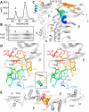
Crystal structure of the complex between the periplasmic domains of E. coli FtsB and FtsQ. (A) Size exclusion profile showing elution of the FtsQB complex. (B) SDS-PAGE of the FtsQB complex. The complex elutes as two peaks (see Fig. 5A and Fig. S6A for multiple angle light scattering [SEC-MALS] and analytic ultracentrifugation [AUC] data on the same complex) that both produced crystals and are most likely related to oligomerization or dimerization of FtsB. mAU, milli-arbitrary units. (C) Crystal structure of the complex determined to 2.6-Å resolution by molecular replacement. Crystallographic data are listed in Table 1. Only residues 64 to 87 of FtsB are resolved in the structure. FtsB forms a short helix, a connecting loop, and a β-sheet that aligns in an antiparallel orientation with the last strand of the β domain of FtsQ. (D) Stereo plot of FtsB 64–87 in stick representation, showing key residues involved in interactions with FtsQ and also internal contacts that are important for FtsB to adopt this particular structure. The structure is colored from the N terminus (blue) to the C terminus (red). The inset shows the same orientation as a ribbon plot. (E) In the crystals, FtsB forms a tight dimer that buries hydrophobic residues, including methionine 77. To be certain about the register of the amino acids of FtsB, we replaced M77 with selenomethionine (SeMet) and performed a single-wavelength anomalous diffraction (SAD) experiment (Table 1). The resulting phased anomalous difference density highlights the only methionine in FtsB in the correct location, validating our interpretation.
FIG 3
FIG 3
Validation of the crystal structure. (A) SPR experiments showing that FtsB mutants R72A and F84A display significantly compromised binding to FtsQ, as does FtsQ mutant Y248W to FtsB, which showed no binding. The crystal structure of the complex of FtsB and FtsQ implicates FtsB F84 and FtsQ Y248 in forming the binding interface. FtsB R72 is involved in a key salt bridge with FtsB E82, and its interruption seems to abrogate binding between FtsB and FtsQ as well. Note that these and other mutants were also investigated in vivo as described in the legends to Fig. 4 and Fig. S3 to S5 in the supplemental material. (B) SPR experiments investigating the role of the FtsB residues that were not resolved in the crystal structure, as only residues 64 to 87 were visible (residues 22 to 63 were not visible). FtsB binding to FtsQ requires only amino acids from residue 64 onwards until residue 87, and it is to be concluded that the remainder of the protein in the crystals is disordered. (C) Summary table quantifying the SPR data in panels A and B. (D) Corroborating the point that only FtsB residues 64 to 87 are needed for the interaction between FtsB and FtsQ, a fully synthetic peptide was produced (see Fig. S2), and its binding to FtsQ was investigated by fluorescence polarization, as the peptide also carried an FITC moiety at the N terminus. Fluorescence polarization (mP) is shown on the y axis. Fitting of the binding curve yielded a Kd of 9.5 µM, similar to the values obtained with recombinant FtsB 22–103 and 64–88 in panels B and C. (E) In fact, using a fully synthetic peptide of FtsB 22–87 (without FITC) produced crystals that, although in a different space group, show exactly the same structure and arrangement as observed before when adding the entire periplasmic domain of FtsB (22–103) to the FtsQ periplasmic domain. (F) Superposition of unbound E. coli FtsQ periplasmic domain (PDB ID 2VH1) (19) and our FtsQ structure in complex with FtsB. Overall, there are only small deviations, but Y248 dramatically changes its side chain conformer (and the entire loop 247–252 changes conformation slightly), as does W256.
FIG 4
FIG 4
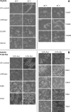
Mutating residues implicated in FtsB and FtsQ complex formation impairs cell division. (A) FtsQ mutants. The FtsQ temperature-sensitive E. coli strain LMC531 (40), harboring a plasmid for the expression of mutant SH8FtsQ was grown for 5 h at the permissive temperature (28°C) or at the nonpermissive temperature (42°C). Cells were analyzed by phase-contrast microscopy. EV, empty vector/plasmid. (B) FtsB mutants. The E. coli FtsB depletion strain NB946 (41), harboring a plasmid for the expression of mutant FtsB-HA (hemagglutinin-tagged FtsB) derivatives was grown under nondepleting conditions in the presence of 0.2% l-arabinose (Ara+) and under depleting conditions in the presence of 0.2% l-glucose (Glu+). Cells were analyzed by phase-contrast microscopy. EV, empty vector/plasmid. The mutated residues are highlighted in Fig. 2D. Bars, 10 µm.
FIG 5
FIG 5
Model of the periplasmic complex between E. coli FtsB and FtsQ. (A) Size exclusion chromatography with multiple angle light scattering (SEC-MALS) of the complex of FtsB and FtsQ. FtsB forms large oligomers on its own. FtsQ is monomeric alone, but together, FtsB and FtsQ most likely form a 2 + 2 complex. Analytic ultracentrifugation (AUC) was also used to investigate the same complex with very similar results (Fig. S6). Relative molecular weight (Mr) (in daltons) is shown on the left-hand y axis. Refractive index is shown on the right-hand y axis in arbitrary units (a.u.). (B) Residues 22 to 64 of FtsB were shown to not interact with FtsQ (Fig. 3B and C). A previous crystal structure (PDB ID 4IFF) (30) showed that residues 28 to 60 are able to form a coiled-coil arrangement with each other (although through artificial dimerization), and it is possible that this interaction leads to the dimerization of the FtsB and FtsQ complex into the observed 2 + 2 stoichiometry. Residues 22 to 64 within FtsB link its single transmembrane helix to the FtsQ-interacting domain in FtsB and also produce the putative dimer as shown by forming a homodimeric coiled coil. It was previously shown that a region around R75 in FtsQ links to FtsL (24). FtsQ is shown in surface representation with sequence conservation color coded (most conserved shown in blue and least conserved shown in red). It is clear from the plot that the FtsB binding region (residues 64 to 87) covers most of the highly conserved patch on the β domain of FtsQ.

Similar articles

Cited by

References

    1. den Blaauwen T, de Pedro MA, Nguyen-Distèche M, Ayala JA. 2008. Morphogenesis of rod-shaped sacculi. FEMS Microbiol Rev 32:321–344. doi:10.1111/j.1574-6976.2007.00090.x. - DOI - PubMed
    1. den Blaauwen T, Hamoen LW, Levin PA. 2017. The divisome at 25: the road ahead. Curr Opin Microbiol 36:85–94. doi:10.1016/j.mib.2017.01.007. - DOI - PMC - PubMed
    1. Du S, Lutkenhaus J. 2017. Assembly and activation of the Escherichia coli divisome. Mol Microbiol 105:177–187. doi:10.1111/mmi.13696. - DOI - PMC - PubMed
    1. Egan AJ, Vollmer W. 2013. The physiology of bacterial cell division. Ann N Y Acad Sci 1277:8–28. doi:10.1111/j.1749-6632.2012.06818.x. - DOI - PubMed
    1. Natale P, Pazos M, Vicente M. 2013. The Escherichia coli divisome: born to divide. Environ Microbiol 15:3169–3182. doi:10.1111/1462-2920.12227. - DOI - PubMed

Publication types

MeSH terms

LinkOut - more resources