. 2018 Nov;210(3):1011-1037.
doi: 10.1534/genetics.118.301421. Epub 2018 Sep 11.

Multiple Mechanisms Inactivate the LIN-41 RNA-Binding Protein To Ensure a Robust Oocyte-to-Embryo Transition in Caenorhabditis elegans

Affiliations

Multiple Mechanisms Inactivate the LIN-41 RNA-Binding Protein To Ensure a Robust Oocyte-to-Embryo Transition in Caenorhabditis elegans

Caroline A Spike et al. Genetics. 2018 Nov.

Abstract

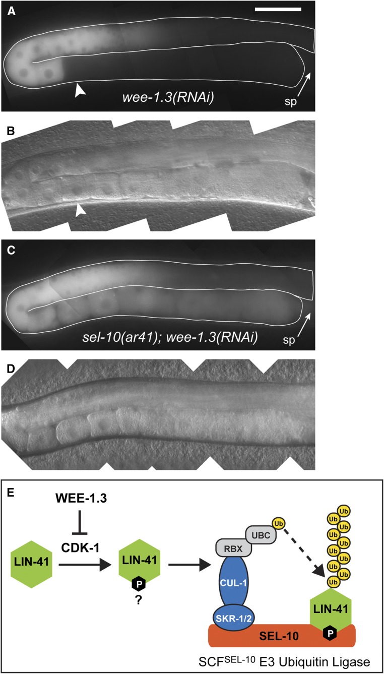
In the nematode Caenorhabditis elegans, the conserved LIN-41 RNA-binding protein is a translational repressor that coordinately controls oocyte growth and meiotic maturation. LIN-41 exerts these effects, at least in part, by preventing the premature activation of the cyclin-dependent kinase CDK-1 Here we investigate the mechanism by which LIN-41 is rapidly eliminated upon the onset of meiotic maturation. Elimination of LIN-41 requires the activities of CDK-1 and multiple SCF (Skp1, Cul1, and F-box protein)-type E3 ubiquitin ligase subunits, including the conserved substrate adaptor protein SEL-10/Fbw7/Cdc4, suggesting that LIN-41 is a target of ubiquitin-mediated protein degradation. Within the LIN-41 protein, two nonoverlapping regions, Deg-A and Deg-B, are individually necessary for LIN-41 degradation; both contain several potential phosphodegron sequences, and at least one of these sequences is required for LIN-41 degradation. Finally, Deg-A and Deg-B are sufficient, in combination, to mediate SEL-10-dependent degradation when transplanted into a different oocyte protein. Although LIN-41 is a potent inhibitor of protein translation and M phase entry, the failure to eliminate LIN-41 from early embryos does not result in the continued translational repression of LIN-41 oocyte messenger RNA targets. Based on these observations, we propose a model for the elimination of LIN-41 by the SEL-10 E3 ubiquitin ligase and suggest that LIN-41 is inactivated before it is degraded. Furthermore, we provide evidence that another RNA-binding protein, the GLD-1 tumor suppressor, is regulated similarly. Redundant mechanisms to extinguish translational repression by RNA-binding proteins may both control and provide robustness to irreversible developmental transitions, including meiotic maturation and the oocyte-to-embryo transition.

Keywords: RNA-binding proteins; oocyte meiotic maturation; oocyte-to-embryo transition; translational regulation; ubiquitin-mediated protein degradation.


Figures

Figure 1
Figure 1
GFP::LIN-41 is eliminated during the first meiotic division. (A and B) Composite GFP (A) and DIC (B) images of a lin-41(tn1541[gfp::tev::s-tag::lin-41]) adult hermaphrodite. GFP::LIN-41 is apparent in the middle and proximal regions of the germline (solid outline, A), with reduced levels in the −1 oocyte immediately adjacent to the spermatheca (sp). The positions of some embryos (dashed outlines, A) and oocytes are indicated relative to the spermatheca in B; a fertilized embryo in the spermatheca would be at the zero position. These labels and naming conventions are used throughout. 100 ms GFP exposures; Bar, 50 μm. (C–G) Time-lapse images of GFP::LIN-41 (white) and mCHERRY::HISTONE-labeled chromosomes (red) were acquired in a living lin-41(tn1541); itIs37[pie-1p::mCherry:::H2B::pie-1 3′UTR, unc-119(+)] adult hermaphrodite by confocal microscopy. Images are shown for select time points (t) prior to meiotic maturation (C, t= −4.5 min), at ovulation (D, t = 0 min), and during the first meiotic division (E, t= +4 min; F, t = +11.8 min; G, t= +16.9 min) as an individual oocyte (C, solid outline) progresses from the −1 to the +1 position and through the OET (D–G, dashed outlines). Bar, 50 μm. Movie S1, worm 1, shows the complete time-lapse series from which the still images were taken. (H) Five oocytes were imaged as they progressed from the −1 position through meiotic divisions; the relative amount of background-corrected GFP::LIN-41 with respect to distal oocytes is shown on the graph at each time point. Three of the oocytes were also imaged at earlier stages as they moved from a more distal location [−2 oocyte (red) or −3 oocyte (green) position] into the −1 oocyte position (blue), as indicated. Timing on the x-axis is relative to ovulation (t = 0). Bars indicate the SD for different meiotic events. Ana, anaphase; Met, metaphase; NEBD, nuclear envelope breakdown.
Figure 2
Figure 2
LIN-41 Deg domains are required for the elimination of GFP::LIN-41 upon the onset of meiotic maturation. (A) The exon–intron structure and deletion analysis of lin-41(tn1541). Colored boxes indicate exonic regions that encode GFP (green) or LIN-41 protein domains (see B). Deletions made in the context of lin-41(tn1541) are drawn as lines, labeled with a deletion-specific allele name, beneath the LIN-41-encoding exons and introns (exons labeled 1–15). GFP::LIN-41 can be detected in the germline of most deletion mutants (solid lines), with one exception (tn1628, dotted line). Deletions in red prevent the elimination of GFP::LIN-41 from early embryos. The vertical dashed lines delimit the beginning of Deg-A and the end of Deg-B, respectively. (B) The previously described [RING (yellow), B-box (gray), BBC (orange), Ig/filamin (purple), NHL (blue)] and newly identified [Deg (red)] protein domains of LIN-41. The vertical dashed line in B indicates the two parts of Deg-B, B1 and B2, which are individually removed in lin-41(tn1541tn1635) and lin-41(tn1541tn1622), respectively. The position of two lin-41 missense alleles (tn1767[T83A] and tn1487ts[D1125N]) generated in the endogenous lin-41 locus are indicated. (C) The amino acid sequences of Deg-A, Deg-B1, and Deg-B2. Many of the amino acids are serines and threonines (underlined) and some are potential targets of proline-directed serine/threonine [S/T] kinases (bold). The indicated alleles change an [S/T] residue to an alanine (colored and bold) in the context of lin-41(tn1541). The tn1645[T83A] mutation in Deg-A results in the persistence of GFP::LIN-41[T83A] in embryos (red), whereas the other changes do not (indicated in blue font). (D–G) GFP::LIN-41 is eliminated from the early embryos (dashed outlines) of lin-41(tn1541) (D, control) and lin-41(tn1541tn1630) (E, RING deleted) homozygous mutants but persists in the early embryos of lin-41(tn1541tn1638) (F, Deg-A deleted) and lin-41(tn1541tn1645) (G, LIN-41[T83A]) homozygous mutants. The position of the spermatheca (sp) is indicated, for reference. 100 ms GFP exposures; Bar, 20 μm. (H) The rate of ovulation is slightly reduced in mutants with a compromised LIN-41 Deg-A domain. Ovulation rate is expressed as the number of ovulations per gonad arm per hr and was measured in at least 25 day 2 adults. The three gfp-tagged alleles are in the top portion of the graph with green shading. Significance was determined using a one-way ANOVA test with Tukey’s post hoc test to compare the means; ** P < 0.01, *** P < 0.001, and **** P < 0.0001. itIs37[pie-1p::mCherry:::H2B::pie-1 3′UTR, unc-119(+)] was also present in each of the GFP::LIN-41-expressing strains; it is not expected to alter the ovulation rate.
Figure 3
Figure 3
LIN-41 degradation domains when implanted into mNG::OMA-2 promote its rapid elimination during meiosis. (A–C) The exon–intron structures of oma-2(cp145[mng::tev::3xflag::oma-2]), oma-2(tn1760[mng::tev::3xflag::deg-a::oma-2]), and oma-2(tn1764[mng::tev::3xflag::deg-a::deg-b::oma-2]). Boxes represent exonic regions that encode mNeonGreen (green), the tobacco etch virus cleavage site (TEV; dark gray), FLAG epitope tags (light gray), LIN-41 Deg-A and Deg-B domains (red), the likely TAF-4 interaction domain of OMA-2 (dark blue), two OMA-2 CCCH zinc fingers (white), and other OMA-2 coding sequences (cyan). The position of LIN-41 T83 within the LIN-41 Deg-A domain is indicated by an asterisk. (D–K) GFP (D–G) and DIC (H–K) images of oma-2(cp145) (D and H), oma-2(tn1760) (E and I), oma-2(tn1764) (F and J), and oma-2(tn1764) lon-3(e2175) sel-10(ar41) (G and K) one-cell embryos at pronuclear meeting (E and I), or just slightly later, as the pronuclei begin a counterclockwise rotation (D, F–G, H, J, and K) prior to nuclear envelope breakdown and the first mitotic division. Part of a −1 oocyte is visible in F and J, and is indicated for reference. 150 ms GFP exposures; Bar, 10 μm. (L–O) Time-lapse images of mNG::Deg-A,B::OMA-2 (white) and mCHERRY::HISTONE-labeled chromosomes (red) were acquired in a living oma-2(tn1764); itIs37[pie-1p::mCherry:::H2B::pie-1 3′UTR, unc-119(+)] adult hermaphrodite by confocal microscopy. Images are shown for select time points (t) at ovulation (L, t = 0 min), during the first (M, t = +5 min; N, t = +10.5 min) and second meiotic divisions (O, t = +24.5 min) as an embryo (dashed outline) progresses through both meiotic divisions. See Movie S3 for the complete time-lapse sequence. Bar, 50 μm. (P) A visual summary of the dynamic expression patterns of mNG::OMA-2 (cyan), GFP::LIN-41 (red), and mNG::Deg-A,B::OMA-2 (purple). Oocytes are to the left and embryos are to the right of the spermatheca (sp). Meiotic embryos (MI, MII) have completed their respective divisions.
Figure 4
Figure 4
Subunits of the SCFSEL-10 E3 ubiquitin ligase are required for the elimination of GFP::LIN-41 from early embryos. (A–E) Composite images of GFP::LIN-41 in adult rrf-1(pk1417) lin-41(tn1541) hermaphrodites fed control RNAi bacteria (A), and adult hermaphrodites with reduced SCFSEL-10 E3 ubiquitin ligase activity (B–E): lin-41(tn1541); skr-1(RNAi) (B), rrf-1(pk1417) lin-41(tn1541); cul-1(RNAi) (C), lin-41(tn1541); lon-3(e2175) sel-10(ar41) (D), and lin-41(tn1541); sel-10(ok1632) (E); 100 ms GFP exposures, brightened slightly (and equivalently) to better visualize embryonic GFP::LIN-41; Bar, 50 μm. (F–M) Images of two-cell embryos removed from the uterus of hermaphrodites were imaged for GFP (F–I) and DIC (J–M); the genotypes were as follows: lin-41(tn1541); lon-3(e2175) (F and J), lin-41(tn1541); lon-3(e2175) sel-10(ar41) (G and K), lin-41(tn1541) (H and L), and lin-41(tn1541); sel-10(ok1632) (I and M). Arrowheads indicate a few of the GFP::LIN-41 aggregates in the posterior blastomeres of sel-10 mutant embryos, which likely correspond to P granules; 300 ms GFP exposures; Bar, 10 μm. sp, spermatheca.
Figure 5
Figure 5
SEL-10 is required for the WEE-1.3–inhibited degradation of GFP::LIN-41. (A–D) Composite GFP (A and C) and DIC (B and D) images of lin-41(tn1541); lon-3(e2175); wee-1.3(RNAi) (A and B) and lin-41(tn1541); lon-3(e2175) sel-10(ar41); wee-1.3(RNAi) (C and D) animals. GFP::LIN-41 is prematurely eliminated from oocytes by wee-1.3(RNAi) (arrowhead), but persists in abnormal oocytes near the spermatheca (sp; arrow) in sel-10(ar41); wee-1.3(RNAi) animals (C and D), suggesting that SEL-10 is required for this process; 150 ms GFP exposures, brightened slightly; Bar, 50 μm. (E) A simple model for the elimination of LIN-41 (green) that incorporates the known molecular functions of WEE-1.3 kinase, cyclin-dependent kinase (CDK-1) and subunits of the SCFSEL-10 E3 ubiquitin ligase. In brief, we hypothesize that SEL-10 (orange) may recognize phosphorylated LIN-41 (green) and trigger its ubiquitin-mediated degradation in collaboration with the other SCF E3 ubiquitin ligase subunits, SKR-1/2 (blue), and CUL-1 (blue). CUL-1 orthologs bind RING finger proteins (RBX; gray), which recruit a ubiquitin-conjugating enzyme (UBC; gray) that catalyzes the transfer of ubiquitin (yellow) to protein substrates, such as LIN-41. Subsequent recruitment of polyubiquitinated substrates to the proteasome results in degradation (not shown). This model is consistent with the epistatic relationship between wee-1.3(RNAi) and sel-10(ar41) with respect to the elimination of GFP::LIN-41, but other models are also possible.
Figure 6
Figure 6
Persisting LIN-41 or LIN-41[T83A] does not strongly inhibit the expression of LIN-41 targets of translational repression in young embryos. (A–J) Young embryos express similar levels of SPN-4::GFP (A, B, G, and H), GFP::MEG-1 (C, D, I, and J) and mNG::ORC-1 (arrowhead in E and F) when ectopic LIN-41[T83A] [B, D, and F; lin-41(tn1767) mutant embryos], ectopic LIN-41 [H and J; sel-10(ar41) mutant embryos] or normal (undetectable) levels of LIN-41 (A, C, E, G, and I) are present. Exposures were 100 ms for SPN-4::GFP, 200 ms for GFP::MEG-1, and 600 ms for mNG::ORC-1; Bar, 10 μm. (K and L) Quantification of the intensity of SPN-4::GFP expression in wild type, lin-41(tn1767), and sel-10(ar41) genetic backgrounds. Statistical significance was evaluated using an unpaired t-test. The plots show the mean and SD. (K) Comparison of spn-4(tn1699) and lin-41(tn1767); spn-4(tn1699) one- and two-cell embryos; no significant differences were seen (n.s.). (L) Comparison of spn-4(tn1699); lon-3(e2175) and spn-4(tn1699); lon-3(e2175) sel-10(ar41) one- and two-cell embryos. Levels appeared to be slightly lower in the sel-10(ar41) two-cell embryos (P < 0.001). Note that the slightly reduced level of SPN-4::GFP in H relative to G accurately illustrates the very modest magnitude of this difference in expression.
Figure 7
Figure 7
GLD-1 persists at elevated levels in the oocytes of sel-10(ar41) mutants. (A and B) Composite images of GLD-1::GFP in gld-1(q485); lon-3(e2175); ozIs2[gld-1::gfp] (A) and gld-1(q485); lon-3(e2175) sel-10(ar41); ozIs2[gld-1::gfp] (B) adult hermaphrodites. GLD-1::GFP levels remain elevated in the proximal oocytes (e.g., −4 oocytes, arrowheads) of sel-10(ar41) animals (B) relative to controls (A); 17 ms GFP exposures, brightened slightly. (C) Slow-migrating forms of GLD-1 (red arrow) are more abundant in sel-10(lf) adult hermaphrodites than in sel-10(+) controls, where the fast-migrating form of GLD-1 (black arrow) predominates. (D and E) Composite images of GLD-1::GFP in fog-3(q470); lon-3(e2175); ozIs2[gld-1::gfp] (D) and fog-3(q470); lon-3(e2175) sel-10(ar41); ozIs2[gld-1::gfp] females (E). GLD-1::GFP levels are elevated in the proximal oocytes (e.g., −4 oocytes, arrowheads) of sel-10(ar41) females (B) relative to controls (A), although this is not as dramatic as in hermaphrodites. A somewhat longer GFP exposure (35 ms, brightened slightly) was needed than in A and B, likely due to the presence of endogenous GLD-1. (F) Quantification of the intensity of GFP::MEX-3 in the proximal oocytes of lon-3(e2175); mex-3(tn1753) and lon-3(e2175) sel-10(ar41); mex-3(tn1753) hermaphrodites at 25°. No significant differences were seen (n.s.). (G and H) Composite images of lon-3(e2175); pwIs116[rme-2p::rme-2::GFP::rme-2 3′UTR] (G) and lon-3(e2175) sel-10(ar41); pwIs116 [rme-2p::rme-2::GFP::rme-2 3′UTR] (H) hermaphrodites at 22°; 300 ms GFP exposures. Neither target of GLD-1 translational repression (MEX-3, RME-2) was strongly or even marginally reduced in expression in sel-10(ar41) oocytes. (I) Quantification of the intensity of mNG::OMA-2 in the proximal oocytes of oma-2(cp145) lon-3(e2175) and oma-2(cp145) lon-3(e2175) sel-10(ar41) hermaphrodites at 20°. Differences in expression were highly significant (**** P < 0.0001), but relatively modest in magnitude. For example, we measured a 37% reduction in average fluorescence in the −2 oocytes of sel-10(ar41) animals relative to the same oocytes in control animals. Statistical tests (F and I) employed an unpaired t-test. The plots show the mean and SD. All phenotypes were analyzed on the first day of adulthood. Bar, 50 μm (A, B, D, E, G, and H). sp, spermatheca.

Similar articles

Cited by

References

    1. Aeschimann F., Kumari P., Bartake H., Gaidatzis D., Xu L., et al. , 2017. LIN41 post-transcriptionally silences mRNAs by two distinct and position-dependent mechanisms. Mol. Cell 65: 476–489.e4. 10.1016/j.molcel.2016.12.010 - DOI - PubMed
    1. Allen A. K., Nesmith J. E., Golden A., 2014. An RNAi-based suppressor screen identifies interactors of the Myt1 ortholog of Caenorhabditis elegans. G3 (Bethesda) 4: 2329–2343. 10.1534/g3.114.013649 - DOI - PMC - PubMed
    1. Arribere J. A., Bell R. T., Fu B. X., Artiles K. L., Hartman P. S., et al. , 2014. Efficient marker-free recovery of custom genetic modifications with CRISPR/Cas9 in Caenorhabditis elegans. Genetics 198: 837–846. 10.1534/genetics.114.169730 - DOI - PMC - PubMed
    1. Arur S., 2017. Signaling-mediated regulation of meiotic prophase I and transition during oogenesis. Results Probl. Cell Differ. 59: 101–123. 10.1007/978-3-319-44820-6_4 - DOI - PMC - PubMed
    1. Avilés-Pagán E. E., Orr-Weaver T. L., 2018. Activating embryonic development in Drosophila. Semin. Cell Dev. Biol. (in press) 10.1016/j.semcdb.2018.02.019 - DOI - PMC - PubMed

Publication types

MeSH terms

Substances